[Preprint]. 2023 Jun 15:2023.02.01.526652.
doi: 10.1101/2023.02.01.526652.

Missense mutations in CRX homeodomain cause dominant retinopathies through two distinct mechanisms

Affiliations

Missense mutations in CRX homeodomain cause dominant retinopathies through two distinct mechanisms

Yiqiao Zheng et al. bioRxiv. .

Update in

Abstract

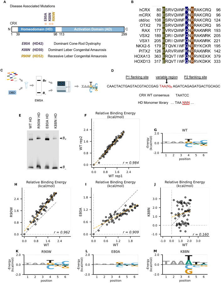
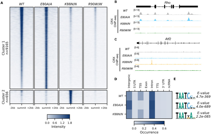
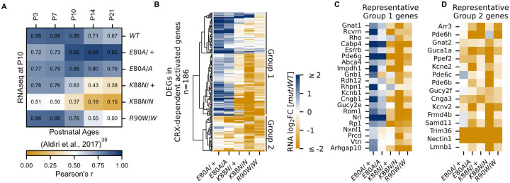
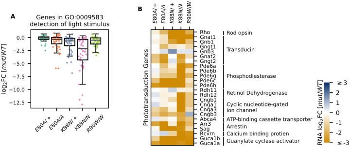
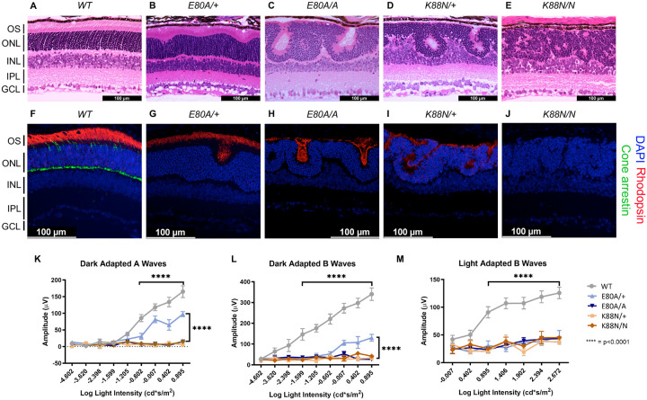
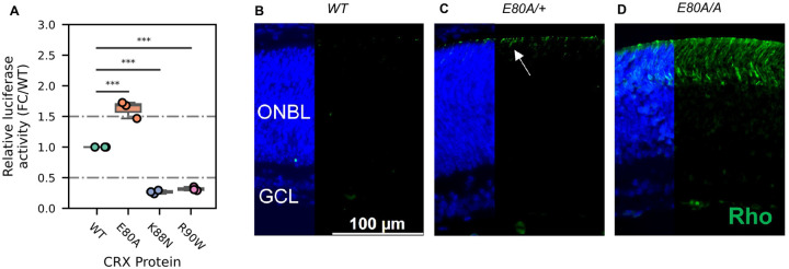
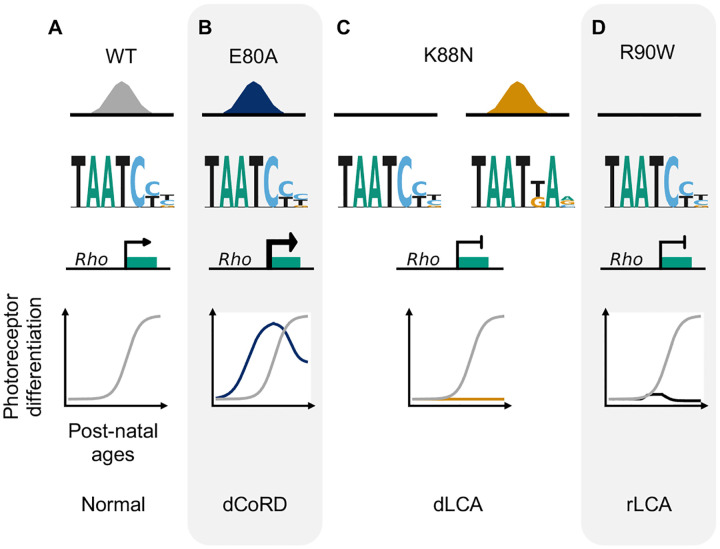
Homeodomain transcription factors (HD TFs) are instrumental to vertebrate development. Mutations in HD TFs have been linked to human diseases, but their pathogenic mechanisms remain elusive. Here we use Cone-Rod Homeobox (CRX) as a model to decipher the disease-causing mechanisms of two HD mutations, p.E80A and p.K88N, that produce severe dominant retinopathies. Through integrated analysis of molecular and functional evidence in vitro and in knock-in mouse models, we uncover two novel gain-of-function mechanisms: p.E80A increases CRX-mediated transactivation of canonical CRX target genes in developing photoreceptors; p.K88N alters CRX DNA-binding specificity resulting in binding at ectopic sites and severe perturbation of CRX target gene expression. Both mechanisms produce novel retinal morphological defects and hinder photoreceptor maturation distinct from loss-of-function models. This study reveals the distinct roles of E80 and K88 residues in CRX HD regulatory functions and emphasizes the importance of transcriptional precision in normal development.

Keywords: CRX mutations; DNA binding; Homeodomain; gene expression; inherited retinal disease; knock-in mouse models; photoreceptor development; transcription factor.


Conflict of interest statement

Declaration of interests The authors declare no competing interests.

Figures

Figure 1.
Figure 1.. Disease associated missense mutations altered CRX HD DNA binding specificity.
(A) Diagram of CRX functional domains: Homeodomain (HD) for DNA-binding and Activation Domain (AD) for target gene transactivation. The three missense mutations in this study are located at the C-terminus of CRX HD and associated with different retinal diseases in human. Number in the parenthesis denotes the CRX HD position of the corresponding mutated residue. (B) Alignments of HD recognition helix sequences for the indicated HD proteins for which HD missense mutations have been associated with inherited diseases. Accession numbers can be found in Supplementary Table S1. Missense variants in this study (highlighted) are located at highly conserved residues across species and different HD TFs. (C) Spec-seq experimental workflow (Methods). (D) Spec-seq library design of monomeric HD binding sites. (E) EMSA gel images of Spec-seq experiments with different CRX HD species. Bx: Bound. B-: Unbound. (F) Relative binding energy comparison from two different experiments with WT HD. (G) Binding energy model for WT CRX HD. (H-J) Relative binding energy comparison between WT HD and R90W HD (H), E80A HD (I), or K88N HD (J). Consensus sequence is defined to have relative binding energy of 0kT (TAATCC for WT, R90W and E80A, TAATTA for K88N). The identity line is represented in grey dash. The orange dashed line shows the best linear fit to the data. (K-M) Binding energy models for R90W HD (K), E80A HD (L), and K88N HD (M). Only sequence variants within two mismatches to the corresponding consensus sequences were used to generate binding models. Negative binding energy is plotted such that bases above the x-axis are preferred bases and bases below the x-axis are unfavorable bases. Constant bases (TAA) carried no information are drawn at arbitrary height in grey.
Figure 2.
Figure 2.. CRX E80A binds to WT sites while CRX K88N occupies novel genomic regions enriched for N88 HD motif in vivo.
(A) Enrichment heat map depicting CRX ChIP-seq normalized reads centered at all possible CRX peaks ± 2kb, sorted by binding intensity in WT samples. Clusters were defined by hierarchical clustering of CRX binding intensity matrix from all genotypes (STAR Methods). (B-C) Genome browser representations of ChIP-seq normalized reads for different CRX species in P14 WT and mutant mouse retinas at Rho and Atf2. (D) Enrichment heatmap showing fraction of CRX ChIP-seq peaks fall in different genomic environments. (E) Logo representations of de novo found short HD motifs under CRX ChIP-seq peaks in WT and mutant mouse retinas with DREME E-value on the right.
Figure 3.
Figure 3.. CRX-dependent activated genes affected in opposite directions in developing CrxE80A and CrxK88N mutant retinas
(A) Heat map showing sample-wise Pearson correlations of the expression of all CRX-dependent activated genes between P10 WT and HD mutant mouse retinas in this study (rows) with post-natal WT retinas from age P3 to P21 (columns, data from GSE87064). (B) Heat map showing the expression changes of DEGs in CRX-dependent activated gene set in HD mutant mouse retinas at P10. (C-D) Heat maps showing expression changes of selected photoreceptor genes from Group 1 and Group 2. Color scale identical to (B).
Figure 4.
Figure 4.. Photoreceptor genes important for phototransduction are down-regulated in all HD mutants
(A) Box plot showing that genes in the detection of light stimulus GO term were down-regulated and affected to various degrees in different adult (P21) HD mutant mouse retinas. (B) Heat map showing that expression of both cone and rod phototransduction genes were down-regulated in adult (P21) HD mutant mouse retinas. Annotation of rod and cone enrichment of each gene is in Supplementary Table S6. See Supplementary Figure S6 for the developmental expression dynamics of these genes.
Figure 5.
Figure 5.. Only CrxE80A/+ retinas maintain photoreceptor OS and residual rod ERG response
(A-E) Hematoxylin-eosin (H&E) staining of P21 retina sections show that photoreceptor OS layer is absent in all mutant retinas except CrxE80A/+. OS: outer segment; ONL: outer nuclear layer; INL: inner nuclear layer; IPL: inner plexiform layer; GCL: ganglion cell layer. Scale bar, 100μm. (F-J) Rhodopsin (RHO, red) immunostaining is present in CrxE80A/+, CrxE80A/A, and CrxK88N/+ retinas and absent in CrxK88N/N retina. Cone arrestin (mCAR, green) immunostaining is absent in all mutant retinas. Nuclei were visualized by DAPI staining (Blue). Scale bar, 100μm. (K-M) The electroretinogram responses (ERG) recorded from 1-month mice. Error bars represent the standard error of the mean (SEM, n ≥ 4). p-value: Two-way ANOVA and Tukey’s multiple comparisons. ****: p ≤ 0.0001. ns: >0.05.
Figure 6.
Figure 6.. CRX E80A hyperactivity underlies precocious photoreceptor differentiation in CrxE80A retinas.
(A) Boxplot showing luciferase reporter activities of different CRX variants. P-values for one-way ANOVA with Turkey honestly significant difference (HSD) test are indicated. p-value: ****: ≤0.0001, ***: ≤0.001, ns: >0.05. (B-D) Rhodopsin (RHO, green) immunostaining is absent in P3 WT retina but detected in CrxE80A/+ and CrxE80A/A retinas. Nuclei are visualized by DAPI staining (Blue). Arrow indicates the sporadic RHO staining in CrxE80A/+ sample. ONBL: outer neuroblast layer; GCL: ganglion cell layer. Scale bar, 100μm.
Figure 7.
Figure 7.
Missense mutations in CRX HD affect photoreceptor gene expression and leads to distinct retinal disease phenotypes through gain- and loss-of-function mechanisms.

References

    1. Mark M., Rijli F.M. & Chambon P. Homeobox Genes in Embryogenesis and Pathogenesis. Pediatric Research 42, 421–429 (1997). - PubMed
    1. Lewis E.B. A gene complex controlling segmentation in Drosophila. Nature 276, 565–570 (1978). - PubMed
    1. Leung R.F. et al. Genetic Regulation of Vertebrate Forebrain Development by Homeobox Genes. Frontiers in Neuroscience 16(2022). - PMC - PubMed
    1. Chi Y.-I. Homeodomain revisited: a lesson from disease-causing mutations. Human Genetics 116, 433–444 (2005). - PMC - PubMed
    1. Zagozewski J.L., Zhang Q., Pinto V.I., Wigle J.T. & Eisenstat D.D. The role of homeobox genes in retinal development and disease. Developmental Biology 393, 195–208 (2014). - PubMed

Publication types