

Diagnostic Contrast-Enhanced Mammography Performed Immediately Prior to Same-Day Biopsy: An Analysis of Index Lesion Enhancement Compared to Histopathology and Follow-up in Patients With Suspicious Ultrasound Findings
- PMID: 36778652
- PMCID: PMC9901423
- DOI: 10.1093/jbi/wbac081
Diagnostic Contrast-Enhanced Mammography Performed Immediately Prior to Same-Day Biopsy: An Analysis of Index Lesion Enhancement Compared to Histopathology and Follow-up in Patients With Suspicious Ultrasound Findings
Abstract
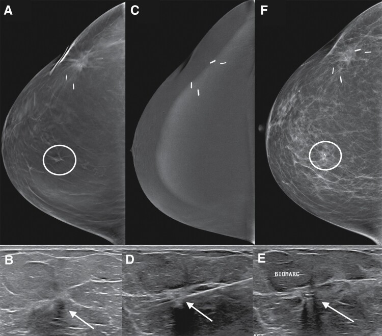
Objective: To measure the diagnostic performance of contrast-enhanced mammography (CEM) for the index lesion when it is performed the same day prior to biopsy in patients with suspicious findings at US.
Methods: This IRB-approved retrospective study compared radiologist original reports of the presence or absence of index lesion enhancement on CEM to biopsy results and follow-up. The most suspicious lesion or the larger of equally suspicious lesions recommended for biopsy by US after a diagnostic workup including mammography was considered the index lesion. CEM exams were performed the same day, immediately prior to the scheduled biopsy, as requested by the radiologist recommending the biopsy. Numeric variables were summarized with means and standard deviations, or medians and the minimum and maximum, where appropriate.
Results: Biopsy demonstrated cancer in 64.7% (200/309) of index lesions. Of these, 197/200 demonstrated enhancement for a sensitivity of 98.5% (95% CI: 95.7%-99.7%) (197/200) and the negative predictive value of CEM for non-enhancing index lesions was 95.1% (58/61; 95% CI: 86.1%-98.4%). The three false negative exams were two grade 1 ER+ HER2- invasive ductal cancers that were 6 mm and 7 mm in size, and a 3-mm grade 2 ductal carcinoma in situ in a complex cystic and solid mass. False positive exams made up 20.6% (51/248) of the positive exams.
Conclusion: Diagnostic CEM showed high sensitivity and specificity for cancer in lesions with suspicious US findings. CEM may reduce the need for some biopsies, and negative CEM may support a true negative biopsy result.
Keywords: biopsy; breast cancer; contrast-enhanced; mammography; ultrasound.
© Society of Breast Imaging 2023. All rights reserved. For permissions, please e-mail: journals.permissions@oup.com.
Figures
References
-
- Fallenberg E, Dromain C, Diekmann F, et al. Contrast-enhanced spectral mammography versus MRI: initial results in the detection of breast cancer and assessment of tumour size. Eur Radiol 2014;24(1):256–264. - PubMed
-
- Chou C, Lewin J, Chiang C, et al. Clinical evaluation of contrast-enhanced digital mammography and contrast enhanced tomosynthesis—comparison to contrast-enhanced breast MRI. Eur J Radiol 2015;84(12):2501–2508. - PubMed
-
- Li L, Roth R, Germaine P, et al. Contrast-enhanced spectral mammography (CESM) versus breast magnetic resonance imaging (MRI): a retrospective comparison in 66 breast lesions. Diagn Interv Imaging 2017;98(2):113–123. - PubMed
Grants and funding
LinkOut - more resources
Full Text Sources
Research Materials
Miscellaneous