. 2023 Jan 27:14:1080369.
doi: 10.3389/fmicb.2023.1080369. eCollection 2023.

Mycobacterial protein PE_PGRS30 induces macrophage apoptosis through prohibitin 2 mitochondrial function interference

Affiliations

Mycobacterial protein PE_PGRS30 induces macrophage apoptosis through prohibitin 2 mitochondrial function interference

Kazunori Matsumura et al. Front Microbiol. .

Abstract

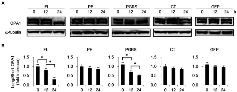
PE_PGRS30 belongs to the PE_PGRS protein family and is characterized by a conserved Pro-Glu (PE) domain and a typically polymorphic GC-rich sequence (PGRS) domain. PE_PGRS30 is a virulence factor of Mycobacterium tuberculosis that induces macrophage cell death. We found that RAW264.7 cells and murine alveolar macrophages underwent apoptosis in response to PE_PGRS30. The host protein prohibitin 2 (PHB2) was identified as a target molecule. PE_PGRS30 and PHB2 interact via the PGRS domain and mitochondrial targeting sequence, respectively. PHB2 overexpression reduced macrophage apoptosis in response to PE_PGRS30. PE_PGRS30 co-localized with PHB2, not in mitochondria, but in lysosomes. The maintenance of mitochondrial structure by PHB2 was impaired in response to the PGRS domain. These results indicated that PE_PGRS30 reduces PHB2 in mitochondria, resulting in mitochondrial dysfunction and cellular apoptosis.

Keywords: PE_PGRS30; PHB2; apoptosis; macrophages; mitochondria; tuberculosis.


Conflict of interest statement

The authors declare that the research was conducted in the absence of any commercial or financial relationships that could be construed as a potential conflict of interest.

Figures

Figure 1
Figure 1
PE_PGRS30-expressing cells undergo apoptosis. (A) Status of cells expressing PE_PGRS protein. Numbers of cells expressing PE_PGRS protein after transfection (upper panel). Numbers of cells expressing PE_PGRS30-Myc, PE_PGRS33-Myc and PE_PGRS62-Myc proteins were counted at the indicated time points. Proportion of PE_PGRS-expressing cells with condensed nuclei (middle panel). Cells were stained with Hoechst 33342 at the indicated time points. Numbers of PE_PGRS-expressing cells with condensed nuclei were counted and are presented as percentage against total PE_PGRS-expressing cell numbers. Proportion of PE_PGRS-expressing cells with dissipated mitochondria (lower panel). Cells were stained with MitoTracker Red CMXRos at the indicated time points. Numbers of PE_PGRS-expressing cells with dissipated mitochondria (mt) were counted and are presented as percentage against total PE_PGRS-expressing cell numbers. (B) Proportions of PE_PGRS-expressing cells with active caspase-3. Cells were stained with anti-active caspase-3 antibody 24 h after transfection. Numbers of PE_PGRS expressing cells with active caspase-3 (CASP3) were counted and are presented as percentages against total PE_PGRS-expressing cell numbers. (C) Flow cytometric analysis of Annexin V+ cells in PE_PGRS-expressing cells. RAW 264.7 cells were transfected with GFP-PE_PGRS30, GFP-PE_PGRS33, GFP-PE_PGRS62, and GFP-expressing vectors. After 24 h, cells were stained with Annexin V-PE/7-AAD. Representative plots of GFP+ cells are presented. (D) Numbers of Annexin V+ cells are presented as percentages against total GFP-PE_PGRS30-, GFP-PE_PGRS33-, GFP-PE_PGRS62, and GFP-expressing cells. (A,B) RAW264.7 cells were transfected with PE_PGRS30, 33 and 62-Myc expression vectors. Cells were stained with anti-Myc-tag antibody at the indicated time points. Images were acquired by confocal microscopy. Means ± SD are shown. *p < 0.05 using one-way ANOVA with Tukey’s multiple comparisons. Data are representative of three independent experiments. More than 100 cells expressing PE_PGRS proteins were counted in each experiment.
Figure 2
Figure 2
PE_PGRS30 binds to PHB2. (A) Host cellular proteins bound to PE_PGRS30. GST-PE_PGRS30 fusion protein or control GST were coupled to beads and incubated with whole cell lysates prepared from RAW264.7 cells. Beads (left 2 lanes) or cell-derived proteins bound to beads (lysates, right 2 lanes) were separated by SDS-PAGE and stained with Coomassie Brilliant Blue. A pulled-down protein (arrow heads) with molecular mass of 33 k Da was excised from the gel and subjected to mass spectrometry (nano-LC–MS/MS), and identified as PHB2 (association number, NP_031557). (B) Pull-down assay. GST-PE_PGRS30-or GST-coupled beads were mixed with whole cell lysates of RAW264.7 cells. The lysate (Lys), beads, and the pulled-down proteins were immunoblotted with anti-GST and anti-PHB2 antibodies. (C) RAW264.7 cells were transfected with PE_PGRS30-Myc and control GFP expression vector as in Figure 1A, and the lysates were immunoprecipitated with anti-PHB2 antibody, then immunoblotted with anti-Myc-tag or anti-GFP antibodies. (D,E) Schematic representation of PE_PGRS30 mutants used in this study. PE, PGRS or CT domain fused with GST at the N-terminus were coupled to beads, then mixed with recombinant His-PHB2 in 0.1% HNTG buffer. Proteins pulled-down with the beads were separated and immunoblotted with anti-GST and anti-PHB2 antibodies. (F,G) Schematic representation of deletion mutants of PHB2. N-terminal region (amino acids 1–50) or C-terminus region (amino acids 51–299) fused with a His-tag at the N-terminus were generated and the recombinant proteins purified, mixed with GST-PE_PGRS30 in 0.1% HNTG buffer, and the bound pulled-down proteins were immunoblotted with anti-His and anti-GST antibodies. (A–G) Data are representative of two independent experiments.
Figure 3
Figure 3
PHB2 overexpression reduces apoptosis of PE_PGRS30-expressing cells. (A) Localization of overexpressed PHB2 in cells. RAW264.7 cells were transfected with PHB2-FLAG expression vector. Cells were stained with Hoechst 33342 (for nuclei), MitoTracker Red CMXRos (for mitochondria) and antibodies (for PHB2) at 24 h post-transfection. Anti-PHB2 or anti-FLAG-tag antibodies were used to detect endogenous PHB2 or overexpressed PHB2-FLAG, respectively. Scale bar, 10 μm. (B) Numbers of PE_PGRS30-expressing cells in PHB2-overexpressing cells. PE_PGRS30-Myc expression vector and PHB2-overexpression or control GFP-expression vector were transfected into RAW264.7 cells simultaneously. Numbers of PE_PGRS30-expresing cells in PHB2-overexpressing cells or in control GFP-expressing cells were counted at the indicated time points. (C) Proportions of PE_PGRS30-expressing cells with condensed nuclei in PHB2-overexpressing cells. Cells were stained with Hoechst 33342 at the indicated time points. Numbers of PE_PGRS30-expressing cells with condensed nuclei in PHB2-overexpressing cells were counted and are presented as percentages against total numbers of PE_PGRS30-expressing cells in PHB2-overexpressing cells. (D) LDH levels in culture supernatants of GFP-and PE_PGRS30-expressing cells, and PHB2-and PE_PGRS30-expressing cells. (A–D) Data are representative of three independent experiments. (B–D) Means ± SD are shown. *p < 0.05 using one-way ANOVA with Tukey’s multiple comparisons. More than 100 cells overexpressing PHB2 were counted in each experiment.
Figure 4
Figure 4
PE_PGRS30 induces apoptosis via PGRS domain. (A) Macrophages undergo apoptosis in response to recombinant PE_PGRS30 proteins. RAW264.7 cells were treated with 10 μg/ml of full-length (FL), PE, PGRS, CT domains and control GFP for 24 h. Cells were stained with Annexin V and analyzed by flow cytometry. Numbers of Annexin V+ cells are presented as percentages against total cells. More than 104 cells were analyzed in each experiment. (B) LDH release by cells treated with PE_PGRS30 proteins. LDH in culture supernatants at 24 h post treatment were detected. (C) Co-localization of PE_PGRS30 with PHB2, mitochondria and LAMP1 in macrophages. RAW264.7 cells were treated as in (A). Cells were stained with a combination of anti-Myc-tag antibody and anti-PHB2 antibody or MitoTracker Red CMXRos or anti-LAMP1 antibodies 16 h after treatment. Arrowheads denote co-localization with PE_PGRS30 and detected molecules. Scale bar, 10 μm. (D–F) Quantification of co-localization of PE_PGRS30 with PHB2 (D), mitochondria (E), and LAMP1 (F) in macrophages. More than 100 cells were analyzed in each experiment. (A–F) Data are representative of three independent experiments. (A,B,D–F) Means ± SD are shown. *p< 0.05 using one-way ANOVA with Tukey’s multiple comparisons.
Figure 5
Figure 5
PHB2 function in mitochondria is impaired in response to PGRS domain. (A) Long and short isoforms of OPA1 in RAW264.7 cells treated with 10 μg/ml FL, PE, PGRS, CT domain or control boiled GFP at the indicated time points. Long and short isoforms of OPA1 in whole cell lysates were detected by immunoblotting. α-tubulin served as loading control. (B) Quantification of long isoform of OPA1 protein in comparison with that of short form OPA1. Ratios of long-to short-isoforms of OPA1 protein measured by band densities are presented. Means ± SD are shown (n = 3). *p < 0.05 using one-way ANOVA with Tukey’s multiple comparisons. (A,B) Data are representative of three independent experiments.
Figure 6
Figure 6
Murine alveolar macrophages undergo apoptosis in response to PE_PGRS30. Bronchoalveolar lavage (BAL) cells were prepared and treated with 10 μg/ml recombinant PE_PGRS30 and control GFP protein for 24 h. (A) Representative plots of Siglec-F+CD11c+ cells (AMs, upper panel) and annexin V+ cells in AMs (lower panel) are presented. (B) Numbers of annexin V+ cells are presented as percentages of total AMs. More than 104 BAL cells were analyzed in each experiment. Data are representative of three independent experiments. Means ± SD are shown. *p < 0.05 using Student’s t-test.

Similar articles

Cited by

References

    1. Abo-Kadoum M. A., Assad M., Ali M. K., Uae M., Nzaou S. A. E., Gong Z., et al. . (2021). Mycobacterium tuberculosis PE17 (Rv1646) promotes host cell apoptosis via host chromatin remodeling mediated by reduced H3K9me3 occupancy. Microb. Pathog. 159:105147. doi: 10.1016/j.micpath.2021.105147, PMID: - DOI - PubMed
    1. Adebayo M., Singh S., Singh A. P., Dasgupta S. (2021). Mitochondrial fusion and fission: the fine-tune balance for cellular homeostasis. FASEB J. 35:e21620. doi: 10.1096/fj.202100067R, PMID: - DOI - PMC - PubMed
    1. Arnett E., Weaver A. M., Woodyard K. C., Montoya M. J., Li M., Hoang K. V., et al. . (2018). PPARγ is critical for Mycobacterium tuberculosis induction of Mcl-1 and limitation of human macrophage apoptosis. PLoS Pathog. 14:e1007100. doi: 10.1371/journal.ppat.1007100, PMID: - DOI - PMC - PubMed
    1. Balaji K. N., Goyal G., Narayana Y., Srinivas M., Chaturvedi R., Mohammad S. (2007). Apoptosis triggered by Rv1818c, a PE family gene from Mycobacterium tuberculosis is regulated by mitochondrial intermediates in T cells. Microbes Infect. 9, 271–281. doi: 10.1016/j.micinf.2006.11.013, PMID: - DOI - PubMed
    1. Basu S., Pathak S. K., Banerjee A., Pathak S., Bhattacharyya A., Yang Z., et al. . (2007). Execution of macrophage apoptosis by PE_PGRS33 of Mycobacterium tuberculosis is mediated by toll-like receptor 2-dependent release of tumor necrosis factor-alpha. J. Biol. Chem. 282, 1039–1050. doi: 10.1074/jbc.M604379200, PMID: - DOI - PubMed