. 2023 Jan 26:17:1112930.
doi: 10.3389/fncel.2023.1112930. eCollection 2023.

Dual role of Apolipoprotein D as long-term instructive factor and acute signal conditioning microglial secretory and phagocytic responses

Affiliations

Dual role of Apolipoprotein D as long-term instructive factor and acute signal conditioning microglial secretory and phagocytic responses

Miriam Corraliza-Gomez et al. Front Cell Neurosci. .

Abstract

Microglial cells are recognized as very dynamic brain cells, screening the environment and sensitive to signals from all other cell types in health and disease. Apolipoprotein D (ApoD), a lipid-binding protein of the Lipocalin family, is required for nervous system optimal function and proper development and maintenance of key neural structures. ApoD has a cell and state-dependent expression in the healthy nervous system, and increases its expression upon aging, damage or neurodegeneration. An extensive overlap exists between processes where ApoD is involved and those where microglia have an active role. However, no study has analyzed the role of ApoD in microglial responses. In this work, we test the hypothesis that ApoD, as an extracellular signal, participates in the intercellular crosstalk sensed by microglia and impacts their responses upon physiological aging or damaging conditions. We find that a significant proportion of ApoD-dependent aging transcriptome are microglia-specific genes, and show that lack of ApoD in vivo dysregulates microglial density in mouse hippocampus in an age-dependent manner. Murine BV2 and primary microglia do not express ApoD, but it can be internalized and targeted to lysosomes, where unlike other cell types it is transiently present. Cytokine secretion profiles and myelin phagocytosis reveal that ApoD has both long-term pre-conditioning effects on microglia as well as acute effects on these microglial immune functions, without significant modification of cell survival. ApoD-triggered cytokine signatures are stimuli (paraquat vs. Aβ oligomers) and sex-dependent. Acute exposure to ApoD induces microglia to switch from their resting state to a secretory and less phagocytic phenotype, while long-term absence of ApoD leads to attenuated cytokine induction and increased myelin uptake, supporting a role for ApoD as priming or immune training factor. This knowledge should help to advance our understanding of the complex responses of microglia during aging and neurodegeneration, where signals received along our lifespan are combined with damage-triggered acute signals, conditioning both beneficial roles and limitations of microglial functions.

Keywords: acute response; amyloid-beta endocytosis; astrocyte-microglia crosstalk; cytokine secretion; immune memory; membrane-binding protein; microglia; myelin phagocytosis.


Conflict of interest statement

The authors declare that the research was conducted in the absence of any commercial or financial relationships that could be construed as a potential conflict of interest.

Figures
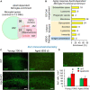
FIGURE 1
FIGURE 1
The expression of Apolipoprotein D (ApoD) alters the microglial response to aging. (A,B) Bioinformatics analysis of microglia-specific gene enrichment within sets dependent on ApoD expression. Microglia-specific genes are enriched in the set of genes that depend on ApoD for their response to aging in the brain. P-values are calculated with a modified Fisher’s exact test (A). Functional enrichment analysis of the set of genes identified in (A), carried out with the DAVID platform (B). (C) Young adult mice lacking ApoD expression increase their number of microglial cells in the hippocampus, a genotype-dependent response that disappears upon physiological aging. Degenerated regions in brain tissue are evident in aged samples. Representative images are shown. (D) Microglial cells labeled by Iba1 antibody and graphical quantification of Iba1-positive cells in young (100 days) (two serial sections; n = 3/genotype) and aged (655 days) (two serial sections; n = 4/genotype) samples. Asterisk points to significant differences between young and aged groups (Student’s t-test). Calibration bars: 100 μm (C); 20 μm (D).
FIGURE 2
FIGURE 2
Microglial cells do not express ApoD but are able to incorporate exogenous ApoD in intracellular compartments. (A) The mouse microglial cell line BV2 is not labeled by a mouse ApoD antibody, while the astrocytic cell line IMA2.1 shows a characteristic ApoD punctate pattern. (B) RT-PCR reveals the absence of ApoD mRNA transcripts in both primary mouse microglia and BV2. PQ 75 μM for 3 h does not induce ApoD expression in BV2 cells. The gene rpl18 was used as a normalizer. RT#1&2 are two separate RT reactions from independent primary cultures. Mouse sciatic nerve expression is used as positive control. An unrelated template is used as negative control. (C) Exogenously added human ApoD (hApoD; 10 nM) is internalized to intracellular compartments of BV2 cells after 3 h exposure. Calibration bars: 20 μm.
FIGURE 3
FIGURE 3
Pre-exposure to purified human ApoD does not rescue BV2 microglial cells from PQ or Aβ-induced cell death. Cell viability analysis of BV2 cells by MTT assay upon PQ (A,B) or Aβ (C,D) oligomers exposure with or without pre-exposure to ApoD. Treatment started after adaptation of cells to serum starvation (5 - 1 - 0% for 24 h experiments; 5 - 0.2 - 0% for 3 h experiments). Addition of ApoD (50 nM) to the culture medium exerts a mild pro-survival effect on BV2 cells in control conditions but is not sustained with increasing doses of PQ or Aβ. All experiments were performed in triplicates. Asterisks point to significant differences in comparison with control conditions evaluated by ANOVA followed by Holm-Sidak pairwise comparisons.
FIGURE 4
FIGURE 4
Cytokine secretion profiles of male primary microglia in control conditions, PQ-induced OS or exposure to Aβ-oligomers. Luminex multiplex assay is used on primary microglia culture media after 18 h incubation under different conditions (control, 1 μM Aβ oligomers, or 25 μM PQ). Concentration of each cytokine secreted by WT microglia (shades of green color), and ApoD-KO microglia (shades of red colors) are represented. Light colors (light green or pink) represent values obtained without exposure to ApoD, and dark colors (dark green or red) represent values obtained when stimuli were preceded by the addition of purified human ApoD (50 nM) to the culture medium. ApoD is maintained during the treatment period. Each profile represents 2–3 technical replicas of conditioned media produced by two independent primary cultures/genotype/sex. Bars represent average ± SEM. Asterisks point to significant differences among groups within each graph, and # point to differences between conditions, with or without ApoD acute exposure, evaluated by ANOVA followed by Holm-Sidak pairwise comparisons.
FIGURE 5
FIGURE 5
Microglial myelin phagocytosis and degradation depends both on myelin ApoD genotype and exogenous ApoD addition. (A) Representative images of differential interference contrast (DIC), DAPI and DiI fluorescence showing that BV2 microglial cells are able to phagocytose DiI-labeled myelin. The right panel shows an orthogonal view derived from a Z-stack to demonstrate the internalized DiI-labeled particles. (B) Temporal pattern of DiI-labeled particle incorporation into BV2 cells exposed to WT or ApoD-KO myelin extracts. Total DiI intensity/number of cells is measured (N = 3 independent experiments, 6–16 pictures/time point/genotype). Based on these experiments we selected a 60 min incubation time for further analysis. (C) Myelin genotype affects the number of phagocytosed myelin particles, their total intensity, the percent area occupied by myelin within the cell, and the mean particle area after 60 min exposure of BV2 microglia to DiI-labeled myelin. A total 398–413 individual cells were measured (N = 3 independent experiments, 4 pictures/experimental condition). Each dot in box plots represents the average value/picture. ApoD-KO myelin shows increased values in the four variables analyzed. The addition of exogenous ApoD (50 nM) 3 h before exposure to myelin reduces these values in an ApoD genotype-dependent manner. (D) Myelin degradation was assayed by immunoblot against Mbp normalized to β-tubulin (N = 3 independent experiments). Four major bands specifically detected by the anti-Mbp antibody were quantified and added to measure the time-course of MBP degradation. Pre-treatment of BV2 cells with exogenous ApoD slows down myelin degradation independently of myelin ApoD genotype. Statistically significant differences were evaluated with ANOVA (two-way in panels B,C; three-way in panel D) followed by Holm-Sidak pairwise comparisons. # indicate differences due to myelin ApoD genotype (p < 0.001). Asterisks indicate differences due to ApoD pre-exposure within each myelin type (p < 0.05). Calibration bars: 10 μm (A, left panels); 20 μm (A, right panel).
FIGURE 6
FIGURE 6
ApoD interacts with microglial cell membranes and is internalized to the late-endosome-lysosomal compartment. (A) Human ApoD interacts with BV2 cell membranes in a dose dependent manner. Equivalent volumes of membrane-bound and unbound (supernatant; Sup) protein extracts were analyzed by immunoblot. The integral plasma membrane protein PMCA is used as positive control. (B) Internalization of exogenous ApoD in BV2 is time and PQ-dependent. (C) After 3 h exposure to 50 nM ApoD, the internalized ApoD steadily decreases with time in BV2 cells. (D) Exogenously added ApoD traffics to the late-endosome-lysosomal compartment of primary mouse microglia in control conditions, but with a lower co-localization index than that of primary astrocytes. N = 2 independent experiments (A) or three independent experiments (B—D). Co-localization index in (D) measured in z-stacks from 12 microglial cells and 8 astrocytes. Asterisks point to significant differences. Calibration bars (B): 20 μm; (C): 50 μm; (D): 10 μm.

References

    1. Askew K., Li K., Olmos-Alonso A., Garcia-Moreno F., Liang Y., Richardson P., et al. (2017). Coupled proliferation and apoptosis maintain the rapid turnover of microglia in the adult brain. Cell Rep. 18 391–405. 10.1016/j.celrep.2016.12.041 - DOI - PMC - PubMed
    1. Bajo-Grañeras R., Ganfornina M. D., Martín-Tejedor E., Sanchez D. (2011a). Apolipoprotein D mediates autocrine protection of astrocytes and controls their reactivity level, contributing to the functional maintenance of paraquat-challenged dopaminergic systems. Glia 59 1551–1566. 10.1002/glia.21200 - DOI - PubMed
    1. Bajo-Grañeras R., Sanchez D., Gutierrez G., González C., Do Carmo S., Rassart E., et al. (2011b). Apolipoprotein D alters the early transcriptional response to oxidative stress in the adult cerebellum. J. Neurochem. 117 949–960. 10.1111/j.1471-4159.2011.07266.x - DOI - PubMed
    1. Bhusal A., Rahman M. H., Lee W.-H., Lee I.-K., Suk K. (2021). Satellite glia as a critical component of diabetic neuropathy: Role of lipocalin-2 and pyruvate dehydrogenase kinase-2 axis in the dorsal root ganglion. Glia 69 971–996. 10.1002/glia.23942 - DOI - PubMed
    1. Blasi E., Barluzzi R., Bocchini V., Mazzolla R., Bistoni F. (1990). Immortalization of murine microglial cells by a v-raf/v-myc carrying retrovirus. J. Neuroimmunol. 27 229–237. 10.1016/0165-5728(90)90073-v - DOI - PubMed