

Electron microscopy analysis of astrocyte-synapse interactions shows altered dynamics in an Alzheimer's disease mouse model
- PMID: 36779013
- PMCID: PMC9908992
- DOI: 10.3389/fncel.2023.1085690
Electron microscopy analysis of astrocyte-synapse interactions shows altered dynamics in an Alzheimer's disease mouse model
Abstract
Introduction: Astrocyte-synapse bi-directional communication is required for neuronal development and synaptic plasticity. Astrocytes structurally interact with synapses using their distal processes also known as leaflets or perisynaptic astrocytic processes (PAPs). We recently showed that these PAPs are retracted from hippocampal synapses, and involved in the consolidation of fear memory. However, whether astrocytic synaptic coverage is affected when memory is impaired is unknown.
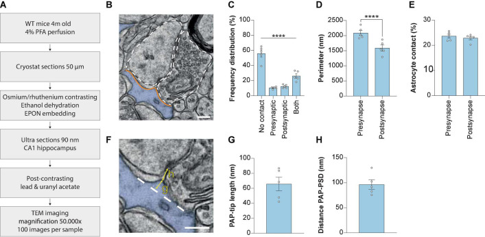
Methods: Here, we describe in detail an electron microscopy method that makes use of a large number of 2D images to investigate structural astrocyte-synapse interaction in paraformaldehyde fixed brain tissue of mice.
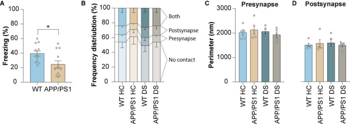
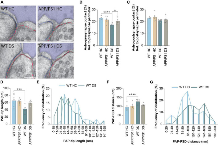
Results and discussion: We show that fear memory-induced synaptic activation reduces the interaction between the PAPs and the presynapse, but not the postsynapse, accompanied by retraction of the PAP tip from the synaptic cleft. Interestingly, this retraction is absent in the APP/PS1 mouse model of Alzheimer's disease, supporting the concept that alterations in astrocyte-synapse coverage contribute to memory processing.
Keywords: APP/PS1; glia; leaflet; memory consolidation; perisynaptic astrocytic processes; synapse; tripartite synapse.
Copyright © 2023 Kater, Badia-Soteras, van Weering, Smit and Verheijen.
Conflict of interest statement
The authors declare that the research was conducted in the absence of any commercial or financial relationships that could be construed as a potential conflict of interest.
Figures
Similar articles
-
Retraction of Astrocyte Leaflets From the Synapse Enhances Fear Memory.Biol Psychiatry. 2023 Aug 1;94(3):226-238. doi: 10.1016/j.biopsych.2022.10.013. Epub 2022 Oct 29. Biol Psychiatry. 2023. PMID: 36702661
-
Synapses lacking astrocyte appear in the amygdala during consolidation of Pavlovian threat conditioning.J Comp Neurol. 2014 Jun 15;522(9):2152-63. doi: 10.1002/cne.23523. J Comp Neurol. 2014. PMID: 24338694 Free PMC article.
-
Necl2/3-mediated mechanism for tripartite synapse formation.Development. 2023 Feb 15;150(4):dev200931. doi: 10.1242/dev.200931. Epub 2023 Feb 22. Development. 2023. PMID: 36458527
-
Astrocyte-synapse interactions and cell adhesion molecules.FEBS J. 2023 Jul;290(14):3512-3526. doi: 10.1111/febs.16540. Epub 2022 Jun 19. FEBS J. 2023. PMID: 35647709 Review.
-
Anatomical aspects of glia-synapse interaction: the perisynaptic glial sheath consists of a specialized astrocyte compartment.J Physiol Paris. 2002 Apr-Jun;96(3-4):177-82. doi: 10.1016/s0928-4257(02)00004-9. J Physiol Paris. 2002. PMID: 12445894 Review.
Cited by
-
Ultrastructural characterization of peri-synaptic astrocytic processes around cerebellar Purkinje spines under resting and stimulated conditions.Mol Brain. 2025 Mar 31;18(1):28. doi: 10.1186/s13041-025-01198-7. Mol Brain. 2025. PMID: 40165219 Free PMC article.
-
Scaled Complexity of Mammalian Astrocytes: Insights From Mouse and Macaque.J Comp Neurol. 2024 Aug;532(8):e25665. doi: 10.1002/cne.25665. J Comp Neurol. 2024. PMID: 39235147 Free PMC article.
-
The Role of Astrocytes in Synaptic Dysfunction and Memory Deficits in Alzheimer's Disease.Biomolecules. 2025 Jun 20;15(7):910. doi: 10.3390/biom15070910. Biomolecules. 2025. PMID: 40723783 Free PMC article. Review.
-
Comparative assessment of the effects of DREADDs and endogenously expressed GPCRs in hippocampal astrocytes on synaptic activity and memory.Front Cell Neurosci. 2023 Mar 27;17:1159756. doi: 10.3389/fncel.2023.1159756. eCollection 2023. Front Cell Neurosci. 2023. PMID: 37051110 Free PMC article. Review.
-
Early-life stress and amyloidosis in mice share pathogenic pathways involving synaptic mitochondria and lipid metabolism.Alzheimers Dement. 2024 Mar;20(3):1637-1655. doi: 10.1002/alz.13569. Epub 2023 Dec 6. Alzheimers Dement. 2024. PMID: 38055782 Free PMC article.
References
LinkOut - more resources
Full Text Sources
Molecular Biology Databases
Research Materials