

Optical Coherence Tomography Angiography Parameters of the Retina in SARS-CoV-2 Recovered Subjects
- PMID: 36779162
- PMCID: PMC9907863
- DOI: 10.7759/cureus.33548
Optical Coherence Tomography Angiography Parameters of the Retina in SARS-CoV-2 Recovered Subjects
Abstract
Introduction: This study aims to evaluate retinochoroidal optical coherence tomography angiography (OCTA) parameters in patients recovered from severe acute respiratory syndrome coronavirus 2 (SARS-CoV-2).
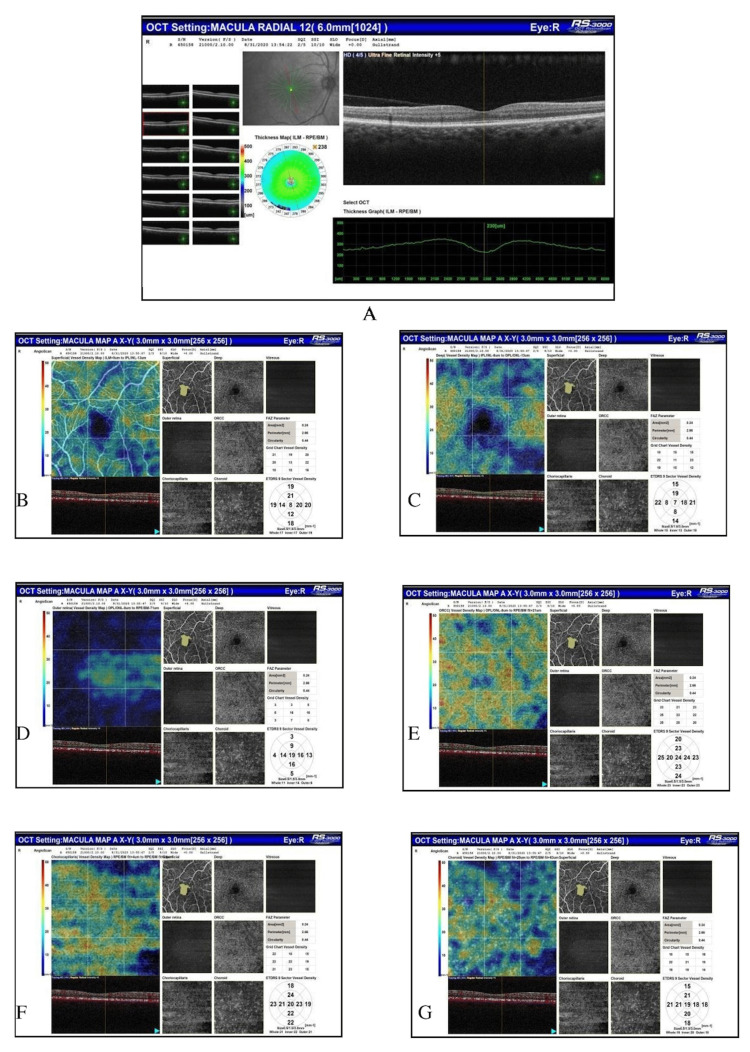
Methods: This study was an observational study that included 80 subjects being discharged after having negative reports on the reverse transcription-polymerase chain reaction (RT-PCR) test for SARS-CoV-2 to evaluate OCTA parameters of the retina. The subjects underwent an ophthalmic evaluation that included best-corrected visual acuity (BCVA), intraocular pressure (IOP), color vision (CV), contrast sensitivity (CS), and optical coherence tomography (OCT) parameters. OCTA was done for all patients and was evaluated for foveal avascular zone (FAZ) area, perimeter, and circularity index, and vessel density (VD) in superficial capillary plexus (SCP), deep capillary plexus (DCP), outer retina (OR), outer retina chorio-capillaries (ORCC), chorio-capillaries (CC), and choroid (C) using 3 x 3 mm scans. The OCTA parameters were compared with normative data of the Indian population for various parameters in question.
Results: The subjects included 54/80 (67.5%) males and 26/80 (32.5%) females having a mean age of 52.40 ± 15.71 (18-60) years. The systemic evaluation revealed 38.75% of subjects had hypertension, 30% had diabetes, 20% had kidney disease, 5% had tuberculosis, and 3.75% had coronary artery disease. The mean distance BCVA was logarithm of the minimum angle of resolution (LogMAR) (1.17 ± 0.22), mean IOP was 17.0 ± 4.0 mmHg, mean CS was 2.13 ± 0.36, 50.62% of subjects had normal CV on Farnsworth test while 47% had tritanopia, and none of the subjects had red-green CV defect on Ishihara plates. The OCT scan was normal in 90% of eyes while the posterior vitreous detachment was seen in 4% of eyes, broad vitreomacular adhesion in 2.5% of eyes, and the globally adherent epiretinal membrane was seen in 2.5% of eyes. The mean central macular thickness (CMT) measured 245.14 ± 28.41 micrometers. The mean FAZ area measured 0.37 ± 0.15 mm2, the perimeter was 3.28 ± 1.08 mm, and the circularity index measured 0.41 ± 0.10. The average VD in SCP measured 16.06 ± 12.29, in DCP measured 9.11 ± 8.75, in OR measured 6.38 ± 7.37, in ORCC measured 42.53 ± 12.46, in CC measured 25.83 ± 16.31, and in C measured 25.52 ± 17.49. The VD in coronavirus disease 2019 (COVID-19) subjects was significantly lesser than that in the healthy Indian population in all layers except ORCC.
Conclusions: The SARS-CoV-2 recovered subjects have a reduced VD in retinochoroidal layers from COVID-19, an underlying systemic disease, or both. The CS values fall within normal limits. Several subjects show tritanopia on the Farnsworth test but no red-green CV defect on Ishihara plates.
Keywords: choroid; covid-19; foveal avascular zone; indian population; optical coherence tomography angiography (oct-a); retina; sars-cov-2; vessel density.
Copyright © 2023, Sodhi et al.
Conflict of interest statement
The authors have declared that no competing interests exist.
Figures
Similar articles
-
Evaluating the Quantitative Foveal Avascular Zone and Retino-Choroidal Vessel Density Using Optical Coherence Tomography Angiography in a Healthy Indian Population.Cureus. 2022 Aug 4;14(8):e27669. doi: 10.7759/cureus.27669. eCollection 2022 Aug. Cureus. 2022. PMID: 36072178 Free PMC article.
-
Optical Coherence Tomography Angiography Parameters in Indian Patients With Central Serous Chorioretinopathy.Cureus. 2023 Oct 4;15(10):e46467. doi: 10.7759/cureus.46467. eCollection 2023 Oct. Cureus. 2023. PMID: 37927676 Free PMC article.
-
The Correlation of Optical Coherence Tomography Angiography Parameters With Serum Vascular Endothelial Growth Factor-A (VEGF-A) Levels in Diabetic Macular Edema.Cureus. 2025 Jul 15;17(7):e88026. doi: 10.7759/cureus.88026. eCollection 2025 Jul. Cureus. 2025. PMID: 40821158 Free PMC article.
-
Retinal microvascular impairment in COVID-19 patients: A meta-analysis.Immun Inflamm Dis. 2022 Jun;10(6):e619. doi: 10.1002/iid3.619. Immun Inflamm Dis. 2022. PMID: 35634955 Free PMC article. Review.
-
Reduced Vessel Density and Enlarged Foveal Avascular Zone in the Macula as a Result of Systemic Hypoxia Caused by SARS-CoV-2 Infection.J Pers Med. 2023 May 31;13(6):926. doi: 10.3390/jpm13060926. J Pers Med. 2023. PMID: 37373915 Free PMC article. Review.
Cited by
-
Spectrum of retinal microvascular ischemia in patients with COVID-19 based on multimodal imaging.Heliyon. 2024 Sep 28;10(19):e38535. doi: 10.1016/j.heliyon.2024.e38535. eCollection 2024 Oct 15. Heliyon. 2024. PMID: 39430536 Free PMC article.
-
Microvascular Alteration in COVID-19 Documented by Nailfold Capillaroscopy.Diagnostics (Basel). 2023 May 29;13(11):1905. doi: 10.3390/diagnostics13111905. Diagnostics (Basel). 2023. PMID: 37296759 Free PMC article. Review.
References
-
- Expression and function of Mas-related G protein-coupled receptor D and its ligand alamandine in retina. Zhu P, Verma A, Prasad T, Li Q. Mol Neurobiol. 2020;57:513–527. - PubMed
-
- Hu K, Patel J, Swiston C, Patel BC. Treasure Island, FL: StatPearls Publishing; 2022. Ophthalmic Manifestations of Coronavirus (COVID-19) - PubMed
LinkOut - more resources
Full Text Sources
Miscellaneous