. 2023 Apr 25;14(2):e0259022.
doi: 10.1128/mbio.02590-22. Epub 2023 Feb 13.

Capsular Polysaccharide Is Essential for the Virulence of the Antimicrobial-Resistant Pathogen Enterobacter hormaechei

Affiliations

Capsular Polysaccharide Is Essential for the Virulence of the Antimicrobial-Resistant Pathogen Enterobacter hormaechei

Amelia St John et al. mBio. .

Abstract

Nosocomial infections caused by multidrug-resistant (MDR) Enterobacter cloacae complex (ECC) pathogens are on the rise. However, the virulence strategies employed by these pathogens remain elusive. Here, we study the interaction of ECC clinical isolates with human serum to define how this pathogen evades the antimicrobial action of complement, one of the first lines of host-mediated immune defense. We identified a small number of serum-sensitive strains, including Enterobacter hormaechei strain NR3055, which we exploited for the in vitro selection of serum-resistant clones. Comparative genomics between the serum-sensitive NR3055 strain and the isolated serum-resistant clones revealed a premature stop codon in the wzy gene of the capsular polysaccharide biosynthesis locus of NR3055. The complementation of wzy conferred serum resistance to NR3055, prevented the deposition of complement proteins on the bacterial surface, inhibited phagocytosis by human neutrophils, and rendered the bacteria virulent in a mouse model of peritonitis. Mice exposed to a nonlethal dose of encapsulated NR3055 were protected from subsequent lethal infections by encapsulated NR3055, whereas mice that were previously exposed to unencapsulated NR3055 succumbed to infection. Thus, capsule is a key immune evasion determinant for E. hormaechei, and it is a potential target for prophylactics and therapeutics to combat these increasingly MDR human pathogens. IMPORTANCE Infections caused by antimicrobial resistant bacteria are of increasing concern, especially those due to carbapenem-resistant Enterobacteriaceae pathogens. Included in this group are species of the Enterobacter cloacae complex, regarding which there is a paucity of knowledge on the infection biology of the pathogens, despite their clinical relevance. In this study, we combine techniques in comparative genomics, bacterial genetics, and diverse models of infection to establish capsule as an important mechanism of Enterobacter pathogens to resist the antibacterial activity of serum, a first line of host defense against bacterial infections. We also show that immune memory targeting the Enterobacter capsule protects against lethal infection. The further characterization of Enterobacter infection biology and the immune response to infection are needed for the development of therapies and preventative interventions targeting these highly antibiotic resistant pathogens.

Keywords: Enterobacter; antimicrobial resistance; capsule; complement; pathogenesis; serum; serum resistance.


Conflict of interest statement

The authors declare no conflict of interest.

Figures
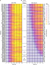
FIG 1
FIG 1
Serum susceptibility of Enterobacter spp. clinical isolates. 96 Enterobacter spp. clinical isolates were cultured in either 50% TSB + 50% PBS (A) or 50% TSB + 50% commercial human serum (B). The OD600 of each culture was measured every 30 min for 24 h. Isolates are sorted according to the time it took the cultures to reach an OD600 of 0.1 in serum and are grouped according to the 4 h time frame in which this optical density was reached by the culture (B). The most serum-resistant isolates appear at the top of the figure, whereas the most sensitive isolates are at the bottom. Readings of the OD600 value over time are mapped to the color and thickness of the horizontal line segments representing the individual strains. The thin, purple line segments represent low OD600 values, whereas the thick, yellow line segments represent high OD600 values. Each strain is represented by two biological replicates that were averaged for this figure. The strain names are listed to the left, and the capsule types are listed to the right. NA, capsule type could not be determined. The figure was plotted using R v. 4.1.1 and ggplot v. 3.3.5.
FIG 2
FIG 2
Serum bactericidal activity against 62 Enterobacter spp. clinical isolates (A and B) Enterobacter clinical isolates were cultured in either 50% TSB + 50% PBS (A) or 50% TSB + 50% commercial serum (B). The cultures were serially diluted in PBS and plated on TSA for CFU counts every 2 h for 8 h. Four highly serum-sensitive strains are shown in purple. The serum-sensitive E. hormaechei strain NR3055 is shown in purple, with purple squares at each time point. Each strain is represented by two biological replicates. The limit of detection for this assay is 100 CFU/mL (dotted lines). (C) NR3055 was cultured in 50% TSB + 50% PBS, 50% TSB + 50% pooled human serum, or 50% TSB + 45% PBS + 5% fresh pooled human serum. The cultures were serially diluted in PBS and plated on TSA for CFU counts every 2 h for 8 h. Each growth condition is represented by three biological replicates. The limit of detection for this assay is 100 CFU/mL (dotted lines). The error bars indicate the standard error of the mean (SEM). Statistical significance was determined via a two-way ANOVA with Dunnett’s multiple-comparison test. Asterisks represent 50% TSB + 50% PBS compared to 50% TSB + 50% pooled human serum, and pound signs represent 50% TSB + 50% PBS compared to 50% TSB + 45% PBS + 5% fresh human serum (****/####, P ≤ 0.0001; ***/###, P ≤ 0.001; **/##, P ≤ 0.01).
FIG 3
FIG 3
In vitro selection of NR3055 serum-resistant clones. (A) PBS and 100% commercial pooled human serum were spotted on a TSA plate on which NR3055 cells were spread. Bacteria grew uninhibited where PBS was spotted, whereas the spotted serum created a zone of clearance. Colonies that arose within the zone of clearance were selected and screened a second time for serum resistance, and those that grew uninhibited were labeled as serum-resistant (SR) clones. The arrow indicates a selected, possibly serum-resistant clone. (B) The eight in vitro-selected NR3055 SR clones and NR3055 were cultured in 50% TSB + 45% PBS + 5% fresh pooled human serum. The cultures were serially diluted in PBS and plated on TSA for CFU counts every 2 h for 8 h. Each clone is represented by three biological replicates. The limit of detection for this assay is 100 CFU/mL (dotted line). The error bars indicate the SEM. (C) The eight in vitro-selected NR3055 SR clones and NR3055 were used to infect whole human blood. The cultures were serially diluted in PBS and plated on TSA for CFU counts every hour for 6 h. Each clone is represented by two biological replicates with two blood donors per replicate. The limit of detection for this assay is 90 CFU/mL (dotted line). The error bars indicate the SEM.
FIG 4
FIG 4
Transmission electron microscopy identified a capsule-like structure surrounding NR3055 SR2. TEM images of NR3055, which exhibited minimal production of the capsule-like structure (A), and NR3055 SR2, which exhibited a thicker zone of putative CPS surrounding the cell (B). The images are pseudocolored based on pixel density and contrast, with yellow indicating the putative capsule and green indicating the cell body. The images are representative of at least 12 cells per strain. Additional images can be found in Fig. S2. (C) Quantification of the width of the capsule-like structures, relative to the diameter of the cell bodies, of 12 NR3055 cell images and 12 NR3055 SR2 cell images. The error bars indicate the SEM. Statistical significance was determined via a Mann-Whitney nonparametric t test (****, P ≤ 0.0001).
FIG 5
FIG 5
The cps locus variants in ST78 E. hormaechei isolates. Phylogeny of 97 ST78 E. hormaechei assemblies (left). 79 of the phylogenies were obtained from NCBI RefSeq, and 18 stem from the present study. All 97 isolates carry a cps locus of type Enterobacter-NL68, which is represented by the gene map on the top. The genetic variants and their locations within the cps locus of these assemblies are represented by dots in the middle panel. Variant types are indicated by different colors. Gene boundaries within the central panel are marked by boxes that span the gene length. Below the gene map, the number of variants found in each gene is tabulated by type. The tree scale relates branch length to the number of substitutions per alignment site. The figure was plotted using R v. 4.1.1 and ggplot v. 3.3.5.
FIG 6
FIG 6
Expression of full-length wzy confers serum resistance to NR3055. (A) Schematic detailing the genetic complementation of wzy in NR3055. Wild-type wzy was expressed from the constitutive PS12 promoter at the neutral attTn7 site on the NR3055 chromosome, downstream of glmS. The natural, truncated wzy gene on the NR3055 chromosome is labeled as wzy*. (B and C) NR3055, NR3055 SR2, and NR3055::wzy were cultured in either 50% TSB + 50% PBS (B) or 50% TSB + 45% PBS + 5% fresh pooled human serum (C). The cultures were diluted in PBS and plated on TSA for CFU every 2 h for 8 h. The limit of detection for this assay is 100 CFU/mL (dotted lines). Each strain is represented by three biological replicates. The error bars indicate the SEM. Statistical significance was determined via a two-way ANOVA with Dunnett’s multiple-comparison test. Asterisks represent NR3055 compared to NR3055 SR2, and pound signs represent NR3055 compared to NR3055::wzy (****/####, P ≤ 0.0001; ***/###, P ≤ 0.001; **/##, P ≤ 0.01). (D and E) NR3055 cells, but not NR3055 SR2 or NR3055::wzy cells, sediment in liquid culture. Sedimentation is demonstrated visually (D) and quantitatively (E). The percent cell sedimentation was calculated using the inverse ratio of the OD600 of the mixed culture to the OD600 of the same culture after a 4 h period of stagnant incubation. Strains are represented by four biological replicates. The error bars indicate the SEM. Statistical significance was determined via a one-way ANOVA with Dunnett’s multiple-comparison test (****, P ≤ 0.0001). (F) The extracellular polysaccharide content from NR3055, NR3055 SR2, NR3055::wzy, and NR3033 was separated using Tris-acetate protein gels and polysaccharides stained with a Pierce Glycoprotein Staining Kit. NR3033 is a ST78, cps type NL68 E. hormaechei strain representing “wild-type” CPS production for ST78. Arrows demark the pink high molecular weight bands that are representative of CPS. A representative gel is shown. The extracellular polysaccharide content of the three other identified serum sensitive strains can be seen in Fig. S3.
FIG 7
FIG 7
The wzy-dependent virulence of NR3055 in a murine peritonitis model. (A) Mice were infected intraperitoneally (i.p.) with 108 CFU of NR3055, NR3055 SR2, or NR3055::wzy and monitored for survival. The morbidity of the animals was monitored for 4 days. Statistical analysis was performed via a log-rank (Mantel-Cox) test that was corrected for multiple comparisons. The data are from at least two independent experiments with a total of 10 mice per group (***, P < 0.001). (B–H) Mice were infected i.p. with 108 CFU of NR3055 or NR3055::wzy, and at 2 and 16 h postinfection (hpi), blood was collected via cardiac puncture (B). Peritoneal lavages were also collected (C), and the indicated organs were harvested (D–H). The organs were homogenized, and the blood, peritoneal lavage fluid, and organ homogenates were serially diluted in PBS and plated for CFU. The limit of detection for blood bacterial burdens is 20 CFU/mL, and that of all other organs and peritoneal lavage fluid is 100 CFU/mL (dotted lines). Red-filled circles indicate no recovered CFU. The error bars indicate the SEM. Each circle represents an individual mouse. The data are from two independent experiments, with a total of 10 mice per group. Statistical significance was determined via a one-way ANOVA with Šidák’s multiple-comparison test (****, P ≤ 0.0001; ***, P ≤ 0.001).
FIG 8
FIG 8
The wzy-dependent evasion of innate immune defenses. (A) NR3055, NR3055 SR2, and NR3055::wzy (2 × 10⁷ CFU/mL) were inoculated in whole human blood, and cultures were serially diluted in PBS and plated for CFU every hour for 6 h. Each strain is represented by two biological replicates, with two separate human blood donors per replicate. The limit of detection for this assay is 90 CFU/mL (dotted line). The error bars indicate the SEM. Statistical significance was determined via a two-way ANOVA with Dunnett’s multiple-comparison test. Asterisks represent NR3055 compared to NR3055 SR2, and pound signs represent NR3055 compared to NR3055::wzy (****/####, P ≤ 0.0001; ***/###, P ≤ 0.001; **/##, P ≤ 0.01; */#, P ≤ 0.1). (B) C3b deposition on the surface of NR3055, NR3055 SR2, and NR3055::wzy was analyzed by measuring the mean fluorescence intensity (MFI) via flow cytometry, using an anti-complement C3b/iC3b antibody. The data are from three experimental days of either one, three, or five biological replicates. The error bars indicate the SEM. Statistical significance was determined via a one-way ANOVA with Dunnett’s multiple-comparison test. All of the experimental groups are compared to NR3055 (****, P ≤ 0.0001). (C) Primary human neutrophils were infected at a multiplicity of infection (MOI) of 10 with either opsonized or nonopsonized GFP-producing NR3055 or NR3055 SR2, or buffer alone as a control. The bacterial uptake over time is represented by the MFI and was determined via flow cytometry. The data are representative of two biological replicates with two human blood donors per replicate. The error bars indicate the SEM. Statistical significance was determined via a two-way ANOVA with Dunnett’s multiple-comparison test (****, P ≤ 0.0001; *, P ≤ 0.1). All of the experimental groups are compared to NR3055 opsonized with 2% serum. Asterisks represent all comparisons at each time point. (D) 20 wild-type (5 male and 15 female) and 15 C3-deficient (6 male and 9 female) 8-week-old C57BL/6J mice were infected with NR3055. All male mice were infected with 2.5 × 10⁸ CFU, and all female mice were infected with 108 CFU. All mice were monitored for morbidity and survival for 4 days. The statistical analysis was performed via a log-rank (Mantel-Cox) test that was corrected for multiple comparisons. The data are from at least two independent experiments with a total of n = 15 to 20 mice per group (****, P ≤ 0.0001).
FIG 9
FIG 9
CPS-producing NR3055::wzy provides immunity against future infections. (A) Diagram of the experimental design. Mice were infected intraperitoneally (i.p.) with 106 CFU of NR3055 or NR3055::wzy. Three weeks postinfection, the mice were reinfected i.p. with 106 CFU of NR3055 or NR3055::wzy. Two weeks post the second exposure, all mice were challenged with 2.5 × 108 CFU of NR3055::wzy and monitored for morbidity and survival for four days. (B) The mice were infected and monitored as detailed in panel A. The statistical analysis was performed via a log-rank (Mantel-Cox) test. The data are from two independent experiments, with a total of 10 mice per group (***, P < 0.001). (C) Diagram of the experimental design. The mice that survived the lethal challenge with NR3055::wzy (B) were terminally bled, and their sera were collected and pooled. Naive mice were then passively immunized with either serum from the NR3055::wzy-infected mice or with serum from uninfected naive control mice. The mice were then infected 24 h post passive transfer with 108 CFU of NR3055::wzy and monitored for morbidity and survival for four days. (D) The mice were infected and monitored as detailed in panel C. The statistical analysis was performed via a log-rank (Mantel-Cox) test. The data are from two independent experiments. with a total of 10 mice per group (*, P ≤ 0.1). Panels A and C were created using BioRender.com.

Similar articles

Cited by

References

    1. Davin-Regli A, Lavigne JP, Pagès JM. 2019. Enterobacter spp.: update on taxonomy, clinical aspects, and emerging antimicrobial resistance. Clin Microbiol Rev 32. doi:10.1128/CMR.00002-19. - DOI - PMC - PubMed
    1. Nishijima KA, Alvarez AM, Hepperly PR, Shintaku MH, Keith LM, Sato DM, Bushe BC, Armstrong JW, Zee FT. 2004. Association of Enterobacter cloacae with rhizome rot of edible ginger in Hawaii. Plant Dis 88:1318–1327. doi:10.1094/PDIS.2004.88.12.1318. - DOI - PubMed
    1. Aubron C, Poirel L, Ash RJ, Nordmann P. 2005. Carbapenemase-producing Enterobacteriaceae, U.S. rivers. Emerg Infect Dis 11:260–264. doi:10.3201/eid1102.030684. - DOI - PMC - PubMed
    1. Ludden C, Reuter S, Judge K, Gouliouris T, Blane B, Coll F, Naydenova P, Hunt M, Tracey A, Hopkins KL, Brown NM, Woodford N, Parkhill J, Peacock SJ. 2017. Sharing of carbapenemase-encoding plasmids between Enterobacteriaceae in UK sewage uncovered by MinION sequencing. Microb Genom 3:e000114. doi:10.1099/mgen.0.000114. - DOI - PMC - PubMed
    1. Taghavi S, van der Lelie D, Hoffman A, Zhang YB, Walla MD, Vangronsveld J, Newman L, Monchy S. 2010. Genome sequence of the plant growth promoting endophytic bacterium Enterobacter sp. 638. PLoS Genet 6:e1000943. doi:10.1371/journal.pgen.1000943. - DOI - PMC - PubMed

Publication types

Supplementary concepts

LinkOut - more resources