. 2023 Feb 13;11(2):e0518922.
doi: 10.1128/spectrum.05189-22. Online ahead of print.

Baculovirus Actin Rearrangement-Inducing Factor 1 Can Remodel the Mammalian Actin Cytoskeleton

Affiliations

Baculovirus Actin Rearrangement-Inducing Factor 1 Can Remodel the Mammalian Actin Cytoskeleton

Anika Steffen et al. Microbiol Spectr. .

Abstract

The actin rearrangement-inducing factor 1 (Arif-1) of Autographa californica multiple nucleopolyhedrovirus (AcMNPV) is an early viral protein that manipulates the actin cytoskeleton of host insect cells. Arif-1 is conserved among alphabaculoviruses and is responsible for the accumulation of F-actin at the plasma membrane during the early phase of infection. However, the molecular mechanism underlying Arif-1-induced cortical actin accumulation is still open. Recent studies have demonstrated the formation of invadosome-like structures induced by Arif-1, suggesting a function in systemic virus spread. Here, we addressed whether Arif-1 is able to manipulate the actin cytoskeleton of mammalian cells comparably to insect cells. Strikingly, transient overexpression of Arif-1 in B16-F1 mouse melanoma cells revealed pronounced F-actin remodeling. Actin assembly was increased, and intense membrane ruffling occurred at the expense of substrate-associated lamellipodia. Deletion mutagenesis studies of Arif-1 confirmed that the C-terminal cytoplasmic region was not sufficient to induce F-actin remodeling, supporting that the transmembrane region for Arif-1 function is also required in mammalian cells. The similarities between Arif-1-induced actin remodeling in insect and mammalian cells indicate that Arif-1 function relies on conserved cellular interaction partners and signal transduction pathways, thus providing an experimental tool to elucidate the underlying mechanism. IMPORTANCE Virus-induced changes of the host cell cytoskeleton play a pivotal role in the pathogenesis of viral infections. The baculovirus Autographa californica multiple nucleopolyhedrovirus (AcMNPV) is known for intervening with the regulation of the host actin cytoskeleton in a wide manner throughout the infection cycle. The actin rearrangement-inducing factor 1 (Arif-1) is a viral protein that causes actin rearrangement during the early phase of AcMNPV infection. Here, we performed overexpression studies of Arif-1 in mammalian cells to establish an experimental tool that allows elucidation of the mechanism underlying the Arif-1-induced remodeling of actin dynamics in a well-characterized and genetically accessible system.

Keywords: AcMNPV; Arif-1; B16-F1 cells; F-actin remodeling; TN368 cells; actin dynamics.


Conflict of interest statement

The authors declare no conflict of interest.

Figures
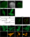
FIG 1
FIG 1
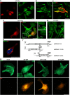
Actin rearrangement in TN368 cells after AcMNPV infection or Arif-1 expression. (a) After seeding TN368 cells on poly-d-lysine-coated coverslips, cells were infected with AcMNPV at a multiplicity of infection of 10 PFU/cell. Representative superresolution images (n = 3 independent experimental settings) are shown after adding the virus suspension (0 h p.i.) and 6 h p.i. Phalloidin staining was used to visualize the actin filament network at 0 h p.i. and the actin accumulations at 6 h p.i. Scale bar, 5 μm. (b) After mock infection and at 6 h p.i., α-tubulin immunostainings indicated a comparable microtubule network. Infected cells were detected by IE2 staining (not shown). (c) Schematic illustrating the Arif-1HA-tagged constructs expressing full-length arif-1 ORF or truncated versions under the control of the hr5/ie1 promoter. (d) TN368 cells seeded on poly-d-lysine-coated coverslips were transfected with the construct pIE1Arif-1HA (arif-1HA) for 48 h (n > 3). The overlay of a confocal bottom section with HA (red) and phalloidin (green) staining depicts the accumulation of actin clusters, which colocalized with Arif-1 at the plasma membrane. (e) After transfection of Arif-1 constructs, cells expressing the N-terminal Arif-1 (arif-1 3′Δ HA) or C-terminal Arif-1 (arif-1cytHA) showed an actin cytoskeleton (green) comparable to that of mock-transfected cells, while cells expressing full-length Arif-1 (arif-1HA) demonstrated the loss of actin stress fibers and accumulation of F-actin clusters. HA stainings (orange) allowed visualization of localization of full-length and N-terminal Arif-1 (arif-1 3′ΔHA) at the plasma membrane, while the C-terminal Arif-1 (arif-1cytHA) localized in the cytoplasm (n = 3). Scale bars, 20 μm.
FIG 2
FIG 2
Arif-1-induced actin rearrangement in B16-F1 cells. B16-F1 cells transfected with Myc-, HA-, or Flag-tagged Arif-1 constructs for 24 h were seeded on laminin-coated coverslips. (a) Phalloidin staining (green) showed membrane ruffles at the entire cell periphery and the loss of lamellipodia and stress fibers in an Arif-1-expressing cell visualized by Myc immunostaining (red) (n > 3). (b) The confocal bottom section depicts colocalization of HA-tagged Arif-1 (red) and F-actin (green) at the plasma membrane (n = 2). (c) Flag immunostaining (red) allowed visualization of Arif-1 in the perinuclear space and at the plasma membrane, while phalloidin staining (green) depicted a rather jagged cell periphery with multiple filopodia (magnified image, arrowheads) compared to the untransfected cell and actin clusters in the cytoplasm (magnified image, arrow) (n > 3). (d) Myc-tagged Arif-1-expressing cells (asterisks) showed altered actin (green) cytoskeleton architecture, including reduced stress fibers, cytoplasmic actin clusters, and membrane ruffles (n > 3). (e) Confocal section from the middle, showing partial colocalization of cis-Golgi structures visualized by GM130 staining (green) and perinuclear HA-tagged Arif-1 (red) (n = 3). (f) Schematic illustrating the Arif-1HA-tagged constructs expressing the full-length arif-1 ORF or truncated versions under the control of the CMV promoter. (g) Strong actin (green) accumulations were visible after full-length Arif-1 expression, while the N and C termini of Arif-1 showed an actin network comparable to that in mock-transfected cells. A mock-transfected cell with characteristically actin-dense lamellipodium is shown. HA stainings (orange) allowed visualization of localization of full-length and N-terminal Arif-1 (arif-1 3′ΔHA) at the plasma membrane, while the C-terminal Arif-1 (arif-1cytHA) localized in the cytoplasm (n = 3). Scale bars, 20 μm.
FIG 3
FIG 3
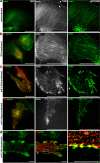
Superresolution microscopy of Arif-1-induced actin rearrangement in B16-F1 cells. B16-F1 cells transfected with IE2-, HA-, or Flag-tagged Arif-1 constructs for 24 h were seeded on laminin-coated coverslips. HA or Flag immunostainings allowed visualization of Arif-1, F-actin was stained with phalloidin, and IE2 was detected with anti-IE2 antiserum (n = 3). (a) The untransfected cell showed the fiber network with lamellipodium (arrowhead) and lamella (arrow) at the front and stress fibers at the rear. (b) The stress fiber network was maintained in the IE2-expressing cell. (c) Filopodia (arrow) and strong actin clusters were visible at the plasma membrane, while smaller clusters and dots were detected in the cytoplasm. (d) The magnified merged dSTORM image shows discontinuous colocalization of Arif-1 aggregates and actin clusters at the plasma membrane and partial colocalization of Arif-1 and actin dots in the cytoplasm. Scale bars, 20 μm (a, b, and c) or 5 μm (d).
FIG 4
FIG 4
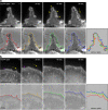
Arif-1-induced actin dynamics in B16-F1 cells. Time-lapse images of a B16-F1 cell seeded on laminin-coated coverslips and coexpressing EGFP-actin and Myc-tagged Arif-1 or expressing EGFP-actin alone. The upper panel shows wide-field fluorescence images of the time-lapse movie at the indicated time points, and the lower panel shows the corresponding phase-contrast images. The cell periphery is outlined with colored dashed lines and overlaid in the contour plot. (a) An Arif-1-expressing cell showed dynamic ruffles (yellow arrowheads) and filopodia (yellow arrow). The very dynamic ruffle formation did not lead to a net translocation, as shown by the contour plot. (b) Characteristics of the EGFP-actin-transfected cell included a broad, effectively protruding lamellipodial network (magenta arrowhead) harboring embedded bundles termed microspikes, some of which occasionally developed into filopodia (yellow arrows). The contour plot illustrates the continuous protrusion of the cell edge. Scale bars, 2 μm.

References

    1. Taylor MP, Koyuncu OO, Enquist LW. 2011. Subversion of the actin cytoskeleton during viral infection. Nat Rev Microbiol 9:427–439. doi:10.1038/nrmicro2574. - DOI - PMC - PubMed
    1. Charlton CA, Volkman LE. 1991. Sequential rearrangement and nuclear polymerization of actin in baculovirus-infected Spodoptera frugiperda cells. J Virol 65:1219–1227. doi:10.1128/JVI.65.3.1219-1227.1991. - DOI - PMC - PubMed
    1. Charlton CA, Volkman LE. 1993. Penetration of Autographa californica nuclear polyhedrosis virus nucleocapsids into IPLB Sf 21 cells induces actin cable formation. Virology 197:245–254. doi:10.1006/viro.1993.1585. - DOI - PubMed
    1. Ohkawa T, Volkman LE. 1999. Nuclear F-actin is required for AcMNPV nucleocapsid morphogenesis. Virology 264:1–4. doi:10.1006/viro.1999.0008. - DOI - PubMed
    1. Ohkawa T, Rowe AR, Volkman LE. 2002. Identification of six Autographa californica multicapsid nucleopolyhedrovirus early genes that mediate nuclear localization of G-actin. J Virol 76:12281–12289. doi:10.1128/jvi.76.23.12281-12289.2002. - DOI - PMC - PubMed

LinkOut - more resources