. 2023 Mar 30;97(3):e0146322.
doi: 10.1128/jvi.01463-22. Epub 2023 Feb 13.

Single Particle Analysis of H3N2 Influenza Entry Differentiates the Impact of the Sialic Acids (Neu5Ac and Neu5Gc) on Virus Binding and Membrane Fusion

Affiliations

Single Particle Analysis of H3N2 Influenza Entry Differentiates the Impact of the Sialic Acids (Neu5Ac and Neu5Gc) on Virus Binding and Membrane Fusion

Yu-An Annie Chien et al. J Virol. .

Abstract

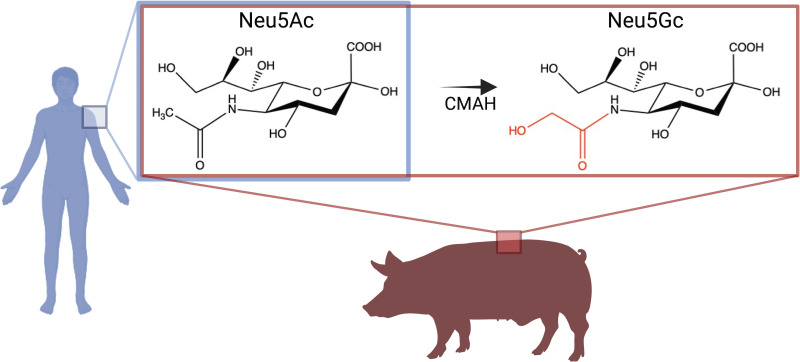
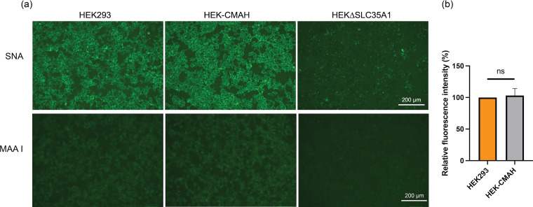
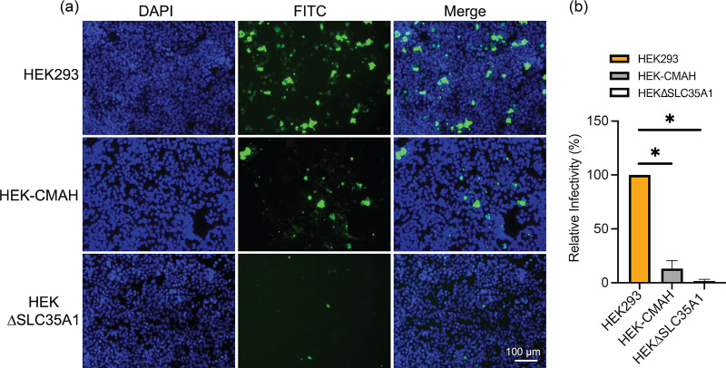
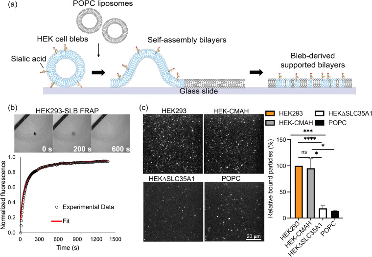
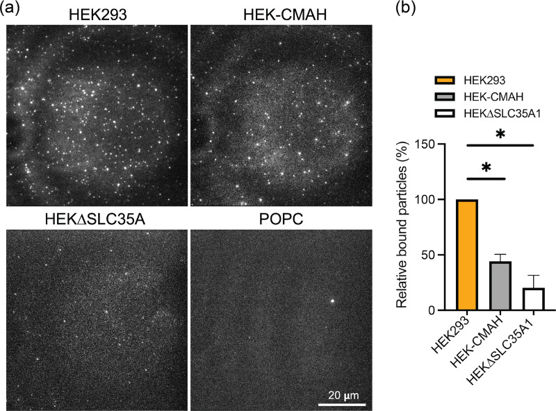
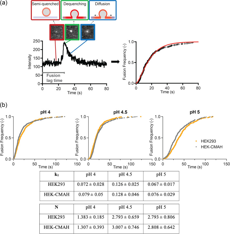
Entry of influenza A viruses (IAVs) into host cells is initiated by binding to sialic acids (Sias), their primary host cell receptor, followed by endocytosis and membrane fusion to release the viral genome into the cytoplasm of the host cell. Host tropism is affected by these entry processes, with a primary factor being receptor specificity. Sias exist in several different chemical forms, including the hydroxylated N-glycolylneuraminic acid (Neu5Gc), which is found in many hosts; however, it has not been clear how modified Sias affect viral binding and entry. Neu5Gc is commonly found in many natural influenza hosts, including pigs and horses, but not in humans or ferrets. Here, we engineered HEK293 cells to express the hydoxylase gene (CMAH) that converts Neu5Ac to Neu5Gc, or knocked out the Sia-CMP transport gene (SLC35A1), resulting in cells that express 95% Neu5Gc or minimal level of Sias, respectively. H3N2 (X-31) showed significantly reduced infectivity in Neu5Gc-rich cells compared to wild-type HEK293 (>95% Neu5Ac). To determine the effects on binding and fusion, we generated supported lipid bilayers (SLBs) derived from the plasma membranes of these cells and carried out single particle microscopy. H3N2 (X-31) exhibited decreased binding to Neu5Gc-containing SLBs, but no significant difference in H3N2 (X-31)'s fusion kinetics to either SLB type, suggesting that reduced receptor binding does not affect subsequent membrane fusion. This finding suggests that for this virus to adapt to host cells rich in Neu5Gc, only receptor affinity changes are required without further adaptation of virus fusion machinery. IMPORTANCE Influenza A virus (IAV) infections continue to threaten human health, causing over 300,000 deaths yearly. IAV infection is initiated by the binding of influenza glycoprotein hemagglutinin (HA) to host cell sialic acids (Sias) and the subsequent viral-host membrane fusion. Generally, human IAVs preferentially bind to the Sia N-acetylneuraminic acid (Neu5Ac). Yet, other mammalian hosts, including pigs, express diverse nonhuman Sias, including N-glycolylneuraminic acid (Neu5Gc). The role of Neu5Gc in human IAV infections in those hosts is not well-understood, and the variant form may play a role in incidents of cross-species transmission and emergence of new epidemic variants. Therefore, it is important to investigate how human IAVs interact with Neu5Ac and Neu5Gc. Here, we use membrane platforms that mimic the host cell surface to examine receptor binding and membrane fusion events of human IAV H3N2. Our findings improve the understanding of viral entry mechanisms that can affect host tropism and virus evolution.

Keywords: Neu5Ac; Neu5Gc; fusion kinetics; influenza; membrane fusion; membranes; sialic acid; single particle tracking; supported lipid bilayer; tropism; virus entry.


Conflict of interest statement

The authors declare no conflict of interest.

Figures

FIG 1
FIG 1
The predominant types of sialic acids found in humans and pigs. Humans present Neu5Ac of Sias, whereas pigs present both Neu5Ac and Neu5Gc of Sias, due to the presence of the CMAH enzyme. This enzyme catalyzes the hydroxylation of the acetyl group on Neu5Ac. Image was created with BioRender.com.
FIG 2
FIG 2
The distribution of Sias in HEK293, HEK-CMAH, and HEKΔSLC35A1 cells. (a) Live cells were stained with 1:5,000 fluorescein-labeled SNA or MAA I for 30 min and then imaged using fluorescence microscopy. Both HEK293 and HEK-CMAH express substantial amounts of α2,6-Sias, including Neu5Ac and Neu5Gc, while HEKΔSLC35A1 cells show little to no expression of Sias. (b) Relative fluorescence level of SNA staining in each cell line was quantified using ImageJ, and data were analyzed using PRISM software. The fluorescence intensity in HEKΔSLC35A1 cells is not shown as the signal is below the detection limit.
FIG 3
FIG 3
H3N2 (X-31) virus infection is reduced in Neu5Gc-rich HEK-CMAH cells. (a) The distribution of influenza virus NP proteins in Neu5Ac-rich HEK293, Neu5Gc-rich HEK-CMAH, and HEKΔSLC35A1 cells at 18 hpi was determined by immunofluorescence assay (IFA). (b) Relative infectivity was determined by quantifying the number of NP positive cells in relation to the total number of cells in the field of view using ImageJ. Data were analyzed using PRISM software.
FIG 4
FIG 4
HEK blebs-derived SLBs formed are mobile and retain available Sias. (a) Illustration of cell bleb-SLB formation. Chemically induced cell blebs are absorbed on a glass surface, then ruptured by fusogenic liposomes to form bilayers (created from Adobe illustrator). (b) The mobility of formed bleb-SLBs was determined by fluorescence recovery after photobleaching (FRAP). The images correspond to various times along the fluorescence recovery process of HEK293-SLB. (c) Left: Sias distribution on SLBs was determined by SNA lectin staining. SNA lectin, which recognizes α2-6 linked Sias, binds well to both Neu5Ac and Neu5Gc. Right: Sias incorporation was quantified by the number count of fluorescent particles in the field of view using ImageJ. Data were analyzed using PRISM software.
FIG 5
FIG 5
Decreased H3N2 (X-31) binding on HEK-CMAH bleb-derived SLBs containing predominantly Neu5Gc. (a) SLBs were incubated with R18-labeled virions and rinsed to remove unbound particles. Bound virus particles on HEK293, HEK-CMAH, and HEKΔSLC35A1 bleb-derived SLBs, as well as on POPC SLBs, were imaged using TIRF microscopy. (b) Quantitative particle counts were determined by ImageJ. Data were analyzed using PRISM software.
FIG 6
FIG 6
H3N2 (X-31) virus fusion kinetics using single particle tracking. (a) Overview of a single H3N2 influenza virus fusion event. The system is acidified to trigger fusion which leads to a spike in R18 intensity due to fluorescence dequenching. Fluorescence intensity then decreases as the fluorophores diffuse out from the site. The lag time between acidification and fusion is the fusion lag time, which is collected from multiple events and plotted as a cumulative distribution curve. Data are fitted to an equation to obtain fusion kinetics, k1 and N. (b) Cumulative distribution curves and fusion kinetics of H3N2 in HEK293- and HEK-CMAH-SLBs at a pH range from 4 to 5. Data were analyzed using MATLAB and Prism software.

References

    1. Cozza V, Campbell H, Chang HH, Iuliano AD, Paget J, Patel NN, Reiner RC, Troeger C, Viboud C, Bresee JS, Fitzner J. 2021. Global seasonal influenza mortality estimates: a comparison of 3 different approaches. Am J Epidemiol 190:718–727. 10.1093/aje/kwaa196. - DOI - PMC - PubMed
    1. Skehel JJ, Wiley DC. 2000. Receptor binding and membrane fusion in virus entry: the influenza hemagglutinin. Annu Rev Biochem 69:531–569. 10.1146/annurev.biochem.69.1.531. - DOI - PubMed
    1. Luo M. 2012. Influenza virus entry, p 201–221. In Rossmann MG, Rao VB (eds), Viral molecular machines: advances in experimental medicine and biology, Springer US, Boston, MA, Vol 726. 10.1007/978-1-4614-0980-9_9. - DOI - PMC - PubMed
    1. Sun X, Whittaker GR. 2006. Entry of influenza virus. In Pöhlmann S, Simmons G (eds), viral entry into host cells. Advances in Experimental Medicine and Biology, vol 790. Springer, New York, NY. 10.1007/978-1-4614-7651-1_4. - DOI - PubMed
    1. Lewis AL, Chen X, Schnaar RL, et al. 2022. Sialic acids and other nonulosonic acids. In Varki A, Cummings RD, Esko JD, et al. Essentials of glycobiology [Internet]. 4th ed. Cold spring harbor (NY): Cold spring harbor laboratory press. Chapter 15. https://www.ncbi.nlm.nih.gov/books/NBK579976/.

Publication types

MeSH terms

LinkOut - more resources