. 2023 Mar;46(1):377-393.
doi: 10.1007/s13246-023-01231-w. Epub 2023 Feb 13.

Open-source, fully-automated hybrid cardiac substructure segmentation: development and optimisation

Affiliations

Open-source, fully-automated hybrid cardiac substructure segmentation: development and optimisation

Robert N Finnegan et al. Phys Eng Sci Med. 2023 Mar.

Abstract

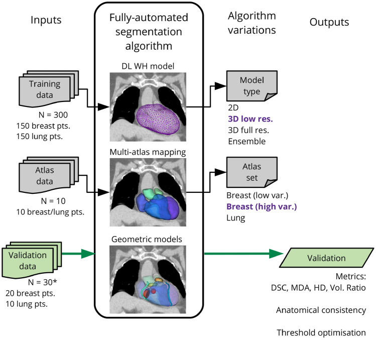
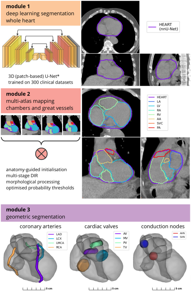
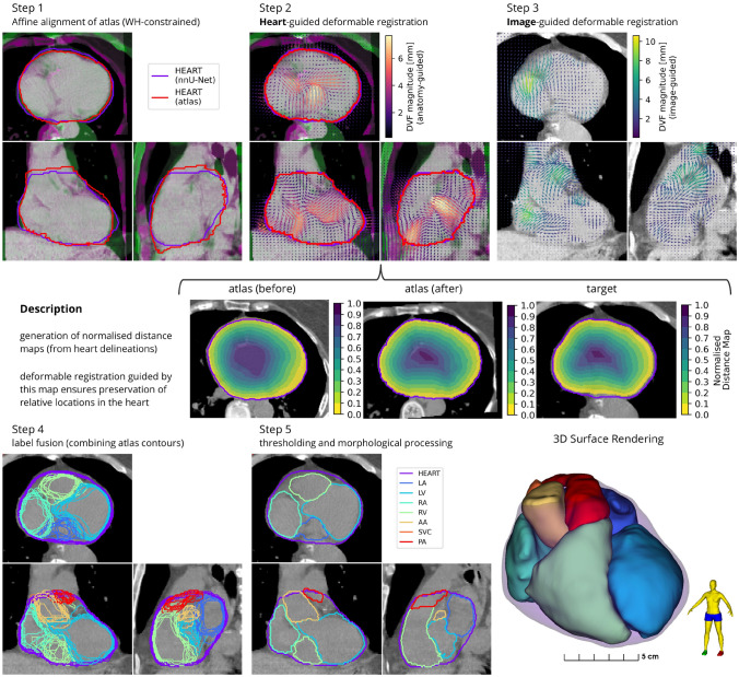
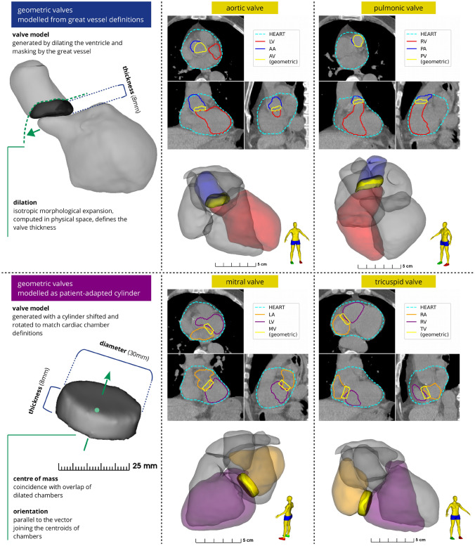
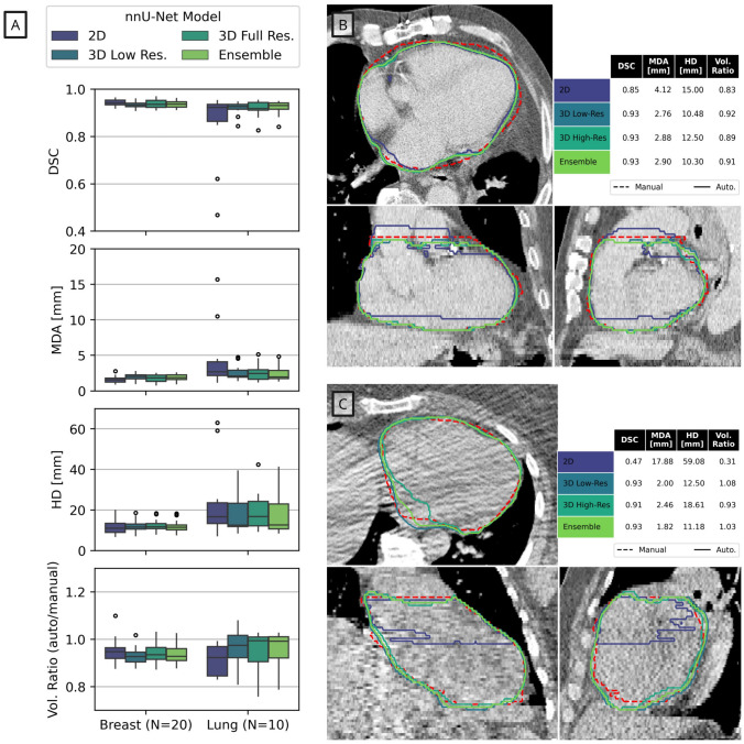
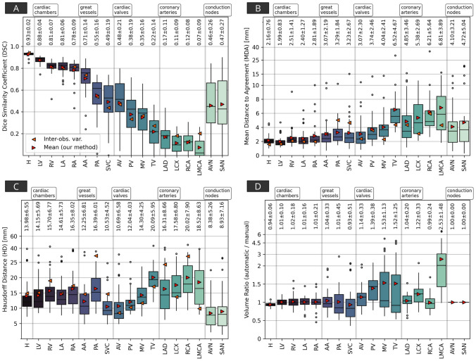
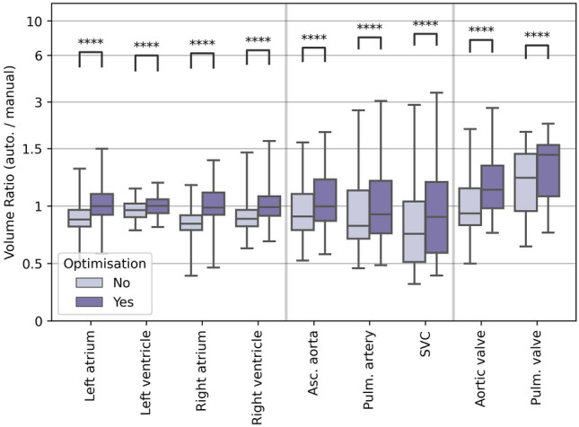
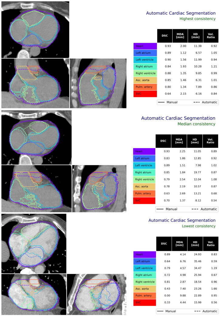
Radiotherapy for thoracic and breast tumours is associated with a range of cardiotoxicities. Emerging evidence suggests cardiac substructure doses may be more predictive of specific outcomes, however, quantitative data necessary to develop clinical planning constraints is lacking. Retrospective analysis of patient data is required, which relies on accurate segmentation of cardiac substructures. In this study, a novel model was designed to deliver reliable, accurate, and anatomically consistent segmentation of 18 cardiac substructures on computed tomography (CT) scans. Thirty manually contoured CT scans were included. The proposed multi-stage method leverages deep learning (DL), multi-atlas mapping, and geometric modelling to automatically segment the whole heart, cardiac chambers, great vessels, heart valves, coronary arteries, and conduction nodes. Segmentation performance was evaluated using the Dice similarity coefficient (DSC), mean distance to agreement (MDA), Hausdorff distance (HD), and volume ratio. Performance was reliable, with no errors observed and acceptable variation in accuracy between cases, including in challenging cases with imaging artefacts and atypical patient anatomy. The median DSC range was 0.81-0.93 for whole heart and cardiac chambers, 0.43-0.76 for great vessels and conduction nodes, and 0.22-0.53 for heart valves. For all structures the median MDA was below 6 mm, median HD ranged 7.7-19.7 mm, and median volume ratio was close to one (0.95-1.49) for all structures except the left main coronary artery (2.07). The fully automatic algorithm takes between 9 and 23 min per case. The proposed fully-automatic method accurately delineates cardiac substructures on radiotherapy planning CT scans. Robust and anatomically consistent segmentations, particularly for smaller structures, represents a major advantage of the proposed segmentation approach. The open-source software will facilitate more precise evaluation of cardiac doses and risks from available clinical datasets.

Keywords: Breast cancer; Cardiac substructures; Cardiotoxicity; Deep learning; Image segmentation; Lung cancer; Radiotherapy.


Conflict of interest statement

The authors have no relevant financial or non-financial interests to disclose.

Figures

Fig. 1
Fig. 1
Overview of the study design. Variations to several components of the fully-automated segmentation algorithm were evaluated to find optimal configurations, and the overall process was validated using 30 cases. For definitions of acronyms please see the text. *This was performed using a leave-one-out analysis: to generate automatic segmentations for cases in the optimal atlas set this case was excluded
Fig. 2
Fig. 2
Outline of the proposed hybrid segmentation approach. The automatic cardiac substructure segmentation method comprises three modules that are used sequentially to fit a detailed model of the heart to individual patient CT imaging. First, a U-Net-based deep learning model delineates the whole heart. Second, the whole heart volume is used to guide a novel multi-atlas mapping process used to delineate the four cardiac chambers (LA, LV, RA, RV) and the bases of three cardiac vessels (AA, SVC, PA). Third, geometric modelling is used to define the coronary arteries (LAD, LCX, LMCA, RCA), heart valves (AV, MV, PV, TV), and conduction nodes (AVN, SAN). Acronyms: H - (whole) heart, LV - left ventricle, RV - right ventricle, LA - left atrium, RA - right atrium, AA - ascending aorta, PA - pulmonary artery, SVC - superior vena cava, AV - aortic valve, PV - pulmonic valve, MV - mitral valve, TV - tricuspid valve, LAD - left anterior descending coronary artery, LCX - left circumflex artery, RCA - right coronary artery, LMCA - left main coronary artery, AVN - atrioventricular node, SAN - sinoatrial node. *The nnU-Net framework [26] was used in this study
Fig. 3
Fig. 3
The multi-atlas mapping stage in the proposed cardiac segmentation framework was designed to delineate the heart chambers and great vessels. Three registration steps are used to co-register a set of ten atlases to the target image (top row). This included a novel WH-guided deformable registration process with distance-preserving regularisation (middle row). The atlas contours are combined using label fusion (bottom left), and then processed to produce the final contours (bottom centre and right). Acronyms: WH - whole heart, LV - left ventricle, RV - right ventricle, LA - left atrium, RA - right atrium, AA - ascending aorta, PA - pulmonary artery, SVC - superior vena cava
Fig. 4
Fig. 4
Geometric models developed in this study to define the heart valves, using delineations of other cardiac substructures. This framework includes a method to define the aortic and pulmonic valves (top), and the mitral and tricuspid valves (bottom). Acronyms: LA - left atrium, LV - left ventricle, RA - right atrium, RV - right ventricle, AV - aortic valve, PV - pulmonic valve, MV - mitral valve, TV - tricuspid valve
Fig. 5
Fig. 5
The results of the deep learning-based whole heart segmentation are summarised on the left (5A), using the Dice (DSC), mean distance to agreement (MDA), maximum Hausdorff distance (HD) and volume ratio (Vol. Ratio). Two example patients are shown on the right (5B-C), demonstrating cases where the 2D nnU-Net model failed to provide an accurate segmentation. In Figure 5A the boxes represent the first and third quartiles, the middle bars represent the medians, and the whiskers represent the range excluding any outliers which are defined as any points greater than 1.5 × the inter-quartile range above or below the first or third quartile, respectively (outliers are shown as empty circles)
Fig. 6
Fig. 6
Results of comparisons between manual contours and automatic segmentations of the cardiac substructures included in this quantitative analysis, for the Dice Similarity Coefficient (DSC, 6A), the mean distance to agreement (MDA, 6B), the (maximum) Hausdorff distance (HD, 6C), and volume ratio (computed as automatic/manual, 6D). These grouped results combine both the breast and lung datasets. The measures of inter-observer contouring variability are derived from the three sets of manual contours on the breast dataset. Acronyms: H - (whole) heart, LV - left ventricle, RV - right ventricle, LA - left atrium, RA - right atrium, AA - ascending aorta, PA - pulmonary artery, SVC - superior vena cava, AV - aortic valve, PV - pulmonic valve, MV - mitral valve, TV - tricuspid valve, LAD - left anterior descending coronary artery, LCX - left circumflex artery, RCA - right coronary artery, LMCA - left main coronary artery, AVN - atrioventricular node, SAN - sinoatrial node. The boxes represent the first and third quartiles, the middle bars represent the medians, and the whiskers represent the range excluding any outliers which are defined as any points greater than 1.5 × the inter-quartile range above or below the first or third quartile, respectively (outliers are shown as empty circles). The printed text presents the mean ± standard deviation of results for each metric and substructure
Fig. 7
Fig. 7
Comparison of volume ratio with and without using the probability threshold optimisation method described in Finnegan et al. [21]. The volume ratio was compared with and without using this optimisation, using the Wilcoxon signed-rank test. This optimisation process was not applied to the heart valves, but as their definition depends on structures to which optimisation was applied they are included here. The boxes represent the first and third quartiles, the middle bars represent the medians, and the whiskers represent the range excluding any outliers which are defined as any points greater than 1.5 × the inter-quartile range above or below the first or third quartile, respectively (outliers are shown as empty circles). Legend: **** = p<10-4, Wilcoxon signed-rank test
Fig. 8
Fig. 8
A Surface renderings of manual contours and automatic segmentations for a representative case (median segmentation accuracy). B Axial slices of radiotherapy planning CT scans shown with automatic segmentations for a number of cases with variations in imaging and patient anatomy. Acronyms: LV - left ventricle, RV - right ventricle, LA - left atrium, RA - right atrium, AA - ascending aorta, PA - pulmonary artery, SVC - superior vena cava, AV - aortic valve, PV - pulmonic valve, MV - mitral valve, TV - tricuspid valve, LAD - left anterior descending coronary artery, LCX - left circumflex artery, RCA - right coronary artery, LMCA - left main coronary artery
Fig. 9
Fig. 9
Orthogonal slices for three patient CT scans demonstrating the performance of the proposed automatic segmentation method. These are shown for patient for which the automatic delineations had the highest (top), median (middle) and lowest (bottom) consistency with manual contours. Acronyms: SVC - superior vena cava, DSC - Dice Similarity Coefficient, MDA - mean distance to agreement, HD - Hausdorff distance, Vol. - volume

References

    1. Finnegan R, Lorenzen EL, Dowling J, Jensen I, Berg M, Thomsen MMS, et al. Analysis of cardiac substructure dose in a large, multi-centre Danish breast cancer cohort (the DBCG HYPO trial): Trends and predictive modelling. Radiother Oncol. 2020;153:130–8. doi: 10.1016/j.radonc.2020.09.004. - DOI - PubMed
    1. Wang X, Palaskas NL, Yusuf SW, Abe J, Lopez-Mattei J, Banchs J, et al. Incidence and onset of severe cardiac events after radiotherapy for esophageal cancer. J Thorac Oncol. 2020;15:1682–90. doi: 10.1016/j.jtho.2020.06.014. - DOI - PMC - PubMed
    1. Iorio GC, Salvestrini V, Borghetti P, De Felice F, Greco C, Nardone V, et al. The impact of modern radiotherapy on radiation-induced late sequelae: focus on early-stage mediastinal classical Hodgkin Lymphoma. A critical review by the Young Group of the Italian Association of Radiotherapy and Clinical Oncology (AIRO) Crit Rev Oncol. 2021;161:103326. doi: 10.1016/j.critrevonc.2021.103326. - DOI - PubMed
    1. Meattini I, Poortmans PM, Aznar MC, Becherini C, Bonzano E, Cardinale D, et al. Association of breast cancer irradiation with cardiac toxic effects: a narrative review. JAMA Oncol. 2021;7:924–32. doi: 10.1001/jamaoncol.2020.7468. - DOI - PubMed
    1. Wei T, Cheng Y. The cardiac toxicity of radiotherapy-a review of characteristics, mechanisms, diagnosis, and prevention. Int J Radiat Biol. 2021;97:1333–40. doi: 10.1080/09553002.2021.1956007. - DOI - PubMed

LinkOut - more resources