Editorial
. 2023 Jul 14;29(14):2651-2667.
doi: 10.1158/1078-0432.CCR-21-3521.

ALK Amplification and Rearrangements Are Recurrent Targetable Events in Congenital and Adult Glioblastoma

Affiliations
Editorial

ALK Amplification and Rearrangements Are Recurrent Targetable Events in Congenital and Adult Glioblastoma

Anne-Florence Blandin et al. Clin Cancer Res. .

Abstract

Purpose: Anaplastic lymphoma kinase (ALK) aberrations have been identified in pediatric-type infant gliomas, but their occurrence across age groups, functional effects, and treatment response has not been broadly established.

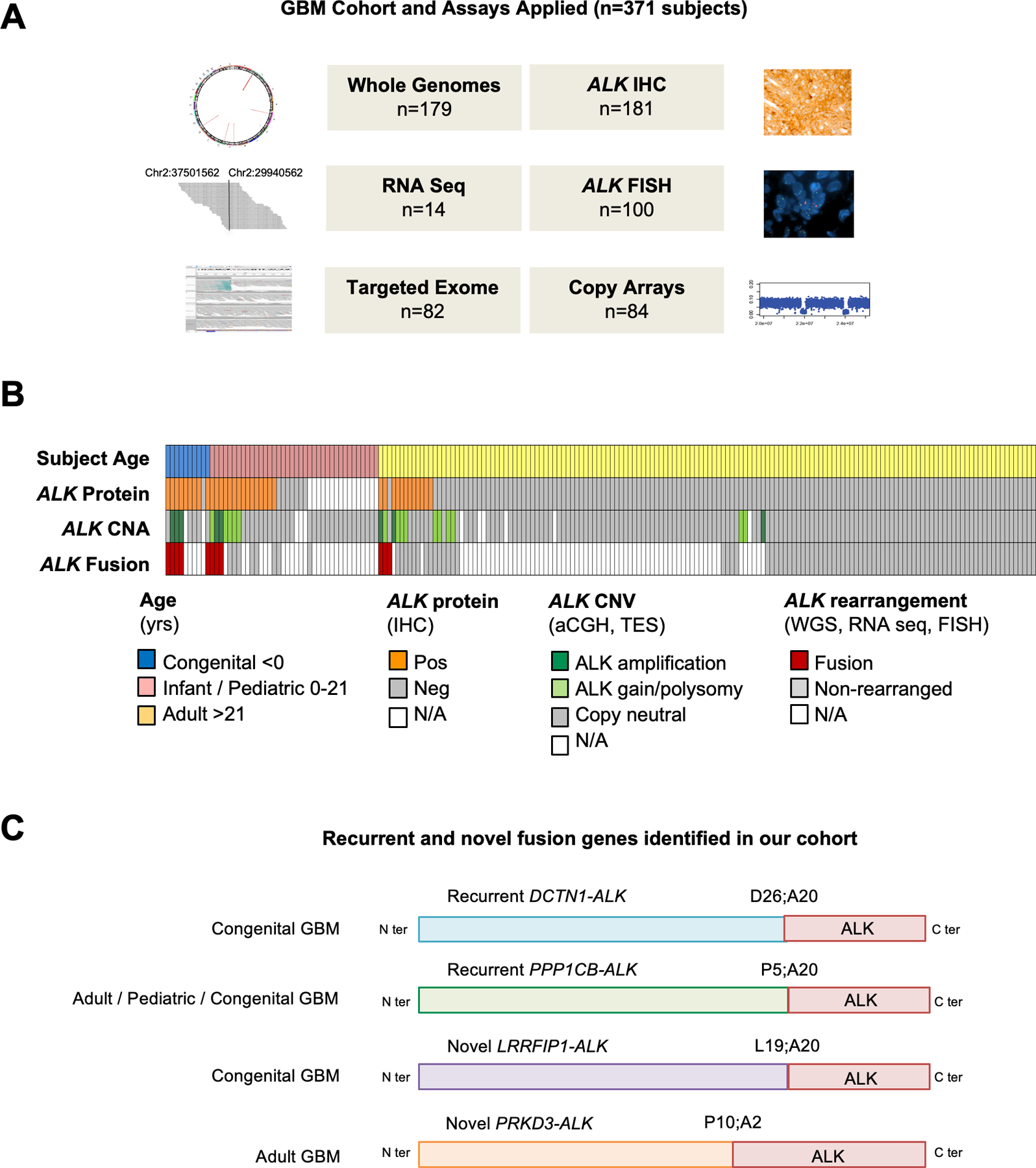
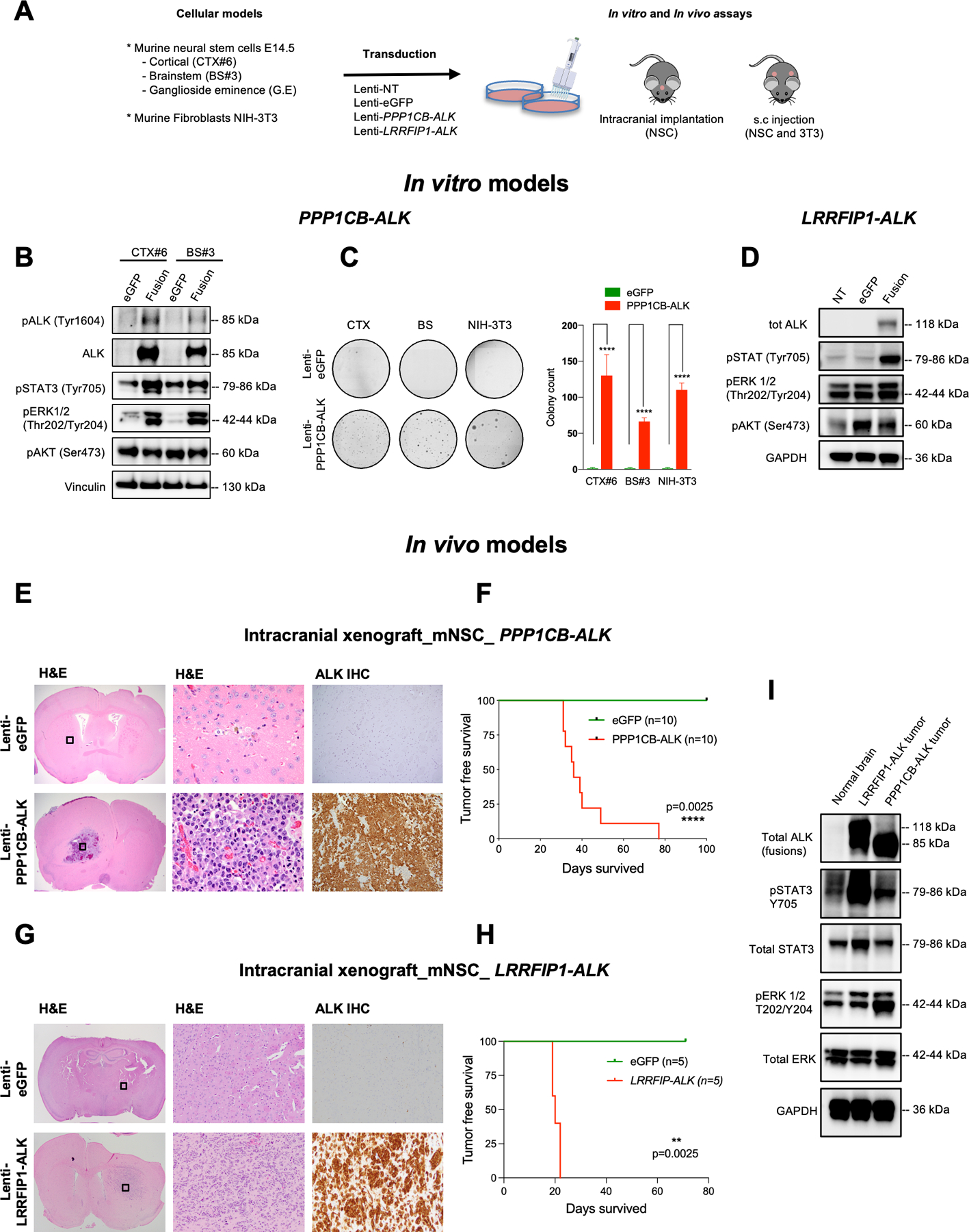
Experimental design: We performed a comprehensive analysis of ALK expression and genomic aberrations in both newly generated and retrospective data from 371 glioblastomas (156 adult, 205 infant/pediatric, and 10 congenital) with in vitro and in vivo validation of aberrations.

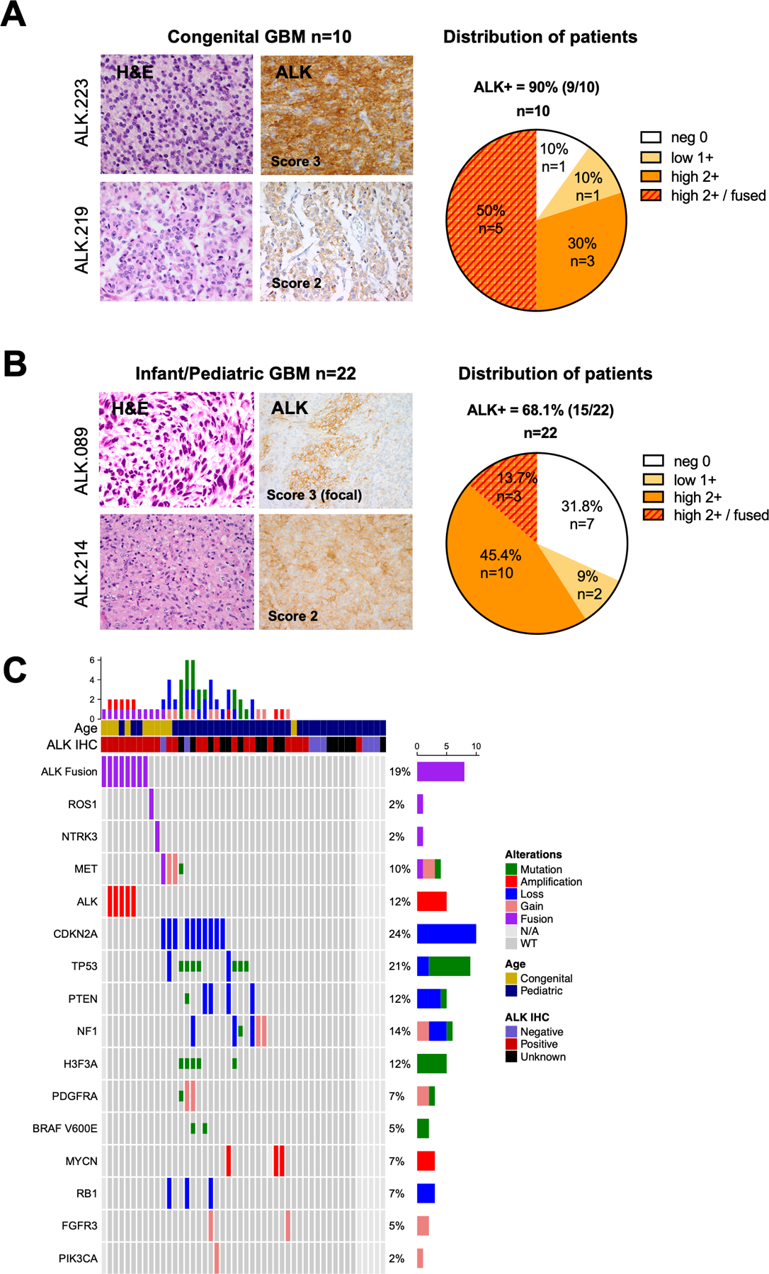
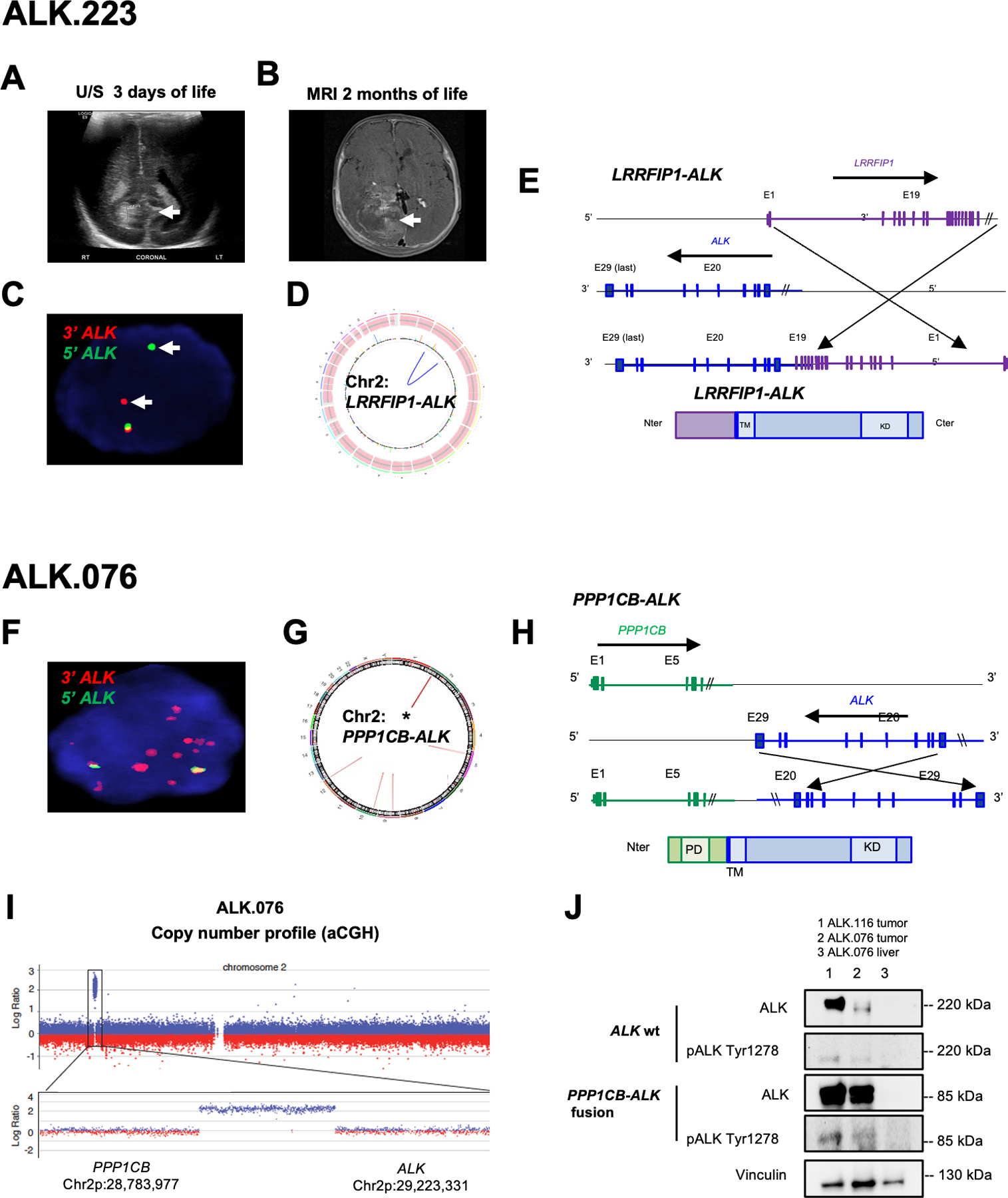
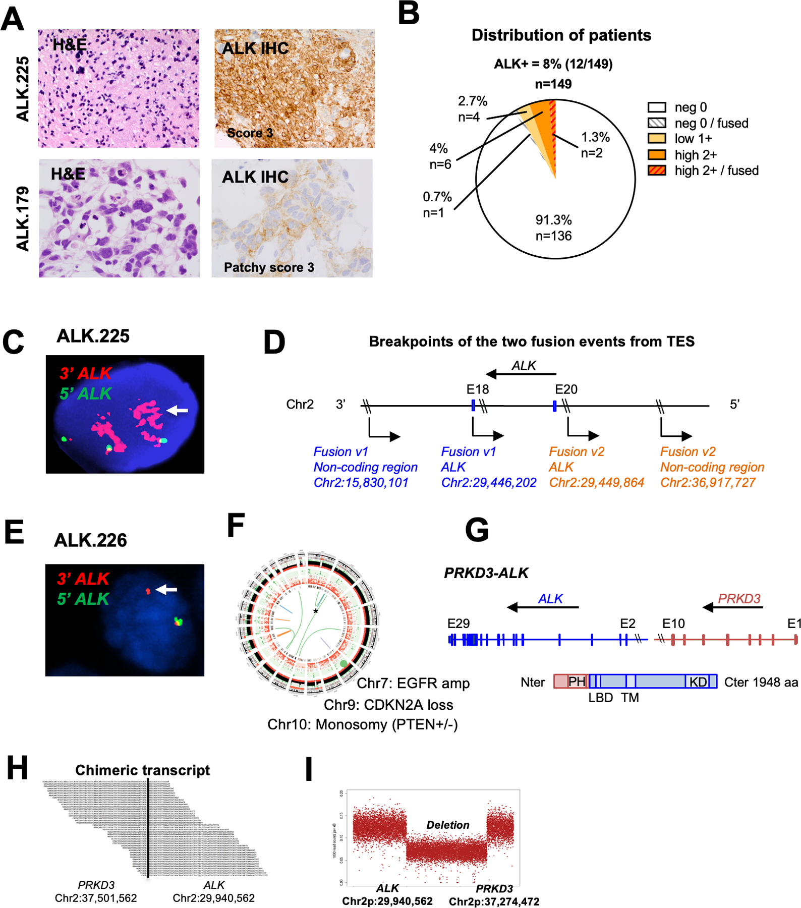
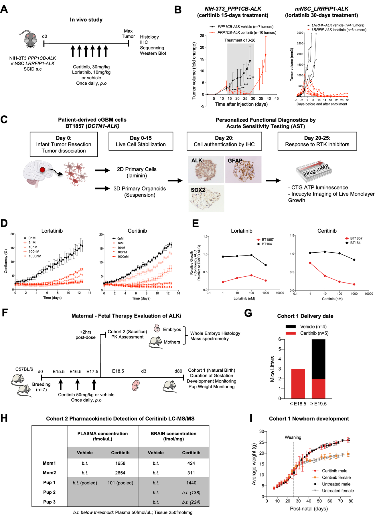
Results: ALK aberrations at the protein or genomic level were detected in 12% of gliomas (45/371) in a wide age range (0-80 years). Recurrent as well as novel ALK fusions (LRRFIP1-ALK, DCTN1-ALK, PRKD3-ALK) were present in 50% (5/10) of congenital/infant, 1.4% (3/205) of pediatric, and 1.9% (3/156) of adult GBMs. ALK fusions were present as the only candidate driver in congenital/infant GBMs and were sometimes focally amplified. In contrast, adult ALK fusions co-occurred with other oncogenic drivers. No activating ALK mutations were identified in any age group. Novel and recurrent ALK rearrangements promoted STAT3 and ERK1/2 pathways and transformation in vitro and in vivo. ALK-fused GBM cellular and mouse models were responsive to ALK inhibitors, including in patient cells derived from a congenital GBM. Relevant to the treatment of infant gliomas, we showed that ALK protein appears minimally expressed in the forebrain at perinatal stages, and no gross effects on perinatal brain development were seen in pregnant mice treated with the ALK inhibitor ceritinib.

Conclusions: These findings support use of brain-penetrant ALK inhibitors in clinical trials across infant, pediatric, and adult GBMs. See related commentary by Mack and Bertrand, p. 2567.


Figures

Figure 1.
Figure 1.. Overview of study and ALK aberrations in GBM.
A. ALK aberration screening was performed on 371 GBMs and HGGs using orthogonal assays including Whole Genome Sequencing (WGS), RNA sequencing (RNAS), Targeted Exome Sequencing (TES), immunohistochemistry (IHC), break apart FISH probes flanking 3’ and 5’ ALK regions, and copy number arrays (array CGH and SNP array). B. Oncoprint summary of ALK alterations in 198 GBMs with sequencing data (WGS or TES). 11/198 GBMs were found to have ALK rearrangements in this cohort. Tumors for which data are unavailable are designated N/A. C. Schematic structure of ALK fusion proteins identified in our GBM cohort.
Figure 2.
Figure 2.. ALK aberrations are common in congenital and pediatric GBMs.
A. H&E staining showing glioma features. ALK immunohistochemistry (IHC) showing typical 3+ (left top) and 2+ (left bottom) staining appearance; pie chart demonstrating the distribution of ALK IHC scores in the congenital GBM cohort (n = 10). See methods for detail scoring schema. B. H&E staining and ALK IHC of two illustrative pediatric GBMs scored as focally IHC 3+ (left top) and 2+ (left bottom); pie chart demonstrating the distribution of ALK IHC scores in the infant/pediatric GBM cohort (n = 22). C. Oncoprint of sequenced GBMs illustrating ALK fusion/amplification is the sole candidate driver alteration in pediatric GBMs. The second lane showed the ALK-positive samples by IHC. Genomic aberrations including fusions (red), focal amplifications (>log 2.0, >10Mb), deletions or mutations (green). The tumors ALK.219, ALK.227 and ALK.228 harbored NTRK3, MET and ROS1 fusions respectively but were negative for ALK fusion.
Figure 3.
Figure 3.. Congenital GBMs have novel and recurrent ALK fusions and amplifications as sole oncogenic drivers
A. Subject #223 head ultrasound at 3 days of life demonstrated periventricular abnormality (arrow) considered as hemorrhage which was monitored. B. MRI brain at 2 months showing large heterogeneous tumor within the right lateral ventricle (arrow) which was resected and diagnosed as GBM. C. ALK FISH analysis of ALK.223 tumor nucleus showing probe break apart indicating rearrangement but no evidence of copy change (red, 3’ ALK; green 5’ ALK; overlapping signals in yellow indicate normal locus; nucleus DAPI blue). D. Circos plot from whole genome sequencing showing intrachromosomal rearrangement of LRRFIP1-ALK and no evidence of other copy aberrations or rearrangements. E. Schematic representation of LRRFIP1-ALK variant which fused in-frame the first 19 exons of LRRFIP1 (N-ter, Chr 2q) to the last nine exons of ALK (C-ter, Chr 2p). F. FISH on showing amplification of the 3’ end of the ALK signal (red) in 5 week-old subject ALK.076 with a congenital GBM. G. Circos plot from WGS identified PPP1CB-ALK fusion as the sole aberration. H. Schematic representation of the PPP1CB-ALK variant which fuses the N-terminal portion of PPP1CB (including the phosphatase domain PD) to the intracellular region of ALK (containing the tyrosine kinase domain KD). I. Copy number analysis showing 116kb focal amplification event involving the 3’ end of ALK and the 5’ end of PPP1CB. J. Western showing PPP1CB-ALK protein expression at the expected molecular weight for the fusion (85 kd) compared to wild type ALK (220 kDa) and the presence of high pALKTyr1278 in fusion positive tumors (column 1 and 2; subjects ALK.116 and ALK.076 respectively). Liver tissue from ALK.076 (comumn 3) did not show expression of ALK wt or ALK fusion.
Figure 4.
Figure 4.. Identification of novel ALK fusions in adult GBMs.
A. H&E staining and ALK IHC of two illustrative adult GBMs scored as IHC 3+ (left top) and 2+ (left bottom); Histology of tumor ALK.225 and ALK.179 showing hallmarks of glioma and strong ALK expression by IHC (score 3+) (ALK.225) and focal moderate staining (ALK.179). B. Distribution of ALK IHC scores in the adult GBM cohort (n = 149). C. ALK FISH of tumor ALK.225 showing rearrangement and amplification of the ALK 3’ region (red). D. Schematic representation of the two ALK-noncoding genomic breakpoints. Fusion variant 1 (blue) occurred between ALK (in intron 20; breakpoint 2:29446202) and a non-coding genomic region on chromosome 2 (breakpoint 2:15830101). Fusion variant 2 (orange) occurred between ALK (exon 18; breakpoint 2:29449864) and a different non-coding region on chromosome 2 (breakpoint 2:36917727). E. ALK FISH of tumor ALK.226 showing evidence of rearrangement (arrow, single 3’ red signal) and one normal appearing allele (yellow). F. Circos plot of WGS for ALK.226 confirming novel PRKD3-ALK fusion co-occuring with classic adult GBM aberrations (EGFR amplification, CDKN2A loss, monosomy 10). G. Schematic representation of the PRKD3-ALK variant, which fuses the N-terminal portion of PRKD3 (including the Pleckstrin homology domain PH implicated in protein membrane recruitment and intracellular trafficking) to the C-terminal portion of ALK starting with exon 2 (including the ligand binding domain LBD, transmembrane TM and kinase KD domains). Amino acid number is indicated on the right. H. RNA sequencing of the PRKD3-ALK fusion demonstrates the novel breakpoints in intron 2 of ALK (chr2:29940562) and intron 10 of PRKD3 (chr2:37501562). I. Copy number analysis shows a 7.5 MB interstitial deletion with breakpoints consistent with fusion of PRKD3 and ALK in-frame.
Figure 5:
Figure 5:. Recurrent PPP1CB-ALK and novel LRRFIP1-ALK fusions are oncogenic drivers in vitro and in vivo.
A. Overview of PPP1CB-ALK assessments in vitro and in vivo using murine fibroblasts and mouse NSCs isolated from cortex (CTX), brainstem (BS) and ganglioside eminence (G.E). B. Representative western blot of PPP1CB-ALK expression and ALK Signaling Pathways in murine neural stem cells (mNSC) isolated from cortex (CTX) and brainstem (BS). Vinculin was used as a loading control. C. Assessment of the anchorage-independent growth ability of murine NSC and murine fibroblasts expressing PPP1CB-ALK fusion. Quantification of colony formation using CellProfiler. Values represent colony counts ± s.d. The mean of three independent replicates is shown. Error bars show standard error of the mean. Significance between eGFP and PPP1CB-ALK cells is determined by the Mann-Whitney test. ****P < 0.01. D. Representative western blot of LRRFIP1-ALK expression and ALK Signaling Pathways in murine neural stem cells (mNSC) isolated from cortex (CTX). GAPDH was used as a loading control. E. H&E and ALK IHC staining of representative mouse brains injected with NSC eGFP (upper row) compared to mNSC PPP1CB-ALK brains (bottom row). H&E boxed areas shown at higher magnification in center. F. Kaplan-Meier survival curves of SCID mice injected intracranially with mNSC CTX-PPP1CB-ALK (red, n = 10) or mNSC CTX-eGFP (green, n = 10). G. H&E and ALK IHC staining of representative mouse brains injected with mNSC eGFP (upper row) compared to mNSC LRRFIP1-ALK grafted brains (bottom row). H&E boxed areas shown at higher magnification in center. H. Kaplan-Meier survival curves of SCID mice injected intracranially with mNSC CTX-PPP1CB-ALK (red, n = 5) or mNSC CTX-eGFP (green, n = 5). I. Western blot of PPP1CB-ALK and LRRFIP1-ALK tumor lysates from mouse intracranial tumors compared to unrelated normal brain control showing ALK activation effects on STAT and ERK signaling. GAPDH was used as a loading control.
Figure 6.
Figure 6.. ALK fusions are sensitive to ALK inhibitors.
A. Study schematic for subcutaneous transplantation of PPP1CB-ALK-NIH-3T3 and LRRFIP-ALK-mNSC in SCID mice. Ceritinib and Lorlatinib treatments were initiated when tumor volume reached 200 +/− 200mm3 and continued for 15 and 30 consecutive days respectively. B. Caliper measurements of tumor volume in mice treated with vehicle (black curves) or ALK inhibitors (red curves). Shaded area denotes treatment range. Data are fold change +/− s.e.m of controls. C. Personalized functional diagnostic approach and acute sensitivity testing of cGBM cells (BT1857), bearing a DCTN1-ALK fusion. Live cells were isolated from the newborn tumor, dissociated, and stabilized for up to 15 days in culture as organoid/spheroid or 2D adherent cultures on laminin prior to authentication by IHC. Cells were tested for sensitivity to ALK and other kinase inhibitors by CellTiterGlo or Incucyte monospheroid/organoid growth assays. D. Incucyte growth profile of adherent cGBM DCTN1-ALK fused cells (BT1857) showing dose response to ALK inhibitors lorlatinib and ceritinib. Incucyte raw growth curves show confluency (%) over ~13 days of continuous imaging. E. DCTN1-ALK fused patient cells (BT1857) sensitivity to ALKi compared to an ALK wild-type GBM cell line (BT164). AUC curves of ALKi-treated cells versus DMSO control. F. Schematic outlining the administration of a brain penetrant ALK inhibitor, Ceritinib, to pregnant C57BL/6 mice to assess pharmacokinetics and neural developmental effects in perinatal period. G. Delivery dates of live pups for pregnant C57BL/6 mice treated with vehicle or ceritinib from E15.5 to E17.5 embryonic development stages. H. Quantification of ceritinib concentrations by LC-MS/MS in maternal, neonatal brain, plasma, and liver. I. Newborn’s weight in control and ceritinib-treated groups.

Comment in

References

    1. El-Ayadi M, Ansari M, Kühnöl CD, Bendel A, Sturm D, Pietsch T, et al. Occurrence of high-grade glioma in Noonan syndrome: Report of two cases. Pediatr Blood Cancer 2019;66:e27625. - PubMed
    1. Korostyshevskaya AM, Savelov AA, Papusha LI, Druy AE, Yarnykh VL. Congenital medulloblastoma: Fetal and postnatal longitudinal observation with quantitative MRI. Clin Imaging 2018;52:172–6. - PubMed
    1. Kostadinov S, de la Monte S. A Case of Congenital Brainstem Oligodendroglioma: Pathology Findings and Review of the Literature. Case Rep Neurol Med [Internet] 2017. [cited 2019 Jul 15];2017. Available from: https://www.ncbi.nlm.nih.gov/pmc/articles/PMC5549497/ - PMC - PubMed
    1. Anestis D, Tsitsopoulos P, Ble C, Tsitouras V, Tsonidis C. Congenital Glioblastoma Multiforme: An Unusual and Challenging Tumor. Neuropediatrics 2017;48:403–12. - PubMed
    1. Hallberg B, Palmer RH. Mechanistic insight into ALK receptor tyrosine kinase in human cancer biology. Nat Rev Cancer 2013;13:685–700. - PubMed

Publication types

Substances