. 2023 Feb 13;18(2):e0281550.
doi: 10.1371/journal.pone.0281550. eCollection 2023.

Impact of blood factors on endothelial cell metabolism and function in two diverse heart failure models

Affiliations

Impact of blood factors on endothelial cell metabolism and function in two diverse heart failure models

Young Song et al. PLoS One. .

Abstract

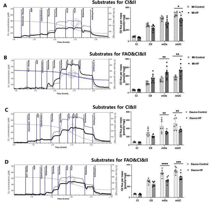
Role of blood-based factors in development and progression of heart failure (HF) is poorly characterized. Blood contains factors released during pathophysiological states that may impact cellular function and provide mechanistic insights to HF management. We tested effects of blood from two distinct HF models on cardiac metabolism and identified possible cellular targets of the effects. Blood plasma was obtained from daunorubicin- and myocardial infarction-induced HF rabbits (Dauno-HF and MI-HF) and their controls (Dauno-Control and MI-Control). Effects of plasma on bioenergetics of myocardial tissue from healthy mice and cellular cardiac components were assessed using high-resolution respirometry and Seahorse flux analyzer. Since endothelial cell respiration was profoundly affected by HF plasma, effects of plasma on endothelial cell barrier function and death were further evaluated. Western-blotting and electron microscopy were performed to evaluate mitochondrial proteins and morphology. Brief exposure to HF plasma decreased cardiac tissue respiration. Endothelial cell respiration was most impacted by exposure to HF plasma. Endothelial cell monolayer integrity was decreased by incubation with Dauno-HF plasma. Apoptosis and necrosis were increased in cells incubated with Dauno-HF plasma for 24 h. Down-regulation of voltage-dependent anion-selective channel (VDAC)-1, translocase of outer membrane 20 (Tom20), and mitochondrial fission factor (MFF) in cells exposed to Dauno-HF plasma and mitochondrial signal transducer and activator of transcription 3 (Stat3) and MFF in cells exposed to MI-HF plasma were observed. Mitochondrial structure was disrupted in cells exposed to HF plasma. These findings indicate that endothelial cells and mitochondrial structure and function may be primary target where HF pathology manifests and accelerates. High-throughput blood-based screening of HF may provide innovative ways to advance disease diagnosis and management.


Conflict of interest statement

The authors have declared that no competing interests exist.

Figures

Fig 1
Fig 1. High-resolution respirometry analysis of naive mouse left ventricular myocardial fibers exposed to heart failure rabbit blood plasma.
Naive mouse left ventricular tissue was incubated with the blood plasma from the heart failure (myocardial infarction- and daunorubicin-induced) rabbits or corresponding control rabbits in the O2K chamber for 10 min and assessed for respiration. The left panels show representative respirometry traces of oxygen flux (grey plots-Control, black plots-HF) in response to sequential administration of substrates and inhibitors. Respiration was measured as oxygen flux (= oxygen consumption rate) per tissue mass calculated by the negative slope of oxygen concentration with time (blue plots). The right panels are quantified respiratory function of complex I (CI) and II, maximum oxidative phosphorylation capacity (mOx), and maximum uncoupled capacity (mUC). Two different protocols of substrates were utilized—(A) and (C); glutamate and malate for CI, (B) and (D); octanoyl-l-carnitine (OCT), malate, and glutamate for CI and fatty acid oxidation (FAO). Assays utilized n = 6–8 plasma samples per group and 3 technical replicates per each plasma sample. Data are presented as means and SEM. Two-way ANOVA with Bonferroni post-hoc test was used. *P < 0.05, **P < 0.01, ***P < 0.001, and ****P < 0.0001 of HF vs. Control. Dauno = Daunorubicin; FCCP = carbonyl cyanide p-trifluoro methoxyphenylhydrazone; HF = heart failure; MI = myocardial infarction.
Fig 2
Fig 2. Mitochondrial stress test to assess oxygen consumption rates in cardiac cell types exposed to heart failure rabbit blood plasma.
Baseline-corrected oxygen consumption rate (OCR) traces of mitochondrial stress tests in the Seahorse extracellular flux analysis system on H9C2 myoblast (A), Ea.hy926 endothelial cells (B), NIH/3T3 fibroblasts (C), and RAW 264.7 macrophage (D) without serum starvation, and Ea.hy926 endothelial cells, 2 h serum-starved before the assay (E). After 3 measures of baseline OCR, the cells were exposed to the blood plasma from the heart failure rabbits or corresponding control rabbits for 30 min with 3 measures of OCR. Then, sequential addition of oligomycin, FCCP, and rotenone, and antimycin A was followed to determine ATP-linked respiration, maximal respiration, and non-mitochondrial respiration, respectively (left panels of each image, normalized to the baseline values). Quantified baseline-corrected OCR upon the plasma exposure, ATP-linked respiration, and maximal respiration at each cell type was compared between the HF and corresponding control group (right panels of each image). Assays utilized n = 6–8 plasma samples per group and 6–7 technical replicates per each plasma sample. Data are presented as means and SEM. Two-way ANOVA with Bonferroni post-hoc test was used. *P < 0.05, **P < 0.01, ***P < 0.001, ****P < 0.0001 of HF vs. Control. ATP = adenosine triphosphate; Dauno = Daunorubicin; FCCP = carbonyl cyanide p trifluoro methoxyphenylhydrazone; HF = heart failure; MI = myocardial infarction; OM = oligomycin; Rot /AMA = rotenone/antimycin A.
Fig 3
Fig 3. Endothelial cell permeability after exposure to daunorubicin heart failure rabbit blood plasma.
Rat heart microvascular endothelial cells were seeded to confluence, and monolayer permeability was measured by electric cell-substrate impedance sensing (ECIS). (A) Representative tracings of cell monolayers exposed to treatments. Cells were equilibrated and then exposed to treatment (arrow indicates at the point the treatment was added), and resistance was measured for ~24h. Representative tracings show the monitoring of the cells for 10h and are presented as the resistance normalized to the point before the addition of treatment (arrow). Both panels were treated with vehicle (1% PBS) or 1U/ml thrombin as negative and positive controls, respectively. Upper panel treatments include MI-control and MI-HF, and bottom panel treatments include Dauno-control and Dauno-HF. (B) Quantification of averaged changes in monolayer resistance at 5 h after treatment in each group. Assays utilized n = 5 plasma samples per group and 3 technical replicates per each plasma sample. Data are presented as means and STD. Unpaired Student’s t-test was used. *P < 0.05, **P < 0.01 of vehicle vs. blood plasma and HF vs. Control. Dauno = Daunorubicin; HF = heart failure; MI = myocardial infarction.
Fig 4
Fig 4. Viability of endothelial cells exposed to heart failure rabbit blood plasma.
Representative scatter plot flow cytometry of Ea.hy926 endothelial cells exposed to rabbit blood plasma for 1 h and 24 h, followed by staining with annexin-V and propidium iodide (PI) to detect apoptosis or necrosis at 1 (A) and 24 h (B) post exposure and comparison of the quantified proportion of early apoptotic cells (Annexin V+/PI-), late apoptotic cells (Annexin V+/PI+), and necrotic cells (Annexin V-/PI+) between those exposed to HF and corresponding control blood plasma. Assays utilized n = 5 plasma samples per group and 3 technical replicates per each plasma sample. Data are presented as means and SEM. Unpaired Student’s t-test was used. *P<0.05 and **P<0.01 in comparing the HF versus Control blood plasma. (C) LDH assay cytotoxicity detected in Ea.hy926 cells exposed to the HF and the corresponding control blood plasma for 1 h and 24 h, respectively. A % cytotoxicity was defined as (Plasma-treated LDH activity—Spontaneous LDH activity)/(Maximum LDH activity-Spontaneous LDH activity) x 100 (%). Assays utilized n = 5 plasma samples per group and 3 technical replicates per each plasma sample. Data are presented as means and SEM. Dauno = Daunorubicin; HF = heart failure; LDH = lactate dehydrogenase; MI = myocardial infarction.
Fig 5
Fig 5. Mitochondrial protein and ultrastructural changes in endothelial cells exposed to heart failure rabbit blood plasma.
Ea.hy926 endothelial cells were grown and serum-starved for 2 h, followed by exposure to the rabbit blood plasma for 1 h. Proteins were purified and subjected to western blot analysis. Representative blots (A) of samples exposed to five blood plasma from each group: MI-HF versus MI-control and Dauno-HF versus Dauno-control. GAPDH and COX IV were used as total protein loading control and mitochondrial loading control, respectively. Band intensity quantification (B) of the western blot normalized to GAPDH and COXIV. Assays utilized n = 5 plasma samples per group and 3 technical replicates per each plasma sample. Data are presented as means and SEM. Unpaired Student’s t-test was used. *P<0.05 and **P<0.01 in comparing the HF versus Control blood plasma. (C) Mitochondrial ultrastructural changes in endothelial cells exposed to HF rabbit blood plasma were determined. Ea.hy926 endothelial cells were grown and serum-starved for 2 h, followed by exposure to the rabbit blood plasma for 1 h. Narrow arrows indicate characteristic mitochondria in the representative cells from each group. Compared to the cells exposed to the MI-Control blood plasma showing normal mitochondrial morphology, those exposed to the MI-HF blood plasma show swollen, balloon-like mitochondria cristae disarrangement, partial cristolysis, and electron-lucent matrix. Cells exposed to the Dauno-Control blood plasma shows normal mitochondrial morphology, while the mitochondria of cells exposed to the Dauno-HF blood plasma shows swelling with loss of cristae and effacement of central architecture with the balloon-like, vesicular structures. Thick black arrows indicate lysed or ruptured outer membrane of mitochondria exposed to the HF blood plasma. Scale bars: 1 μm (left-side images) and 500 nm (right-sided images). Assays utilized n = 5 plasma samples per group and 3 technical replicates per each plasma sample. COXIV = Cytochrome C oxidase; Dauno = Daunorubicin; HF = heart failure. MI = myocardial infarction.

References

    1. Ibrahim NE, Januzzi JL Jr., Established and Emerging Roles of Biomarkers in Heart Failure. Circ Res. 2018; 123:614–629. doi: 10.1161/CIRCRESAHA.118.312706 - DOI - PubMed
    1. Rosca MG, Hoppel CL. Mitochondria in heart failure. Cardiovasc Res. 2010; 88:40–50. doi: 10.1093/cvr/cvq240 - DOI - PMC - PubMed
    1. Tyrrell DJ, Bharadwaj MS, Jorgensen MJ, Register TC, Molina AJA. Blood cell respirometry is associated with skeletal and cardiac muscle bioenergetics: Implications for a minimally invasive biomarker of mitochondrial health. Redox Biology. 2016; 10:65–77. doi: 10.1016/j.redox.2016.09.009 - DOI - PMC - PubMed
    1. Grip J, Jakobsson T, Tardif N, Rooyackers O. The effect of plasma from septic ICU patients on healthy rat muscle mitochondria. Intensive Care Med Exp. 2016; 4:20. doi: 10.1186/s40635-016-0093-2 - DOI - PMC - PubMed
    1. Boulos M, Astiz ME, Barua RS, Osman M. Impaired mitochondrial function induced by serum from septic shock patients is attenuated by inhibition of nitric oxide synthase and poly (ADP-ribose) synthase. Crit Care Med. 2003; 31:353–358. doi: 10.1097/01.CCM.0000050074.82486.B2 - DOI - PubMed

Publication types