. 2023 Feb 21;120(8):e2211703120.
doi: 10.1073/pnas.2211703120. Epub 2023 Feb 13.

Helminth egg derivatives as proregenerative immunotherapies

Affiliations

Helminth egg derivatives as proregenerative immunotherapies

David R Maestas Jr et al. Proc Natl Acad Sci U S A. .

Abstract

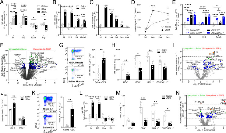
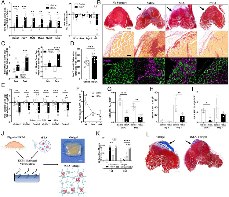
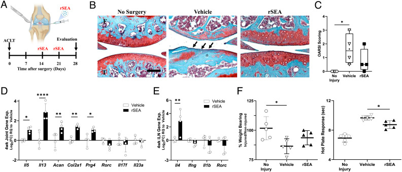
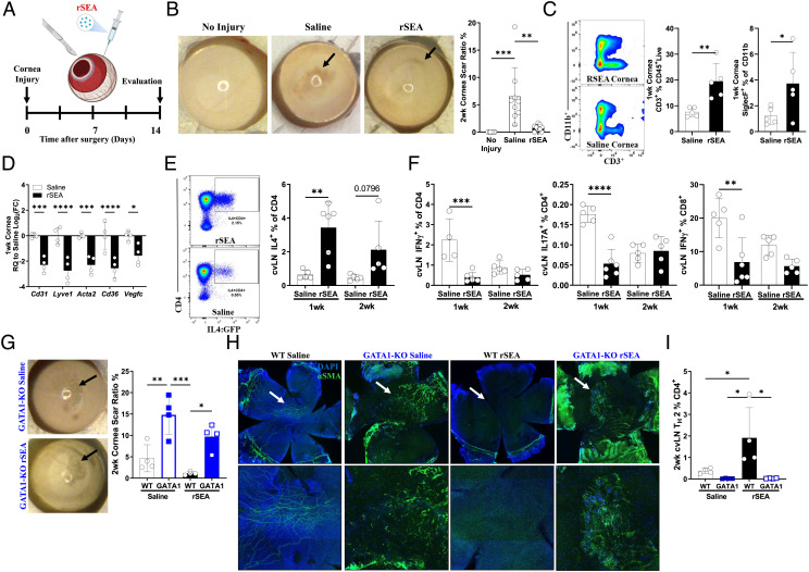
The immune system is increasingly recognized as an important regulator of tissue repair. We developed a regenerative immunotherapy from the helminth Schistosoma mansoni soluble egg antigen (SEA) to stimulate production of interleukin (IL)-4 and other type 2-associated cytokines without negative infection-related sequelae. The regenerative SEA (rSEA) applied to a murine muscle injury induced accumulation of IL-4-expressing T helper cells, eosinophils, and regulatory T cells and decreased expression of IL-17A in gamma delta (γδ) T cells, resulting in improved repair and decreased fibrosis. Encapsulation and controlled release of rSEA in a hydrogel further enhanced type 2 immunity and larger volumes of tissue repair. The broad regenerative capacity of rSEA was validated in articular joint and corneal injury models. These results introduce a regenerative immunotherapy approach using natural helminth derivatives.

Keywords: biomaterials; helminth; immune response; regenerative medicine; tissue engineering.


Conflict of interest statement

The authors have organizational affiliations and stock ownership to disclose, J.H.E. holds equity in Unity Biotechnology and Aegeria Soft Tissue and is a consultant for Tessara. D.M.P. is consultant at Aduro Biotech, Amgen, Astra Zeneca, Bayer, Compugen, DNAtrix, Dynavax Technologies Corporation, Ervaxx, FLX Bio, Immunomic, Janssen, Merck, and Rock Springs Capital. D.M.P. holds equity in Aduro Biotech, DNAtrix, Ervaxx, Five Prime therapeutics, Immunomic, Potenza, Trieza Therapeutics. D.M.P. is a member of the scientific advisory board for Bristol Myers Squibb, Camden Nexus II, Five Prime Therapeutics, and WindMil. D.M.P. is a member of board of directors in Dracen Pharmaceuticals.

Figures

Fig. 1.
Fig. 1.
rSEA treatment promotes a proregenerative type 2 immune microenvironment after muscle injury. Mice had a partial quadriceps resection to create VML injury and received the indicated treatments administered locally at the time of resection. (A and B) Muscle tissue expression of selected TH2 and TH1 genes assessed by qRT–PCR 1 wk after VML and treatment with saline, unfractionated soluble SEA extract and fractionated SEA formulation (regenerative SEA, rSEA). (CIl4 gene expression of iLNs from VML-injured mice at different time points after rSEA treatment. (D) Flow cytometric quantitation of postinjury muscle eosinophils, defined by coexpression of Siglec F and CD11b, over time with rSEA treatment. (E) Muscle expression of selected type 2 genes after saline vs. rSEA treatment of WT and GATA1 KO mice. (F) Volcano plot of differential expression from bulk RNA sequencing of the muscle 1 wk postinjury and rSEA treatment referenced to saline-treated injuries by the EdgeR analysis. (G) Flow cytometry of muscle CD3+CD4+ cells from IL4 reporter (4get) mice treated with either saline or rSEA. (H) Muscle IFNγ+ production by various immune cell types assayed by ICS. (I) Gene expression profile of sorted CD3+ T cells from the muscle 1 wk postinjury comparing saline and rSEA. (J) CD4+Foxp3+ (Tregs) in the muscle 5 and 7 d after treatment with either saline or rSEA. (K) Flow plots of iLNs from 4get mice draining VML treated 1 wk with saline or rSEA. (L) iLN gene expression 1 wk after muscle injury and treatment. (M) iLN IFNγ production by various cell types assayed by ICS. (N) Gene expression profiling of CD11b+F4/80+Hi macrophages sorted from the muscle 1 wk after saline vs. rSEA treatment. Statistical tests represent all biological replicates, and all experiments excluding (F, I, and M) were replicated at least twice. Graphs show mean ± SD (AD, G, H, and JM) and mean ± SEM, n = 3 to 5 (E). *P < 0.05, **P < 0.01, ***P < 0.001, ****P < 0.0001 by unpaired two-tailed Student’s t test (G and K), and two-way ANOVA with Sidak’s multiple comparisons. For volcano plot differential expression (F, I, and N), the EdgeR analysis was performed for bulk sequencing FDR (F), and sorted cell NanoString analysis FDR values determined by the Benjamini–Yekutieli method (I and N). Dashed lines (F) represent -log10(FDR) < 0.05 and 0.01 and in (I and N) represent -log10(adjusted P value) significance determined by nCounter software (adjusted P < 0.5, 0.1, 0.05, and 0.01, respectively).
Fig. 2.
Fig. 2.
rSEA promotes skeletal muscle repair and decreases fibrosis and type 3 immune responses. (A) One-week postinjury expression of genes involved in muscle regeneration and associated with TH17 cells in mice treated with saline or rSEA. (B) Six-week postinjury muscle histology treated with either saline or rSEA and stained with Masson’s trichrome, picrosirius red, and immunofluorescent staining [nuclei = DAPI (purple) and mature muscle = dystrophin (green)]. Arrows indicate site of original muscle resection. (C) Muscle gene expression levels of adipose browning-associated genes at 1 and 3 wk after injury and treatment with saline or rSEA. (D) Treadmill exhaustion functional testing of mice at 6 wk after injury and treatment with saline or rSEA. (E) Gene expression of various fibrosis-associated collagens at 6 wk after injury and treatment with saline or rSEA. (F) Percentage of CD4 cells expressing IL-17A at 1, 3, and 6 wk after injury and treatment with saline or rSEA as determined by ICS. (G) Percentage of CD4 cells expressing IL-17A by ICS in iLN 1-wk postmuscle injury and treatment with saline or rSEA. (H) Percentage of γδ T cells expressing IL-17A in the muscle at 1 wk after injury and treatment. (I) Percentage of γδ T cells expressing IL-17A in iLN at 1 wk after injury and treatment with saline or rSEA. (J) Production summary and gross image of vitrified SIS-ECM combined with rSEA. (K) Gene expression of Il4 in the muscle for 1, 3, and 6 wk after injury and treatment with saline, rSEA, pure vitrigel, or vitrigel formulated with rSEA. (L) Transverse histological sections of muscles stained with Masson’s trichrome 6 wk postinjury and treatment with saline, pure vitrigel, or vitrigel formulated with rSEA. Statistical tests represent all biological replicates, and all experiments were replicated at least twice. Graphs show mean ± SD (A, CI, and K), n = 3 to 4. *P < 0.05, **P < 0.01, ***P < 0.001, and ****P < 0.0001 by two-way ANOVA with Sidak’s multiple comparisons. (Scale bars, 100 μm (B), 1 cm (J), and 1 mm (L).
Fig. 3.
Fig. 3.
rSEA immunotherapy promotes repair in articular joint injuries. (A) ACLT injury model timeline. (B) Representative images from safranin-O stains of uninjured joints (no surgery) or injured joints treated with vehicle or rSEA 4 wk post-ACLT. Arrows indicate where cartilage damage has occurred. (C) OARSI scoring of uninjured joints or injured joints treated with vehicle or rSEA 4 wk post-ACLT. (D), Articular joint gene expression of type 2, type 3 immune genes, and cartilage ECM genes Aggrecan (Acan), type 2 collagen (Col2a1), and Lubricin (Prg4) 4 wk post-ACLT treated with vehicle or rSEA. (E) iLN gene expression of type 2 and type 3 immune genes 4 wk post-ACLT. (F) Hot plate reaction times and weight-bearing assessments in mice without injury or 4 wk post-ACLT treated with vehicle (saline) or rSEA. Statistical tests represent all biological replicates, and all experiments were replicated at least twice. Graphs show mean ± SD (CE), n = 4 to 5, box and whisker plot with median as central line, and “+” designates mean (C). *P < 0.05, **P < 0.01, ***P < 0.001, and ****P < 0.0001 by one-way ANOVA with Tukey’s multiple comparisons (C and F) and two-way ANOVA with Sidak’s multiple comparisons (D and E). (Scale bar, 100 μm (B).)
Fig. 4.
Fig. 4.
rSEA immunotherapy promotes repair in corneal injury. (A) Corneal debridement injury model timeline. (B) Corneal gross images and scar ratio assessment at 2 wk after injury treated with saline vehicle or rSEA, two study replications combined. (C) Flow cytometry populations isolated from the cornea 1 wk postinjury and treatment with saline or rSEA. (D) Corneal expression of genes associated with scar vascularization at 1 wk after injury and treatment with saline or rSEA. (E) Representative flow plots of 2 wk after injury 4get cervical LNs (cvLN) and % TH2 populations at 1 wk vs. 2 wk after injury and treatment with saline or rSEA. (F) ICS from cvLN of TH1, TH17, and IFNγ+CD8+ T cells (%) at 1 and 2 wk after injury and treatment with saline or rSEA. (G) Representative gross images of wounded corneas from GATA1 KO mice treated 2 wk with saline or rSEA and scar area ratio assessment. (H) Immunofluorescent staining of nuclei (DAPI, blue) and α-SMA (green) on corneas 2 wk postinjury and treatment with saline or rSEA in WT vs. GATA1 KO mice. (I) cvLN ICS for IL-4 in WT vs. GATA1 KO mice 2 wk postinjury and treatment with saline or rSEA. Statistical tests represent all biological replicates, and all experiments were replicated at least twice. Graphs show mean ± SD (BF, G, and I), n = 4 to 6. *P < 0.05, **P < 0.01, ***P < 0.001, and ****P < 0.0001 by one-way ANOVA with Tukey’s multiple comparisons (B) and two-way ANOVA with Sidak’s multiple comparisons (DF, G, and I).

References

    1. Krueger T. E. G., Thorek D. L. J., Denmeade S. R., Isaacs J. T., Brennen W. N., Concise review: Mesenchymal stem cell-based drug delivery: The good, the bad, the ugly, and the promise. Stem Cells Transl Med. 7, 651–663 (2018). - PMC - PubMed
    1. Chabannon C., et al. , Hematopoietic stem cell transplantation in its 60s: A platform for cellular therapies. Sci. Transl Med. 10, eaap9630 (2018). - PubMed
    1. Laurent P., et al. , Immune-mediated repair: A matter of plasticity. Front. Immunol. 8, 454 (2017). - PMC - PubMed
    1. Sadtler K., et al. , Developing a pro-regenerative biomaterial scaffold microenvironment requires T helper 2 cells. Science 352, 366–370 (2016). - PMC - PubMed
    1. Heredia J. E., et al. , Type 2 innate signals stimulate fibro/adipogenic progenitors to facilitate muscle regeneration. Cell 153, 376–388 (2013). - PMC - PubMed

Publication types