. 2023 Jun;48(6):1958-1970.
doi: 10.1007/s11064-023-03888-x. Epub 2023 Feb 13.

Anti-inflammatory Action of BT75, a Novel RARα Agonist, in Cultured Microglia and in an Experimental Mouse Model of Alzheimer's Disease

Affiliations

Anti-inflammatory Action of BT75, a Novel RARα Agonist, in Cultured Microglia and in an Experimental Mouse Model of Alzheimer's Disease

Xiuli Zhang et al. Neurochem Res. 2023 Jun.

Abstract

BT75, a boron-containing retinoid, is a novel retinoic acid receptor (RAR)α agonist synthesized by our group. Previous studies indicated that activation of retinoic acid (RA) signaling may attenuate progression of Alzheimer's disease (AD). Presently, we aimed to examine the anti-inflammatory effect of BT75 and explore the possible mechanism using cultured cells and an AD mouse model. Pretreatment with BT75 (1-25 µM) suppressed the release of nitric oxide (NO) and IL-1β in the culture medium of mouse microglial SIM-A9 cells activated by LPS. BMS195614, an RARα antagonist, partially blocked the inhibition of NO production by BT75. Moreover, BT75 attenuated phospho-Akt and phospho-NF-κB p65 expression augmented by LPS. In addition, BT75 elevated arginase 1, IL-10, and CD206, and inhibited inducible nitric oxide synthase (iNOS) and IL-6 formation in LPS-treated SIM-A9 cells, suggesting the promotion of M1-M2 microglial phenotypic polarization. C57BL/6 mice were injected intracerebroventricularly (icv) with streptozotocin (STZ) (3 mg/kg) to provide an AD-like mouse model. BT75 (5 mg/kg) or the vehicle was intraperitoneally (ip) injected to icv-STZ mice once a day for 3 weeks. Immunohistochemical analyses indicated that GFAP-positive cells and rod or amoeboid-like Iba1-positive cells, which increased in the hippocampal fimbria of icv-STZ mice, were reduced by BT75 treatment. Western blot results showed that BT75 decreased levels of neuronal nitric oxide synthase (nNOS), GFAP, and phosphorylated Tau, and increased levels of synaptophysin in the hippocampus of icv-STZ mice. BT75 may attenuate neuroinflammation by affecting the Akt/NF-κB pathway and microglial M1-M2 polarization in LPS-stimulated SIM-A9 cells. BT75 also reduced AD-like pathology including glial activation in the icv-STZ mice. Thus, BT75 may be a promising anti-inflammatory and neuroprotective agent worthy of further AD studies.

Keywords: Boron-containing retinoid; Microglial polarization; Neurodegenerative disease; Neuroinflammation; Nitric oxide; Retinoic acid receptor.


Conflict of interest statement

Competing Interests: The authors have no relevant financial and non-financial interests to disclose.

Figures

Fig. 1.
Fig. 1.
BT75 chemical structure and its synthetic formula.
Fig. 2.
Fig. 2.
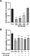
Effects of BT75 on the cytotoxicity and cell viability of SIM-A9 cells stimulated by LPS. (A) Cell viability of SIM-A9 cells treated with BT75 (500, 250, 100 and 50 μM) for 24 h. The results are expressed as the mean ± SD (n = 8). **p < 0.01 as compared with control group. (B) The cell viability of SIM-A9 cells pretreated with BT75 (25, 10, 5 and 1 μM) for 30 min followed by treatment with LPS (1 μg/mL) for another 24 h. Cell viability was assessed using the MTT assay. The results are expressed as the mean ± SD (n = 8). ##p < 0.01 as compared with control group, **p < 0.01 as compared with LPS-treated group.
Fig. 3.
Fig. 3.
Effects of BT75 on NO production and inflammatory factors induced by LPS in SIM-A9 cells. SIM-A9 cells were pretreated with BT75 (25, 10, 5 and 1 μM) for 30 min followed by treatment with LPS (100 ng/mL) for another 24 h. (A) NO production in the cell culture medium was assessed by the Griess method. TNF-α (B), IL-1β (C), and IL-10 (D) levels in the cell culture medium were detected by ELISA. The results are expressed as the mean ± SD (n = 4–5). ##p < 0.01 as compared with control group, *p < 0.05 and **p<0.01 as compared with LPS-treated group.
Fig. 4.
Fig. 4.
BT75 may ameliorate inflammation induced by LPS in SIM-A9 cells via regulation of the AKT/NF-κB pathway. Cells were pretreated with BT75 (10μM) for 30 min before 100 ng/ml LPS treatment for 24 h. Akt/p-AKT and NF-κb/p- NF-κb expression in the cells were detected by Western blot. (A) Represented immunoblot bands for p-Akt, Akt, p- NF-κB, NF-κB, and β-actin. (B and C) Quantitative analysis of immunoblot bands. Protein expression levels were normalized to β-actin. The data were expressed as mean ± SD (n = 3–4). ##p < 0.01 as compared with control group, *p < 0.05 as compared with LPS-treated group.
Fig. 5.
Fig. 5.
BT75 ameliorated inflammation induced by LPS in SIM-A9 cells via promoting M1-M2 phenotypic polarization. Cells were pretreated with BT75 (10 μM) for 30 min before 100 ng/ml LPS treatment for 24 h. iNOS, IL-6, Arg1, and CD206 expression in the cells were detected by Western blot. (A) Representative immunoblots for iNOS, IL-6, Arg-1, CD206, and β-actin. (B to E) Quantitative analysis of immunoblot bands. Protein expression levels were normalized to β-actin. The data were expressed as mean ± SD (n = 3–4). ##p < 0.01 as compared with control group, **p < 0.01 as compared with LPS-treated group.
Fig. 6.
Fig. 6.
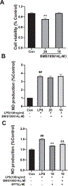
BMS195614, an RARα antagonist, partially blocked the inhibition of NO production by BT75 in LPS-activated SIM-A9 cells. Cells were pretreated with BMS195614 (10 μM) for 30 min and then incubated with BT75 (10 μM) for another 30 min. Then, LPS (100 ng/ml) was added and incubated for 24 h. (A) The cytotoxicity test of BMS195614. (B) Effect of BMS195614 on NO production in LPS-induced SIM-A9 cells. (C) BMS195614 partially blocked the inhibition of NO production by BT75 in LPS-activated SIM-A9 cells. The data were expressed as mean ± SD (n = 4). ##p < 0.01 as compared with control group, **p < 0.01 as compared with LPS-treated group. &&p<0.01 as compared to LPS+BT75 group.
Fig. 7.
Fig. 7.
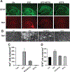
BT75 reduced icv-STZ-induced glial activation in mouse brain. (A) GFAP-positive astrocytes and Iba1-positive microglia in the dorsal hippocampus are shown here. The representative image from each group, Ctr (n=3), STZ (n=3), STZ+BT75 (n=3), and BT75 (n=4) group, is shown here. The bar indicates 500 μm. (B) Iba1-positive microglia in the hippocampus fimbria are shown. The bar indicates 50 μm. (C) Larger round or rod-like cell densities in the fimbria were compared among the control, STZ, STZ+BT75, and BT75 groups. * indicates that the STZ group is different from all other groups significantly (p<0.05). (D) The densities of GFAP-positive cells were compared among the control, STZ, STZ+BT75, and BT75 groups. * indicates that the STZ group is different from all other groups significantly (p<0.05).
Fig. 8.
Fig. 8.
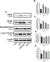
Effect of BT75 on nNOS, GFAP, synaptophysin and p-Tau expression in the hippocampus of icv-STZ-treated mice. (A) Representative immunoblot bands for synaptophysin and p-Tau and β-actin. (B and C) Quantitative analysis of immunoblot bands. Protein expression levels were normalized to β-actin. The data were expressed as mean ± SD (n = 3). #p < 0.05 as compared with control group, *p < 0.01 as compared with STZ-treated group.
Fig. 9.
Fig. 9.
BT75 had no protective effect on STZ-induced Neuro-2a cytotoxicity. (A) Cell viability of Neuro-2a cells treated with BT75 (1mM, 100μm, 10μm and 1 μM) for 24 h. (B) Cell viability of Neuro-2a cells treated with STZ (1mM, 500μm, 100μm, 50μm and 10 μM) for 24 h. (C) The cell viability of Neuro-2a cells pretreated with BT75 (25, 10, 5 and 1 μM) for half an hour followed by treatment with STZ (1 mM) for another 24 h. Cell viability was assessed using the MTT assay. The results are expressed as the mean ± SD (n = 4). **p < 0.01 as compared with control group.

References

    1. Jessen F, Amariglio RE, Buckley RF, van der Flier WM, Han Y, Molinuevo JL, Rabin L, Rentz DM, Rodriguez-Gomez O, Saykin AJ et al.: The characterisation of subjective cognitive decline. Lancet Neurol 2020, 19(3):271–278. - PMC - PubMed
    1. Tiwari S, Atluri V, Kaushik A, Yndart A, Nair M: Alzheimer’s disease: pathogenesis, diagnostics, and therapeutics. Int J Nanomedicine 2019, 14:5541–5554. - PMC - PubMed
    1. Lashley T, Schott JM, Weston P, Murray CE, Wellington H, Keshavan A, Foti SC, Foiani M, Toombs J, Rohrer JD et al.: Molecular biomarkers of Alzheimer’s disease: progress and prospects. Dis Model Mech 2018, 11(5). - PMC - PubMed
    1. Revi M: Alzheimer’s Disease Therapeutic Approaches. Adv Exp Med Biol 2020, 1195:105–116. - PubMed
    1. Ozben T, Ozben S: Neuro-inflammation and anti-inflammatory treatment options for Alzheimer’s disease. Clin Biochem 2019, 72:87–89. - PubMed

MeSH terms