

Genetic analysis of heterogeneous subsets of circulating tumour cells from high grade serous ovarian carcinoma patients
- PMID: 36781954
- PMCID: PMC9925814
- DOI: 10.1038/s41598-023-29416-z
Genetic analysis of heterogeneous subsets of circulating tumour cells from high grade serous ovarian carcinoma patients
Abstract
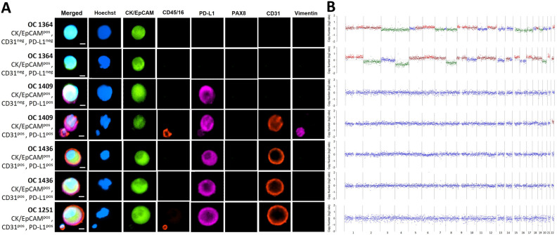
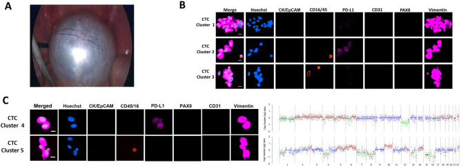
Circulating tumour cells (CTCs) are heterogenous and contain genetic information from the tumour of origin. They bear specific intra- and extra-cellular protein markers aiding in their detection. However, since these markers may be shared with other rare cells in the blood, only genetic testing can confirm their malignancy. Herein, we analyse different CTC subsets using single cell whole genome DNA sequencing to validate their malignant origin. We randomly selected putative CTCs identified by immunostaining that were isolated from 4 patients with high grade serous ovarian cancer (HGSOC) and one with benign cystadenoma. We specifically targeted CTCs positive for epithelial (CK/EpCAMpos), mesenchymal (vimentinpos), and pseudoendothelial (CK/EpCAMpos plus CD31pos) markers. We isolated these cells and performed whole genome amplification (WGA) and low-pass whole-genome sequencing (LP-WGS) for analysis of copy number alterations (CNA). Of the CK/EpCAMpos cells analysed from the HGSOC patients, 2 of 3 cells showed diverse chromosomal CNAs. However, the 4 pseudoendothelial cells (CK/EpCAMpos plus CD31pos) observed in the HGSOC cases did not carry any CNA. Lastly, two of the clusters of vimentin positive cells sequenced from those found in the benign cystadenoma case had CNA. Despite the low number of cells analysed, our results underscore the importance of genetic analysis of putative CTCs to confirm their neoplastic origin. In particular, it highlights the presence of a population of CK/EpCAMpos cells that are not tumour cells in patients with HGSOC, which otherwise would be counted as CTCs.
© 2023. The Author(s).
Conflict of interest statement
The authors declare no competing interests.
Figures
References
-
- Pantel K, Alix-Panabières C. Liquid biopsy and minimal residual disease—Latest advances and implications for cure. Nat. Rev. Clin. Oncol. 2019;16(7):409–424. - PubMed
-
- Pantel K, Brakenhoff RH. Dissecting the metastatic cascade. Nat. Rev. Cancer. 2004;4:448–456. - PubMed
-
- Mittal V. Epithelial mesenchymal transition in tumor metastasis. Annu. Rev. Pathol. 2018;13:395–412. - PubMed
Publication types
MeSH terms
Substances
LinkOut - more resources
Full Text Sources
Medical
Research Materials
Miscellaneous
