. 2023 Feb 13;13(1):2543.
doi: 10.1038/s41598-023-29853-w.

Reversal of aging-associated increase in myelopoiesis and expression of alarmins by angiotensin-(1-7)

Affiliations

Reversal of aging-associated increase in myelopoiesis and expression of alarmins by angiotensin-(1-7)

Kishore Chittimalli et al. Sci Rep. .

Abstract

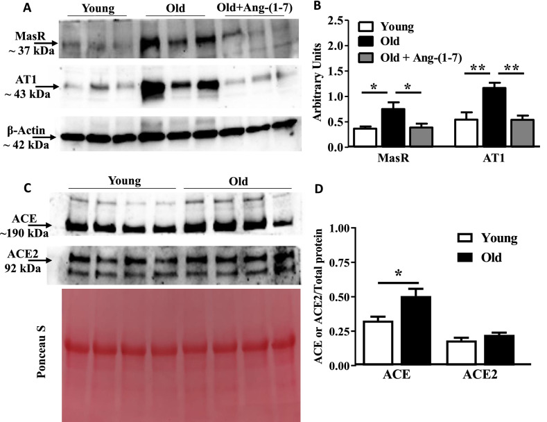
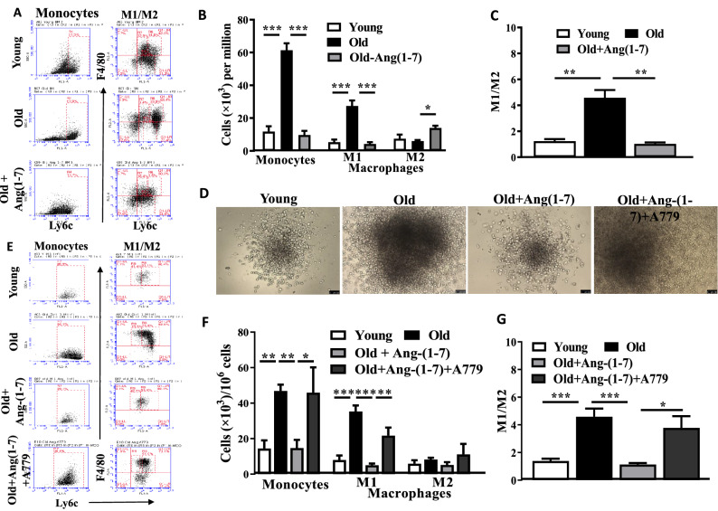
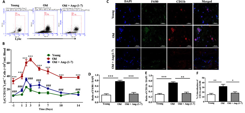
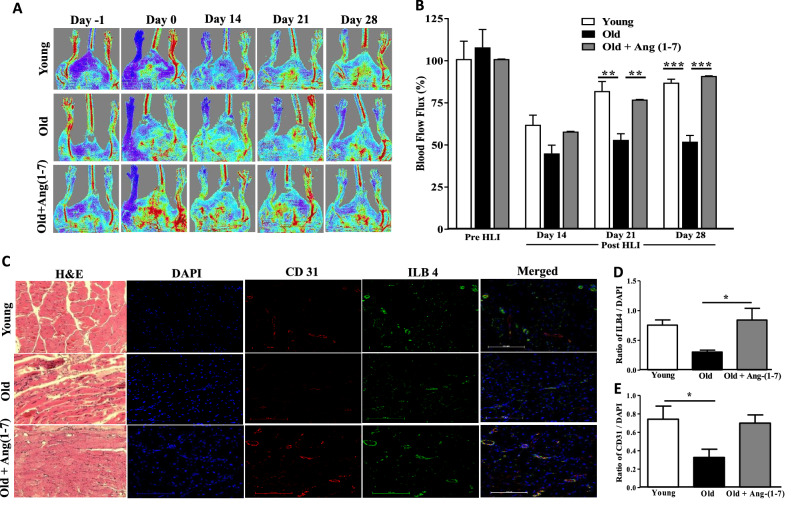
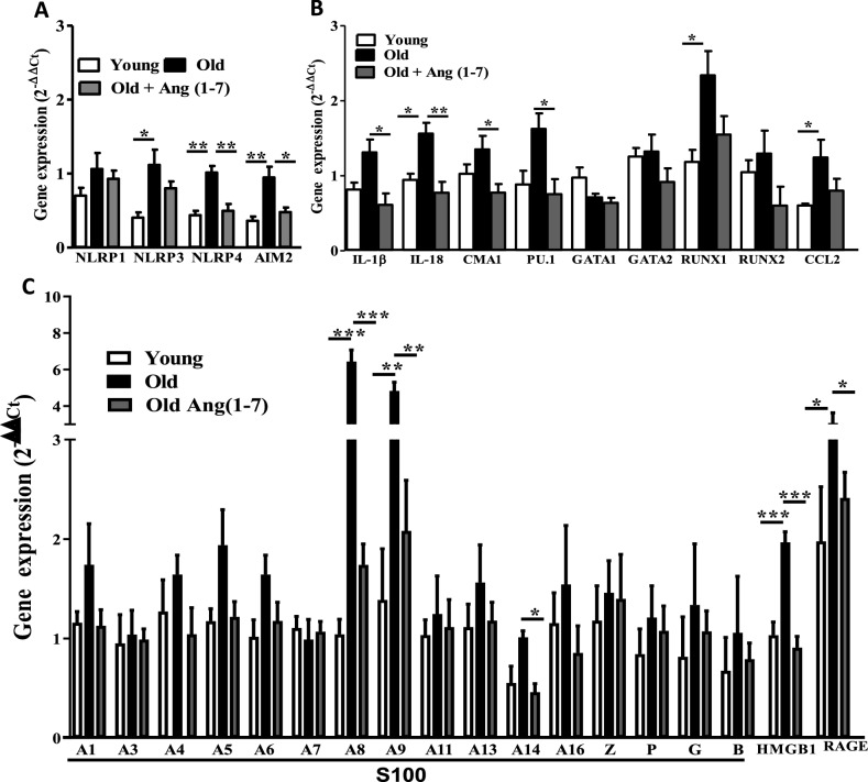
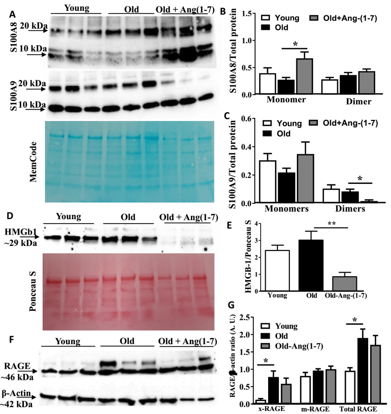
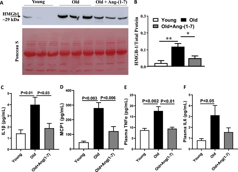
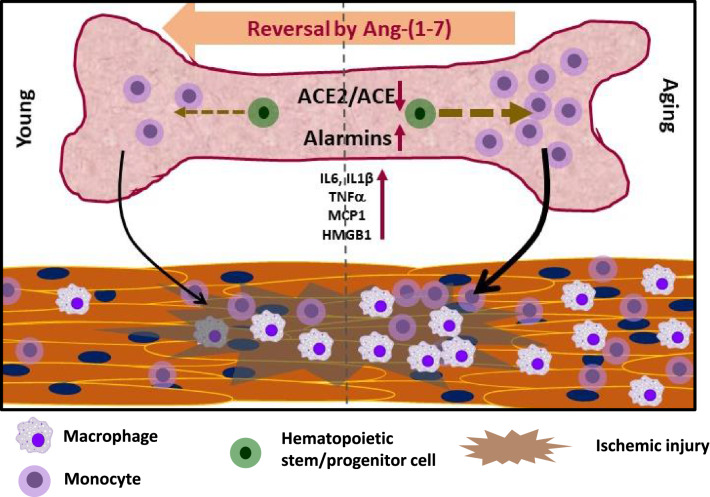
Aging is associated with chronic systemic inflammation largely due to increased myelopoiesis, which in turn increases risk for vascular disease. We have previously shown evidence for the therapeutic potential of Angiotensin-(1-7) (Ang-(1-7)) in reversing vasoreparative dysfunction in aging. This study tested the hypothesis that ischemic vascular repair in aging by Ang-(1-7) involves attenuation of myelopoietic potential in the bone marrow and decreased mobilization of inflammatory cells. Young or Old male mice of age 3-4 and 22-24 months, respectively, received Ang-(1-7) (1 µg/kg/min, s.c.) for four weeks. Myelopoiesis was evaluated in the bone marrow (BM) cells by carrying out the colony forming unit (CFU-GM) assay followed by flow cytometry of monocyte-macrophages. Expression of pro-myelopoietic factors and alarmins in the hematopoietic progenitor-enriched BM cells was evaluated. Hindlimb ischemia (HLI) was induced by femoral ligation, and mobilization of monocytes into the blood stream was determined. Blood flow recovery was monitored by Laser Doppler imaging and infiltration of inflammatory cells was evaluated by immunohistochemistry. BM cells from Old mice generated a higher number of monocytes (Ly6G-CD11b+Ly6Chi) and M1 macrophages (Ly6ChiF4/80+) compared to that of Young, which was reversed by Ang-(1-7). Gene expression of selected myelopoietic factors, alarmins (S100A8, S100A9, S100A14 and HMGb1) and the receptor for alarmins, RAGE, was higher in the Old hematopoietic progenitor-enriched BM cells compared to the Young. Increased expressions of these factors were decreased by Ang-(1-7). Ischemia-induced mobilization of monocytes was higher in Old mice with decreased blood flow recovery and increased infiltration of monocyte-macrophages compared to the Young, all of which were reversed by Ang-(1-7). Enhanced ischemic vascular repair by Ang-(1-7) in aging is largely by decreasing the generation and recruitment of inflammatory monocyte-macrophages to the areas of ischemic injury. This is associated with decreased alarmin signaling in the BM-hematopoietic progenitor cells.


Conflict of interest statement

The authors declare no competing interests.

Figures

Figure 1
Figure 1
Aging alters expression of angiotensin receptors and ACEs in bone marrow cells: (A) Representative western blots for MasR and AT1R protein expressions in the bone marrow Lineage-depleted cells (A) and ACE and ACE2 (C) protein levels in the bone marrow supernatants derived from Young and Old mice. (B) Protein expression of both MasR and AT1R was higher in the Old cells compared to the Young, was decreased to levels similar to that observed in Young by Ang-(1–7) treatment (P < 0.05 and P < 0.01 for MasR and AT1R, respectively, n = 6). Groups were compared by One-way ANOVA followed by Tukey’s post-test. (D) ACE protein levels in the bone marrow supernatant were higher in the Old compared to the Young (P < 0.05, n = 8) while ACE2 levels were unchanged. Statistical differences were tested by using unpaired Students’ ‘t’-test.
Figure 2
Figure 2
Myelopoiesis is increased in aging and reversed by Ang-(1–7): (A) Representative flow-cytometry dot plots for enumeration of monocyte-macrophages in the bone marrow derived from Young, Old or Old mice treated with Ang-(1–7). (B) Monocytes (Ly6G-CD11b+Ly6C+ cells) were significantly higher in the Old bone marrow compared to the Young (P < 0.001, n = 6) and decreased by Ang-(1–7) to the similar levels as Young (P < 0.001). M1 macrophages (Ly6G-CD11b+Ly6ChighF4/80+) were higher in the Old bone marrow (P < 0.001) that was reversed by Ang-(1–7) (P < 0.001, n = 6). M2 macrophages (Ly6G-CD11b+Ly6ClowF4/80+) in Old bone marrow were similar to that observed in the Young but significantly increased by Ang-(1–7) (P < 0.05, n = 6). (C) M1/M2 ratio was higher in the Old bone marrow (P < 0.01, n = 6) that was decreased by Ang-(1–7) (P < 0.01, n = 6). (D) Representative bright field images of CFUs derived from bone marrow cells of Young, Old or Old treated with Ang-(1–7) or Ang-(1–7) and A779. (F) Higher number of monocytes (P < 0.01, n = 6) and M1 macrophages (P < 0.001, n = 6) were generated in the CFU assay by Old bone marrow cells compared to the Young, that were decreased by Ang-(1–7) (P < 0.01 and P < 0.001 for monocytes and M1 macrophages, respectively, n = 6). Concurrent administration of A779 reversed the effect of Ang-(1–7) (P < 0.01 vs Ang-(1–7)-treated group). M2 macrophages were similar in all groups. (G) M1/M2 ratio is higher in CFUs generated by Old bone marrow cells (P < 0.001, n = 6) that was decreased by Ang-(1–7) (P < 0.001, n = 6). This effect was not observed with the simultaneous administration of A779 (P < 0.05 vs Ang-(1–7)-treated group). All groups were tested by One-way ANOVA with Tukey’s post-test for multiple comparisons. Scale bar measures 100 microns.
Figure 3
Figure 3
Mobilization and recruitment of monocyte-macrophages to areas of ischemia are higher in aging mice—Reversal by Ang-(1–7): (A) Shown were representative dot plots of monocytes (Ly6G-CD11b+Ly6C+ cells) from Young, Old and Ang-(1–7)-treated Old mice on Day-2 post-ischemic injury. (B) Time-course of mobilization of monocytes into the blood stream in three different experimental groups. Two-way ANOVA detected significant differences in the time course or mobilization in three groups (P < 0.0001, n = 6). Bonferrroni’s post-test for multiple comparisons identified monocyte levels in the circulation are higher in the Old group compared to Young at all time points (***P < 0.001, n = 6) and Ang-(1–7) treatment decreased the mobilization of monocytes in the Old group at all time points tested (###P < 0.001, n = 6). (C) Shown were representative immunofluorescence images of gastrocnemius muscle sections from ischemic limbs derived from three experimental groups stained for DAPI, F4/80 and CD11b for identifying monocytes (CD11b) and macrophages (F4/80). Scale bar measures 20 microns. (D,E) Quantification of CD11b and F4/80 fluorescence, respectively, in ischemic muscle sections from three different experimental groups. One-way ANOVA detected significant increase in macrophages (P < 0.001, n = 5) and monocytes (P < 0.001, n = 5) compared to the Young in the Old group, which was decreased by Ang-(1–7) (P < 0.001 and P < 0.01, respectively, n = 5) (two sections from each mouse).
Figure 4
Figure 4
Blood flow recovery and vascularization in the areas of ischemia were lower in the Old that was restored by Ang-(1–7): (A) Shown were representative pseudo-color images of blood flow obtained by Laser Doppler imaging in different treatment groups before (day -1) and at day-0, day-14 and day-28 after HLI. (B) Blood flow recovery was lower in the Old group compared to the Young on day-21 an day-28, which was restored by Ang-(1–7) treatment. P < 0.01 and ***P < 0.001, n = 6. Two-way ANOVA with Bonferroni detected significant differences among treatment groups (P < 0.0001). Bonferroni post-test was used to perform multiple comparisons. (C) Shown were representative bright field images of Hematoxylin and Eosin (H&E) staining and fluorescence images with CD31 and isolectin-B4 (IlB4) staining of gastrocnemius muscle sections. (D,E) Intensity of capillary density with CD31 or IlB4-positivity was quantified by using ImageJ software. Capillary density determined by either CD31 or IlB4 was lower in the ischemic areas derived from Old mice compared to the Young, which was increased by Ang-(1–7) treatment. *P < 0.05, One-way ANOVA with Tukey’s post-test for multiple comparisons (n = 6).
Figure 5
Figure 5
Expression of alarmins in the aging bone marrow cells and the effect of Ang-(1–7). (A,B) Gene expression of selected pro-myelopoietic and pro-inflammatory factors were altered in aging and by Ang-(1–7). *P < 0.05 and **P < 0.01, n = 5. One-way ANOVA with Tukey’s test for multiple comparisons was used to test for statistical significance. (C) Gene expression of alarmins. S100A8, S100A9, S100A14 and HMGB1, and the receptor RAGE were higher in the bone marrow cells of Old mice compared to the Young (*P < 0.05, **P < 0.01, ***P < 0.001, n = 5) and it was reversed by Ang-(1–7) compared to the untreated (*P < 0.05, **P < 0.01, ***P < 0.001, n = 5). Groups were compared by One-way ANOVA followed by Tukey’s post-test for multiple comparisons.
Figure 6
Figure 6
Protein levels of selected alarmins in the bone marrow supernatants derived from aging mice and the effect of Ang-(1–7): (A) representative western blots for S100A8 and S100A9 in the bone marrow supernatants from Young, Old and Ang-(1–7)-treated Old mice. Memcode was used to estimate the total protein. (B) S100A8 monomers were similar in Young and Old groups, Ang-(1–7) increased the levels in the Old compared to the untreated Old group (*P < 0.05, n = 6). No significant differences were observed in dimer levels. (C) S100A9 monomers were similar in all three groups. On the other hand, dimer levels were similar in the Young and Old but Ang-(1–7) decreased the dimer levels in the Old group (*P < 0.05, n = 6). (D,E) Representative western blots of HMGB1 in three different groups of bone marrow supernatants and Ponceau S was used for the estimation of total protein. HMGB1 levels were similar in Young and Old groups. Ang-(1–7) decreased the protein in the Old group (**P < 0.01, n = 6). (F,G) Representative western blots of RAGE in the bone marrow cells derived from three experimental groups with β-actin as an internal control. While the protein levels of mRAGE were similar in all three groups, xRAGE and total RAGE were higher in the Old bone marrow cells compared to the Young (*P < 0.05, n = 6). Ang-(1–7) has no effect on RAGE levels in Old group. One-way ANOVA was used for testing the statistical differences among experimental groups for comparing protein levels.
Figure 7
Figure 7
Ang-(1–7) decreased the concentrations of circulating inflammatory factors in the aging mice. (A) Representative western blots of HMGB1 levels in the plasma samples of Young, Old and Ang-(1–7)-treated Old mice. Ponceau S was used for the estimation of total protein. (CF) Old mice have higher levels of HMGB1 in the circulation (**P < 0.01, n = 6) that were decreased by Ang-(1–7) (*P < 0.05, n = 6). IL1β, MCP1, TNFα and IL6 levels were higher in the plasma samples of Old mice compared to the Young. With exception of IL6, Ang-(1–7) decreased the levels of pro-inflammatory factors in the Old mice (n = 6).
Figure 8
Figure 8
Summary of findings. Aging in mice is associated with imbalance in ACE2/ACE in the BM-environment and increased myelopoiesis and alarmin expression in the hematopoietic progenitor cells. Increased generation of monocyte-macrophages resulted in systemic inflammation and tissue infiltration of monocyte-macrophages. Recruitment of macrophages to areas of ischemic injury was higher in the Old mice, which resulted in impaired recovery. Ang-(1–7) via activation of MasR reversed aging-associated changes in alarmin expression, myelopoiesis and recruitment of monocyte-macrophages following ischemic insult.

Similar articles

Cited by

References

    1. Arnold L, et al. Inflammatory monocytes recruited after skeletal muscle injury switch into antiinflammatory macrophages to support myogenesis. J. Exp. Med. 2007;204:1057–1069. doi: 10.1084/jem.20070075. - DOI - PMC - PubMed
    1. Kain V, Prabhu SD, Halade GV. Inflammation revisited: Inflammation versus resolution of inflammation following myocardial infarction. Basic Res. Cardiol. 2014;109:444. doi: 10.1007/s00395-014-0444-7. - DOI - PubMed
    1. Shi Q, et al. Evidence for circulating bone marrow-derived endothelial cells. Blood. 1998;92:362–367. doi: 10.1182/blood.V92.2.362. - DOI - PubMed
    1. Majka M, et al. Numerous growth factors, cytokines, and chemokines are secreted by human CD34(+) cells, myeloblasts, erythroblasts, and megakaryoblasts and regulate normal hematopoiesis in an autocrine/paracrine manner. Blood. 2001;97:3075–3085. doi: 10.1182/blood.v97.10.3075. - DOI - PubMed
    1. Urbich C, et al. Soluble factors released by endothelial progenitor cells promote migration of endothelial cells and cardiac resident progenitor cells. J. Mol. Cell Cardiol. 2005;39:733–742. doi: 10.1016/j.yjmcc.2005.07.003. - DOI - PubMed

Publication types