. 2023 Feb;55(2):426-442.
doi: 10.1038/s12276-023-00937-x. Epub 2023 Feb 13.

ANGPTL4 stabilizes atherosclerotic plaques and modulates the phenotypic transition of vascular smooth muscle cells through KLF4 downregulation

Affiliations

ANGPTL4 stabilizes atherosclerotic plaques and modulates the phenotypic transition of vascular smooth muscle cells through KLF4 downregulation

Dong Im Cho et al. Exp Mol Med. 2023 Feb.

Abstract

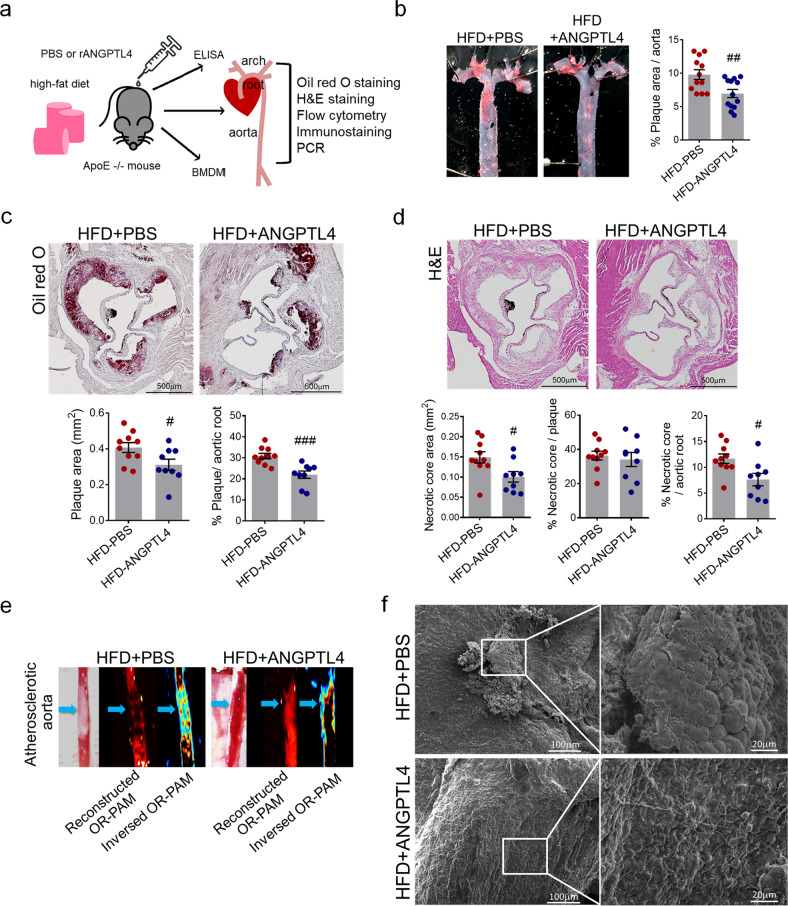
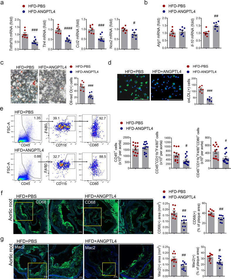
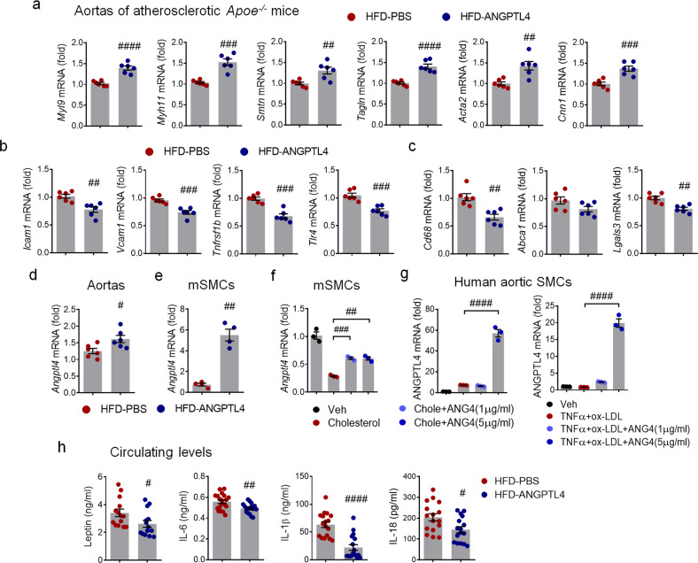
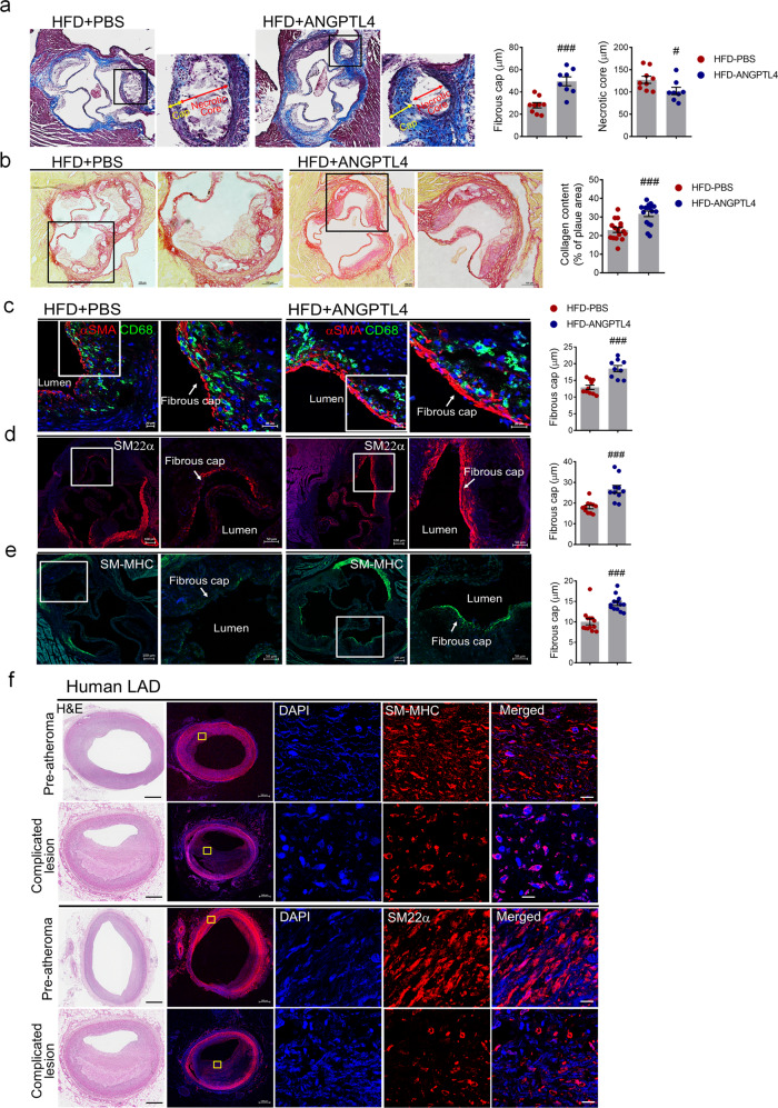
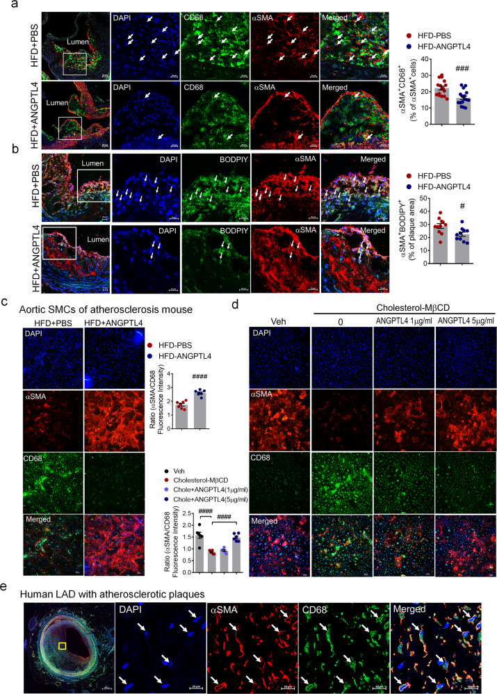
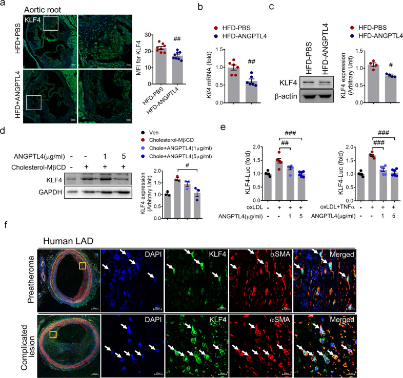
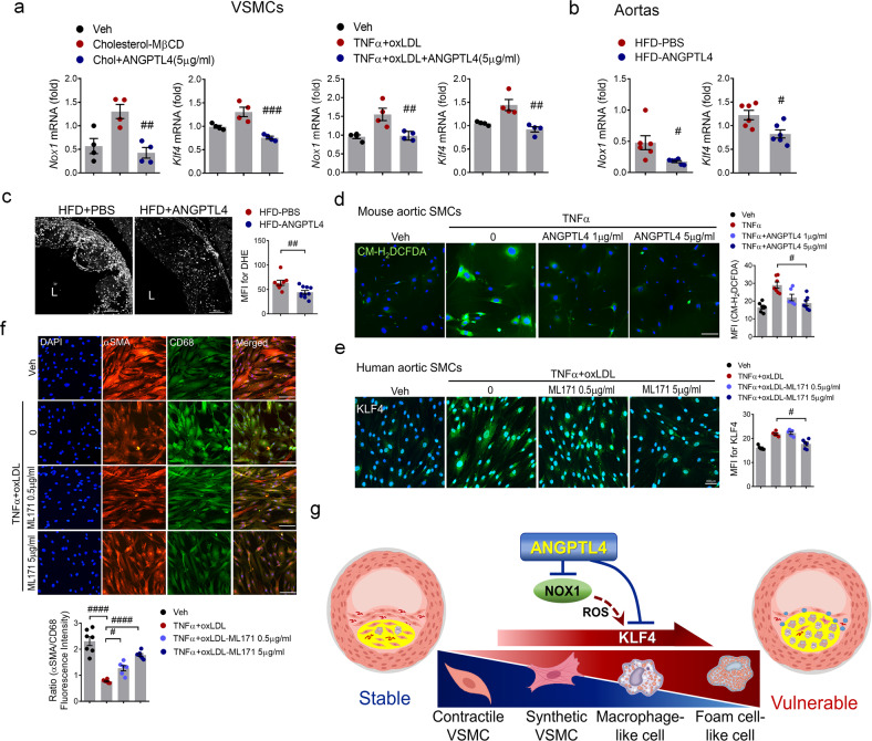
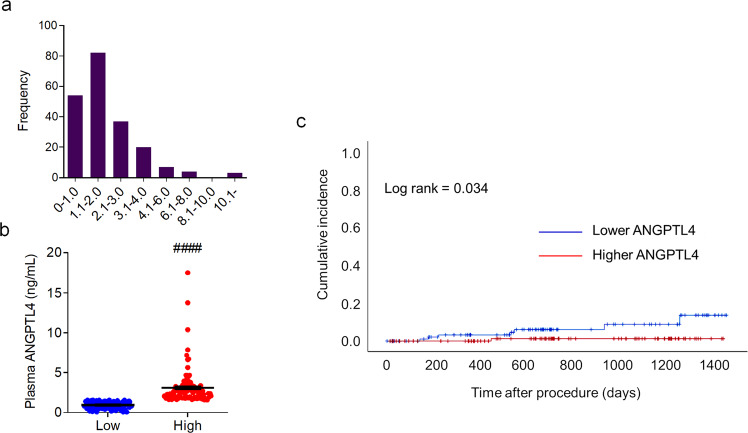
Atherosclerosis, the leading cause of death, is a vascular disease of chronic inflammation. We recently showed that angiopoietin-like 4 (ANGPTL4) promotes cardiac repair by suppressing pathological inflammation. Given the fundamental contribution of inflammation to atherosclerosis, we assessed the role of ANGPTL4 in the development of atherosclerosis and determined whether ANGPTL4 regulates atherosclerotic plaque stability. We injected ANGPTL4 protein twice a week into atherosclerotic Apoe-/- mice and analyzed the atherosclerotic lesion size, inflammation, and plaque stability. In atherosclerotic mice, ANGPTL4 reduced atherosclerotic plaque size and vascular inflammation. In the atherosclerotic lesions and fibrous caps, the number of α-SMA(+), SM22α(+), and SM-MHC(+) cells was higher, while the number of CD68(+) and Mac2(+) cells was lower in the ANGPTL4 group. Most importantly, the fibrous cap was significantly thicker in the ANGPTL4 group than in the control group. Smooth muscle cells (SMCs) isolated from atherosclerotic aortas showed significantly increased expression of CD68 and Krüppel-like factor 4 (KLF4), a modulator of the vascular SMC phenotype, along with downregulation of α-SMA, and these changes were attenuated by ANGPTL4 treatment. Furthermore, ANGPTL4 reduced TNFα-induced NADPH oxidase 1 (NOX1), a major source of reactive oxygen species, resulting in the attenuation of KLF4-mediated SMC phenotypic changes. We showed that acute myocardial infarction (AMI) patients with higher levels of ANGPTL4 had fewer vascular events than AMI patients with lower levels of ANGPTL4 (p < 0.05). Our results reveal that ANGPTL4 treatment inhibits atherogenesis and suggest that targeting vascular stability and inflammation may serve as a novel therapeutic strategy to prevent and treat atherosclerosis. Even more importantly, ANGPTL4 treatment inhibited the phenotypic changes of SMCs into macrophage-like cells by downregulating NOX1 activation of KLF4, leading to the formation of more stable plaques.


Conflict of interest statement

The authors declare no competing interests.

Figures

Fig. 1
Fig. 1. Effects of ANGPTL4 administration on atherosclerotic progression in the atherosclerosis Apoe−/− mouse model.
a Experimental scheme. Apoe−/− mice were fed a high-fat diet (HFD) with an injection of PBS or recombinant ANGPTL4 protein (2 μg per mouse, intraperitoneally twice a week) for 8 weeks. Blood, BMDMs, aortic roots, and aortas were collected for further analyses. b Representative Oil red O-stained aortas from PBS-injected and ANGPTL4-injected Apoe−/− mice fed an HFD for 8 weeks. The relative atherosclerotic plaque area was quantified. c, Lesion area measured in Oil red O-stained cross sections of the aortic roots from the PBS and ANGPTL4 groups. The plaque area was measured and quantified as the relative size of the plaque to the aortic area. Scale bar, 500 μm. d Representative images are shown for H&E-stained aortic roots from the PBS and ANGPTL4 groups. The necrotic core area, qualified by the anucleated area, was measured and quantified as the relative size of the necrotic core to the plaque or aortic root. Scale bar, 500 μm. e Representative images of atherosclerotic aortas were obtained using high-resolution optical resolution photoacoustic microscopy (OR-PAM). Blue arrows indicate atherosclerotic plaques. f Scanning electron microscope images showing the aorta surface isolated from the PBS and ANGPTL4 groups. Boxed areas on the aortic surface depict atherosclerotic plaques at higher magnification. Scale bar, 20 μm. Data were presented as the mean ± SEM. #p < 0.05, ##p < 0.01, ###p < 0.001 (by Student’s t-test).
Fig. 2
Fig. 2. Effects of ANGPTL4 on macrophage function in atherosclerosis.
a, b Proinflammatory and anti-inflammatory gene expression were analyzed by real-time PCR in BMDMs isolated from Apoe−/− mice treated with PBS or ANGPTL4. c BMDMs from the PBS and ANGPTL4 groups were analyzed using Oil red O staining. Scale bar, 200 μm. d BMDMs from the PBS and ANGPTL4 groups were treated with oxLDL-DyLight 488 for 24 h, and then oxidized LDL uptake was analyzed. Scale bar, 200 μm. e Flow cytometry analyses of single-cell aortic suspensions isolated from the PBS and ANGPTL4 groups. Inflammatory macrophages were quantified by the number of CD80+ cells among the CD45+F4/80+CD11b+ population. f, g The content of macrophages in aortic root sections from the two groups was determined by immunohistochemical staining with anti-CD68 antibody (f) and anti-Mac2 antibodies (g). Representative images are shown, and CD68-positive areas (f) and Mac2-positive areas (g) were measured and quantified as a percentage of the plaque area. Data were presented as the mean ± SEM. #p < 0.05, ##p < 0.01, ###p < 0.001, ####p < 0.0001 (by Student’s t-test).
Fig. 3
Fig. 3. The expression patterns of atherogenic mediators from the aortas and plasma of Apoe−/− mice.
Relative expression of genes related to contractility (a), proinflammation (b), and macrophage markers (c) in aortas isolated from the PBS and ANGPTL4 groups. Apoe−/− mice fed a high-fat diet (HFD) were injected with PBS or ANGPTL4 twice a week for 8 weeks (2 μg, i.p.). ANGPTL4 expression in aortas (d) and aortic SMCs (e, f) of Apoe−/− mice and human aortic SMCs (g). Aortic SMCs were stimulated with cholesterol (10 μg/ml) or TNFα (100 ng/ml) and oxLDL (10 μg/ml) with or without ANGPTL4. h Circulating leptin, IL-6, IL-1β, and IL-18 were quantified in the PBS and ANGPTL4 groups. Data were presented as the mean ± SEM. #p < 0.05, ##p < 0.01, ###p < 0.001, ####p < 0.0001 (by Student’s t-test or one-way ANOVA with Bonferroni’s multiple-comparisons test).
Fig. 4
Fig. 4. Effects of ANGPTL4 on the stability of atherosclerotic plaques.
ae Representative histologic analysis of the aortic root from the PBS and ANGPTL4 groups. a Representative images of Masson trichrome staining are shown. Boxed areas in the aortic root depict the fibrous cap and necrotic core at higher magnification, and the fibrous cap and necrotic core were measured as the lesion thickness. Scale bar, 100 μm. b Representative images of picrosirius red staining for collagen, and quantification of the collagen content presented as a percentage of the plaque area. Scale bar, 100 μm. Immunofluorescence staining of atherosclerotic plaques showed α-SMA and CD68 (c), SM22α (d), and SM-MHC (e) in the fibrous caps. Quantification of the fibrous cap thickness is presented in the right panels. Scale bar, 20 μm. f H&E (left panels) and SM-MHC and SM22α-stained confocal images of lesions representing preatheromatous plaques and complicated lesions of the human LAD. Scale bar, 10 μm. Data were presented as the mean ± SEM. #p < 0.05, ###p < 0.001 (by Student’s t-test). LAD left anterior descending artery.
Fig. 5
Fig. 5. ANGPTL4 regulates SMC phenotypic changes in atherosclerosis.
a Immunofluorescence staining of atherosclerotic plaques showing CD68 (green) and α-SMA (red) in the aortic root. Boxed areas show close-up images of CD68+α-SMA+ cells (arrowheads) in atherosclerotic plaques. Quantification of the frequency of double-positive (CD68+α-SMA+) cells among the total α-SMA+ cells within the whole lesion and the fibrous cap (n = 16). Scale bar, 20 μm. b Representative images of atherosclerotic plaques in the aortic root showing lipid droplets stained by BODIPY (green) and α-SMA (red). Arrowheads indicate BODIPY+αSMA+ cells. The percentage of BODIPY+α-SMA+ cells within the plaque area (n = 11). Scale bar, 20 μm. c Aortic SMCs were isolated from atherosclerotic Apoe−/− mice treated with PBS or ANGPTL4 and then stained with α-SMA as an SMC marker and CD68 as a macrophage marker. Quantification of α-SMA/CD68 fluorescence intensity. Scale bar, 100 μm. d Aortic SMCs were stimulated with cholesterol (10 μg/ml) for 72 h with or without ANGPTL4 and stained with α-SMA and CD68. Scale bar, 100 μm. e In human LAD, the atherosclerotic lesion displayed cells double positive for α-SMA and CD68. Scale bar, 10 μm. Data were presented as the mean ± SEM. #p < 0.05, ###p < 0.001, ####p < 0.0001 (by Student’s t-test or one-way ANOVA with Bonferroni’s multiple-comparisons test).
Fig. 6
Fig. 6. Attenuation of KLF4 upregulation by ANGPTL4 in atherosclerosis.
a Expression of KLF4 within the plaque of the aortic root from the two groups was determined by immunofluorescent staining. Data were the mean fluorescence intensity (MFI) of KLF4 (mean ± SEM, n = 8). Scale bar, 20 μm. b, c Aortic SMCs were isolated from atherosclerotic Apoe−/− mice treated with PBS or ANGPTL4, and the level of KLF4 was measured. The levels of KLF4 mRNA (b) and protein (c) were lower in the ANGPTL4 group than in the PBS group. d Western blotting of KLF4 expression in SMCs from Apoe−/− mice pretreated with ANGPTL4 for 24 h and then stimulated with cholesterol (10 μg/ml) for 72 h. e KLF4 promoter activity was increased in human aortic SMCs stimulated by oxLDL (10 μg/ml) or oxLDL and TNFα (100 ng/ml) for 24 h but was inhibited by ANGPTL4 (1, 5 μg/ml) treatment. f Representative images of α-SMA+KLF4+ staining in preatheromatous plaques and complicated lesions of the human LAD. Scale bar, 10 μm. Data were presented as the mean ± SEM. #p < 0.05, ##p < 0.01, ###p < 0.001 (by Student’s t-test or one-way ANOVA with Bonferroni’s multiple-comparisons test).
Fig. 7
Fig. 7. ANGPTL4 modulates SMC phenotypic changes through KLF4 induction by NOX1.
a Aortic SMCs pretreated with ANGPTL4 were stimulated with cholesterol (10 μg/ml) or oxLDL (10 μg/ml) and TNFα (100 ng/ml), and the levels of Nox1 and KLF4 were measured. b Aortas were isolated from atherosclerotic Apoe−/− mice treated with PBS or ANGPTL4, and the levels of Nox1 and KLF4 were measured. c Representative images and quantification of dihydroethidium (DHE) fluorescence in aortic root sections. The aortic root sections were incubated with 5 μM DHE for 10 min in the dark. The data presented are the MFI of DHE (mean ± SEM, n = 9). Scale bar, 100 μm. d Representative images of CM-H2DCFDA staining. Aortic SMCs from atherosclerotic Apoe−/− mice were pretreated with ANGPTL4 and TNFα and exposed to 13 μM CM-H2DCFDA. Quantification of ROS levels by MFI. Scale bar, 100 μm. e, f Human aortic SMCs were stimulated with oxLDL (10 μg/ml) and TNFα (100 ng/ml) with or without ML171, a NOX1 inhibitor (0.5 μg/ml, 5 μg/ml), and were stained with KLF4 (e) and α-SMA and CD68 (f). Scale bar, 100 μm. g Proposed mechanism of action for ANGPTL4 administration in atherosclerosis and plaque stabilization. Data were presented as the mean ± SEM. #p < 0.05, ##p < 0.01, ###p < 0.001, ####p < 0.0001 (by Student’s t-test or one-way ANOVA with Bonferroni’s multiple-comparisons test).
Fig. 8
Fig. 8. Circulating levels of ANGPTL4 in patients with cardiovascular disease.
a Distribution of plasma levels of ANGPTL4 measured in all patients (n = 207). b Plasma levels of ANGPTL4 in the low ANGPTL4 (n = 103) and high ANGPTL4 (n = 104) groups. c Kaplan‒Meier curve illustrating the vascular event incidence of patients during the follow-up period after surgery based on plasma ANGPTL4 levels above (red) and below (blue) the median value. Data were presented as the mean ± SEM. ####p < 0.0001 (by Student’s t-test).

Similar articles

Cited by

References

    1. Muller JE, Tofler GH, Stone PH. Circadian variation and triggers of onset of acute cardiovascular disease. Circulation. 1989;79:733–743. doi: 10.1161/01.CIR.79.4.733. - DOI - PubMed
    1. Arbab-Zadeh A, Fuster V. From detecting the vulnerable plaque to managing the vulnerable patient: JACC state-of-the-art review. J. Am. Coll. Cardiol. 2019;74:1582–1593. doi: 10.1016/j.jacc.2019.07.062. - DOI - PubMed
    1. Bentzon JF, Otsuka F, Virmani R, Falk E. Mechanisms of plaque formation and rupture. Circ. Res. 2014;114:1852–1866. doi: 10.1161/CIRCRESAHA.114.302721. - DOI - PubMed
    1. Karthikeyan VJ, Lip GY. Statins and intra-plaque angiogenesis in carotid artery disease. Atherosclerosis. 2007;192:455–456. doi: 10.1016/j.atherosclerosis.2007.01.018. - DOI - PubMed
    1. Nie P, et al. Atorvastatin improves plaque stability in ApoE-knockout mice by regulating chemokines and chemokine receptors. PLoS ONE. 2014;9:e97009. doi: 10.1371/journal.pone.0097009. - DOI - PMC - PubMed

Publication types

Substances