Review
. 2023 Mar;8(3):375-386.
doi: 10.1038/s41564-022-01314-6. Epub 2023 Feb 13.

Phase separation in fungi

Affiliations
Review

Phase separation in fungi

Mae I Staples et al. Nat Microbiol. 2023 Mar.

Abstract

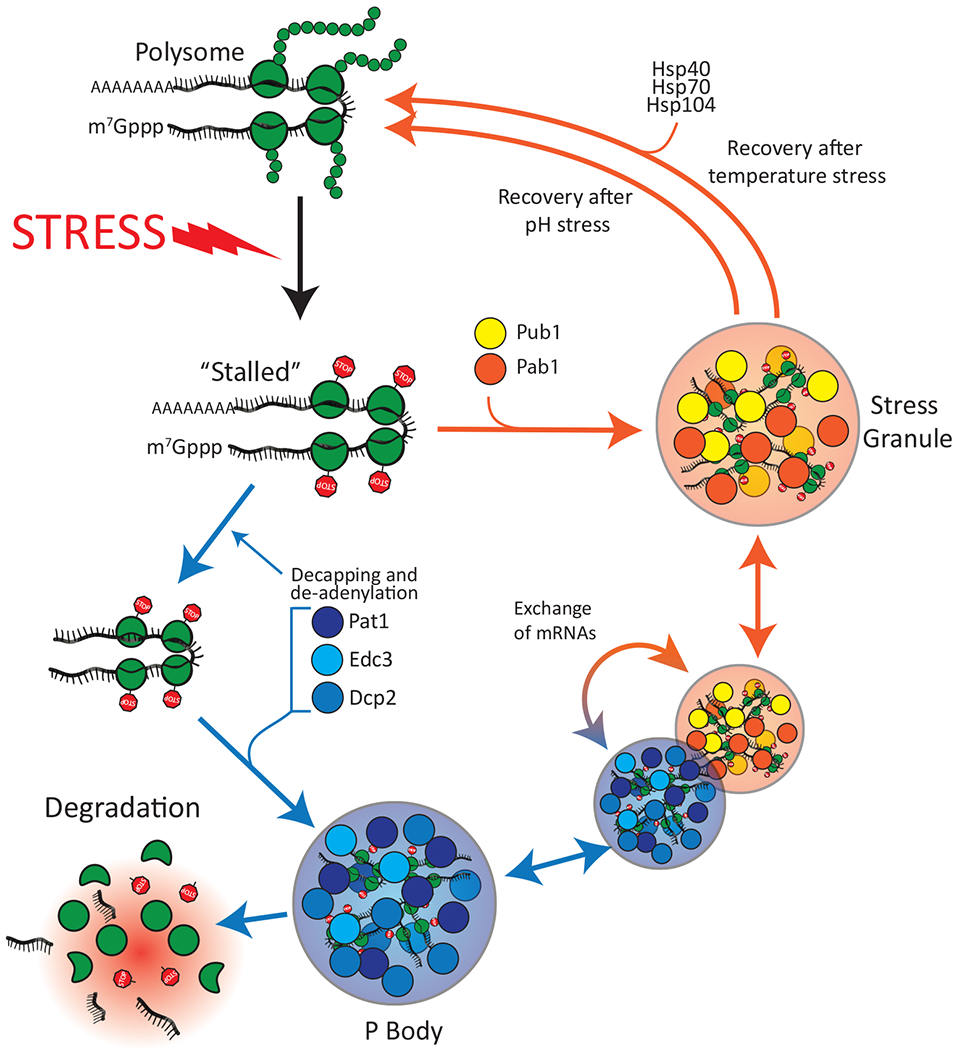
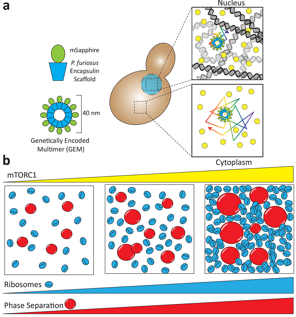
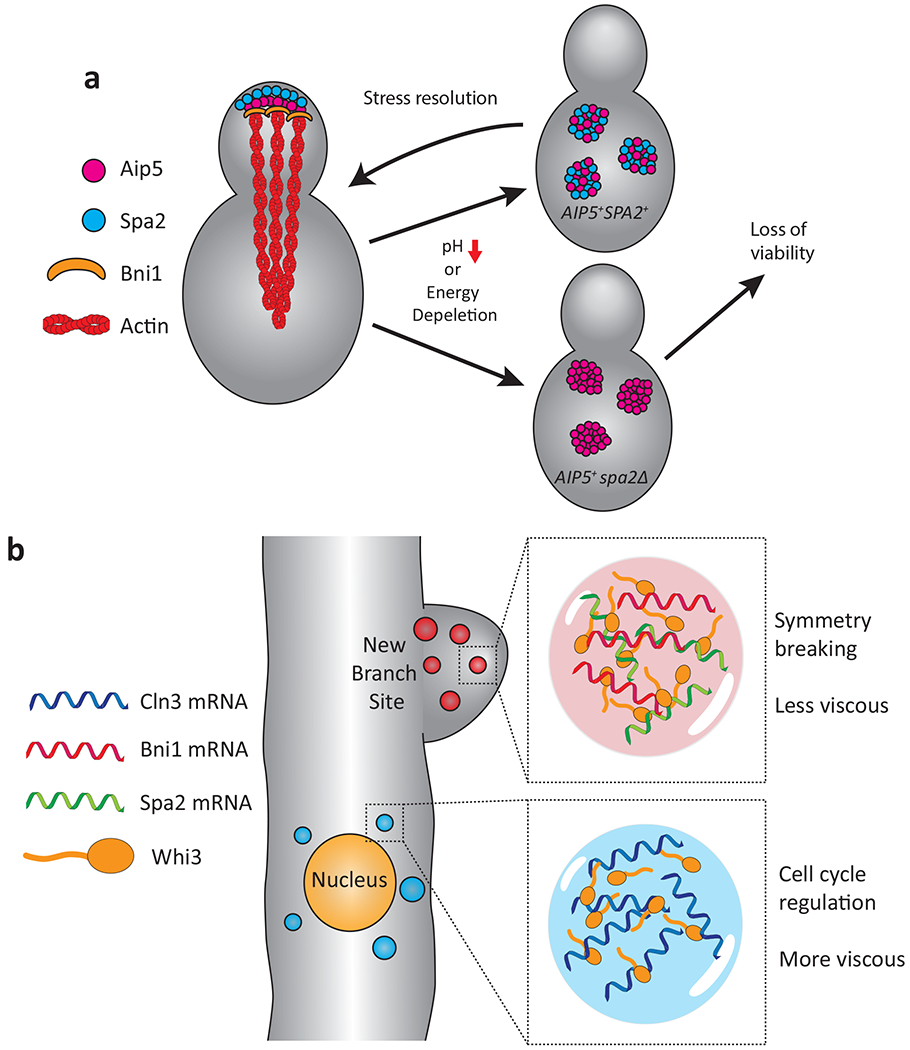
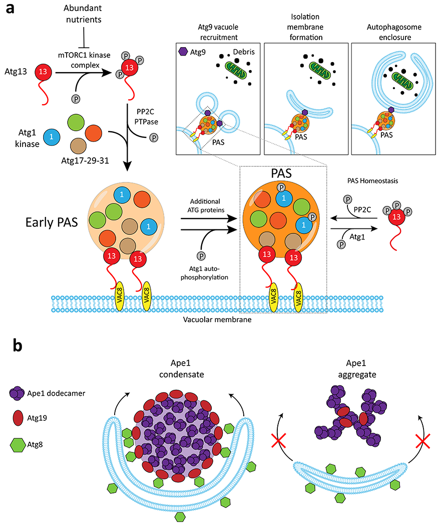
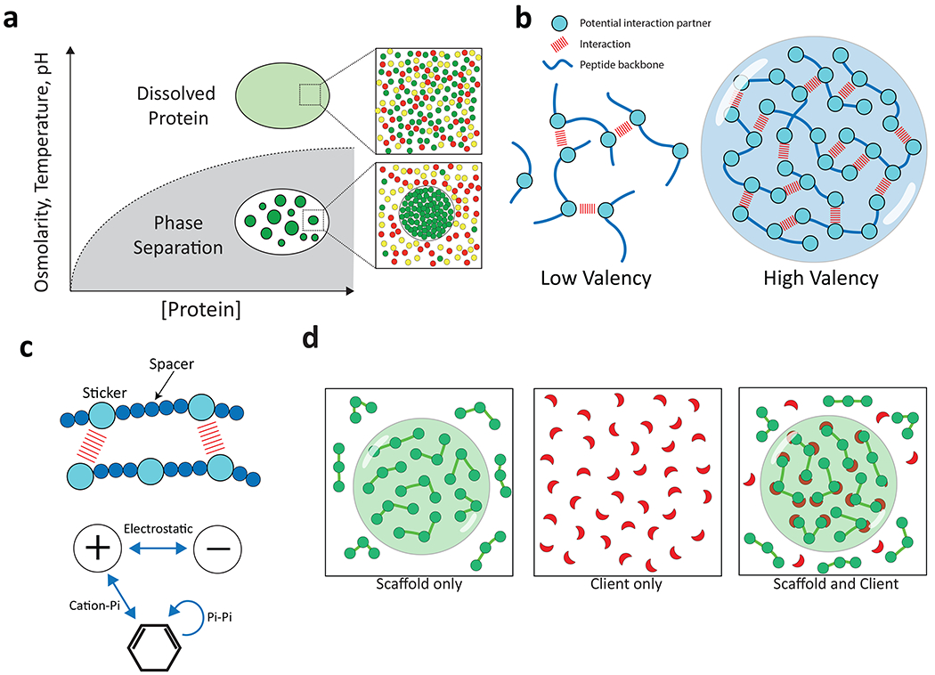
Phase separation, in which macromolecules partition into a concentrated phase that is immiscible with a dilute phase, is involved with fundamental cellular processes across the tree of life. We review the principles of phase separation and highlight how it impacts diverse processes in the fungal kingdom. These include the regulation of autophagy, cell signalling pathways, transcriptional circuits and the establishment of asymmetry in fungal cells. We describe examples of stable, phase-separated assemblies including membraneless organelles such as the nucleolus as well as transient condensates that also arise through phase separation and enable cells to rapidly and reversibly respond to important environmental cues. We showcase how research into phase separation in model yeasts, such as Saccharomyces cerevisiae and Schizosaccharomyces pombe, in conjunction with that in plant and human fungal pathogens, such as Ashbya gossypii and Candida albicans, is continuing to enrich our understanding of fundamental molecular processes.


Conflict of interest statement

Competing interests

NLF is a member of the scientific advisory board of Dewpoint Therapeutics.

Figures

Figure 1.
Figure 1.. Stress granule and P body formation in response to environmental changes.
Extracellular stress such as heat shock or a sudden drop in pH leads to global inhibition of translation and ribosome stalling. Stalled mRNAs are diverted to either P bodies or stress granules. Following pH stress, stress granules dissolve spontaneously, whereas stress granules formed following temperature stress require the assistance of Hsp40, Hsp70 and Hsp104. Subsets of mRNAs that are concentrated in P bodies can either be degraded or exchanged with stress granules.
Figure 2.
Figure 2.. The effect of molecular crowding on phase separation.
a, Microrheology using self-assembling Genetically Encoded Multimers (GEMs) allows measurement of intracellular crowding in the nucleus and the cytoplasm. Individual monomers consist of a Pyrococcus furiosus encapsulin scaffold fused to a fluorescent protein and spontaneously assemble into 40 nm spheres. The nucleus is a more crowded milieu than the cytoplasm and thus the random thermal motion of GEMs is decreased. b, mTORC activation following starvation results in an increased ribosome number. This increase in molecular crowding can increase phase separation of cytoplasmic proteins. Adapted from .
Figure 3.
Figure 3.. Phase separation in polarized growth, cell asymmetry and nuclear divisions.
a, Spa2 localizes Aip5 to the S. cerevisiae bud tip, recruiting Bni1 and nucleating actin filaments. Stresses such as low pH or energy depletion result in Aip5 and Spa5 forming cytoplasmic condensates. When both Aip5 and Spa2 are present, these condensates are rapidly disassembled following removal of the stress. In the absence of Spa2, Aip5 forms more stable condensates that are not readily disassembled and there is a consequent loss of viability following prolonged stress. Adapted from . b, In A. gossypii, mRNA binding protein Whi3 forms distinct protein/mRNA condensates. Whi3 condensates formed with Bni1and Spa2 mRNA are localized to the site of branch formation (symmetry breaking) where they nucleate actin assembly. Whi3 droplets containing Cln3 mRNA form adjacent to the nucleus where they regulate asynchronous nuclear division (cell cycle regulation). Adapted from .
Figure 4.
Figure 4.. Phase separation regulation of bulk and selective autophagy.
a, When nutrients are abundant, Atg13 is hyperphosphorylated by TORC1 kinase. Upon nutrient starvation, Atg13 is dephosphorylated by PP2C phosphatase enabling interactions with the Atg17-29-31 complex and the Atg1 kinase which then coalesce into condensates. The resulting Pre-Autophagosomal Structure (PAS) is tethered to the vacuolar membrane by interactions with Vac8. In the newly formed PAS, Atg1 auto-phosphorylates itself and hyper-phosphorylates Atg13. Cytoplasmic phospho-Atg13 is trafficked back to the PAS following dephosphorylation by PP2C. Thus, Atg1 and PP2C maintain an equilibrium of unphosphorylated and phosphorylated Atg13 which maintains the PAS. The PAS is also the site of Isolation Membrane formation, which in turn will become the mature autophagosome. Inset, Following PAS formation, vacuoles containing ATG9 are recruited and subsequently fuse to become the cupped isolation membrane, which grows to engulf materials destined for destruction in the lysosome. b, Dodecamers of Ape1 undergo phase separation and are degraded by selective autophagy in nutrient rich conditions. The Ape1 condensate is coated by a shell of the Atg9 adapter protein which templates growth of Atg8-decorated isolation membrane. Ape1 mutants which form aggregates are not engulfed. Adapted from .
Figure 5.
Figure 5.. Phase separation regulates transcription and fungal cell fate.
a, Coordinated binding of multiple TFs to regions upstream of their ORFs is often observed even without consensus binding sites for these regulators, suggestive of recruitment by protein-protein interactions. b, A phase separation model of transcription where TFs form condensates together with the transcriptional machinery to regulate the expression of cell identity genes. c, C. albicans switches epigenetically between “white” and “opaque” phenotypic states. d, The white-to-opaque transition is regulated by a TF network whose members bind to their own promoters as well as those of others in the network, as indicated by the arrows. Adapted from ref . e, RNA polymerase II interacts with transcriptional initiation or elongation condensates depending on the phosphorylation state of its C-terminal domain (CTD).
Box 1 Fig.
Box 1 Fig.. Weak protein-protein interactions and multivalency drive phase separation.
a, Phase separation occurs when protein concentration, osmolarity, temperature, and/or pH cross a threshold where intermolecular interactions drive assembly into a dense phase that co-exists with the surrounding dilute phase (adapted from ref ). b, Higher valency due to a higher number of potential interactions between two peptide chains promotes phase separation. c, A “sticker” and “spacer” model for phase separation. Charged and aromatic “sticker” residues (larger balls) are distributed along a polypeptide interspersed with stretches of polar, hydrophilic residues (smaller balls) that act as “spacer” residues. d, “Scaffold” proteins have the capacity to undergo phase separation independent of other factors due to their high valency. Scaffolds can recruit “client” proteins that by themselves are not able to undergo phase separation under the same conditions.

References

    1. Boeynaems S et al. Protein phase separation: A new phase in cell biology. Trends Cell Biol 28, 420–435, doi:10.1016/j.tcb.2018.02.004 (2018). - DOI - PMC - PubMed
    1. Gomes E & Shorter J The molecular language of membraneless organelles. J Biol Chem 294, 7115–7127, doi:10.1074/jbc.TM118.001192 (2019). - DOI - PMC - PubMed
    1. Shin Y & Brangwynne CP Liquid phase condensation in cell physiology and disease. Science 357, doi:10.1126/science.aaf4382 (2017). - DOI - PubMed
    1. Mitrea DM & Kriwacki RW Phase separation in biology; functional organization of a higher order. Cell communication and signaling : CCS 14, 1, doi:10.1186/s12964-015-0125-7 (2016). - DOI - PMC - PubMed
    1. Riback JA et al. Stress-triggered phase separation is an adaptive, evolutionarily tuned response. Cell 168, 1028–1040 e1019, doi:10.1016/j.cell.2017.02.027 (2017). - DOI - PMC - PubMed

Publication types

LinkOut - more resources