

Gastric intramural metastasis caused by needle tract seeding after preoperative fine needle aspiration for pancreatic body cancer subsequently resected by total pancreatectomy: a case report and literature review
- PMID: 36782222
- PMCID: PMC9926546
- DOI: 10.1186/s12957-023-02914-0
Gastric intramural metastasis caused by needle tract seeding after preoperative fine needle aspiration for pancreatic body cancer subsequently resected by total pancreatectomy: a case report and literature review
Abstract
Background: Recently, there has been an increase in the number of reports of needle tract seeding (NTS) of tumor cells after a biopsy as one of the adverse events related to endoscopic ultrasonography-guided fine needle aspiration (EUS-FNA). In most of the previously reported cases of NTS in pancreatic cancer, distal pancreatectomy was performed as the initial surgery, following which metachronous metastasis was discovered in the gastric wall, whose localization matched the puncture route of the EUS-FNA. We report a case of early metastasis from pancreatic cancer in the gastric wall, which was postulated to be caused by NTS. Our patient underwent a total pancreatectomy (TP), and the NTS was resected synchronously.
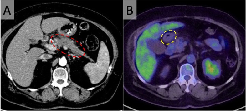
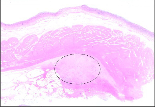
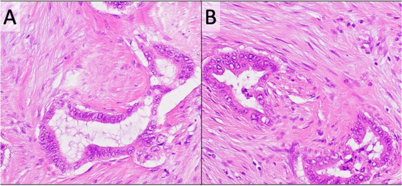
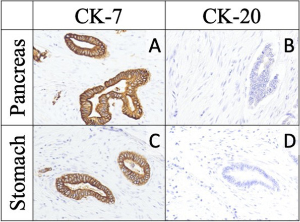
Case presentation: A 70-year-old woman with a diagnosis of pancreatic head-body-tail cancer presented to our department for surgery. Transgastric EUS-FNA and biopsy established the histological diagnosis in her case. We administered neoadjuvant chemotherapy (NAC) to the patient and performed a TP. Histopathological and immunohistochemical examination subsequently confirmed the diagnosis of pT3N1aM1 pancreatic adenocarcinoma and its gastric metastasis, which was caused by NTS. It is postulated that the tumor cells of NTS had progressed to develop the metastatic lesion in the gastric wall during the NAC period. This was also resected during the initial surgery. The patient developed an early postoperative recurrence in the peritoneum 8 months after the surgery.
Conclusion: In pancreatic head cancer cases, the puncture route is often included in the resection area of radical surgery, and NTS is seldom considered as a potential clinical problem. However, NTS can progress rapidly and may be associated with early recurrence of malignancy. Therefore, when transgastrointestinal puncture is performed for the diagnosis of pancreatic cancer, the treatment strategy should be established considering the potential development of NTS.
Keywords: Gastric metastasis; Needle tract seeding; Pancreatic cancer; Total pancreatectomy.
© 2023. The Author(s).
Conflict of interest statement
The authors declare that they have no competing interests.
Figures

References
Publication types
MeSH terms
LinkOut - more resources
Full Text Sources
Medical
