Review
. 2023 Feb 13;11(1):26.
doi: 10.1186/s40478-023-01523-y.

CNS tumor with EP300::BCOR fusion: discussing its prevalence in adult population

Affiliations
Review

CNS tumor with EP300::BCOR fusion: discussing its prevalence in adult population

Arnault Tauziède-Espariat et al. Acta Neuropathol Commun. .

Abstract

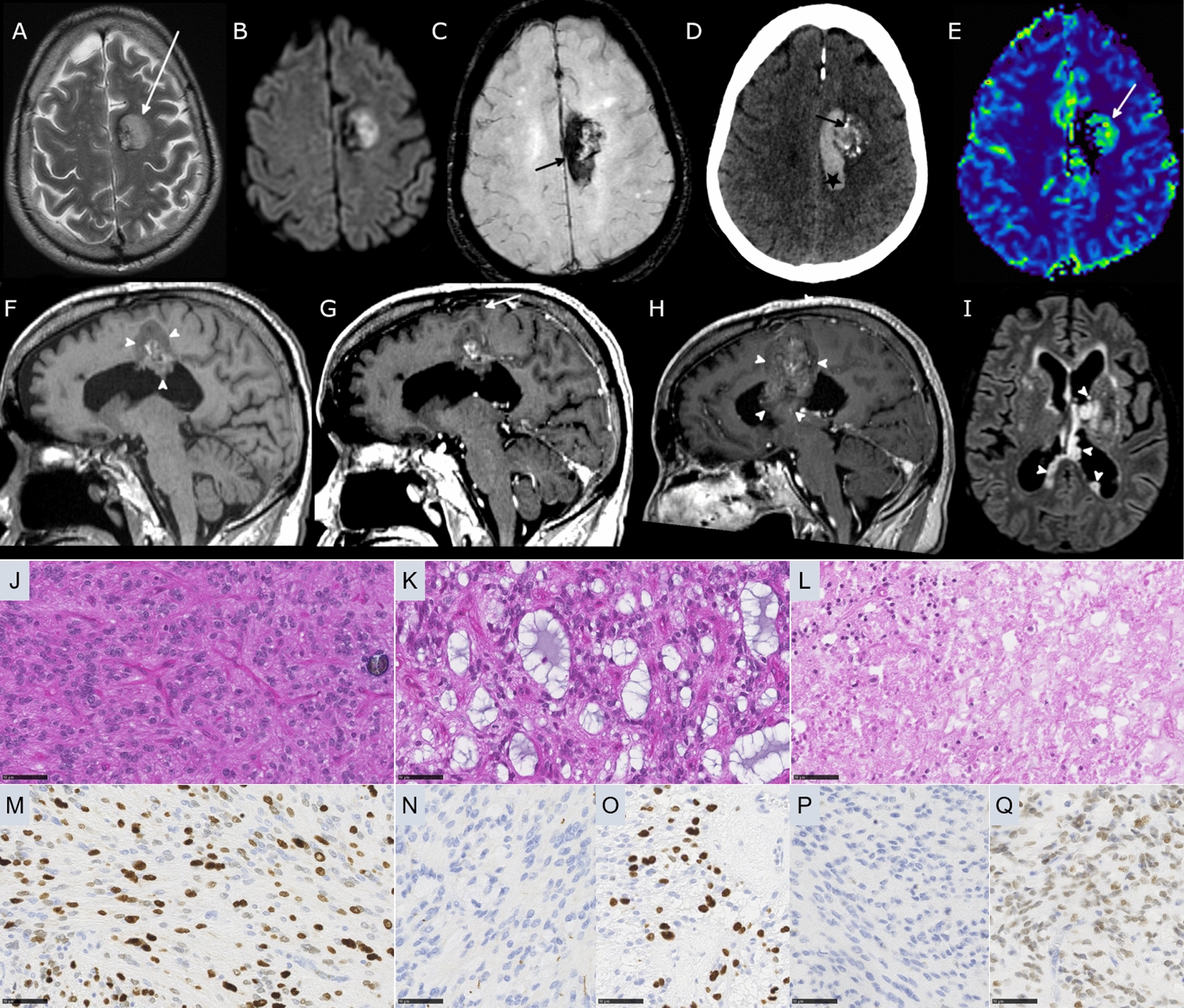
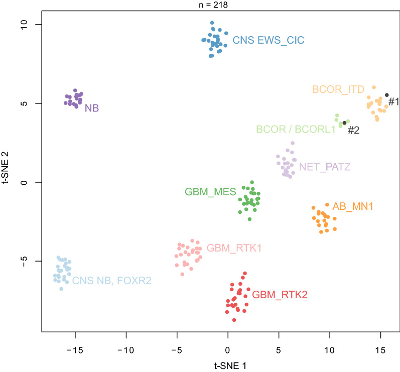
The Central Nervous System (CNS) tumor with BCOR internal tandem duplication (ITD) has recently been added as a novel embryonal histomolecular tumor type to the 2021 World Health Organization (WHO) Classification of CNS Tumors. In addition, other CNS tumors harboring a BCOR/BCORL1 fusion, which are defined by a distinct DNA-methylation profile, have been recently identified in the literature but clinical, radiological and histopathological data remain scarce. Herein, we present two adult cases of CNS tumors with EP300::BCOR fusion. These two cases presented radiological, histopathological, and immunohistochemical homologies with CNS tumors having BCOR ITD in children. To compare these tumors with different BCOR alterations, we performed a literature review with a meta-analysis. CNS tumors with EP300::BCOR fusion seem to be distinct from their BCOR ITD counterparts in terms of age, location, progression-free survival, tumor growth pattern, and immunopositivity for the BCOR protein. CNS tumors from the EP300::BCOR fusion methylation class in adults may be added to the future WHO classification.

Keywords: Adult; BCOR; EP300.


Conflict of interest statement

The authors declare that they have no conflicts of interest directly related to the topic of this article.

Figures

Fig. 1
Fig. 1
Radiological features of Case #1 tumor. a Axial T2-weighted MRI sequence showing a well-delineated left frontal hyperintense lesion (arrow). b Axial diffusion-weighted sequence demonstrating hyperintensity corresponding to hypercellularity of the solid component c Axial susceptibility-weighted sequence showing hypointensity consistent with hemorrhage within the tumor (arrow). d Axial CT showing calcifications (arrow) and acute hemorrhage (star) e Axial perfusion sequence with Cerebral Blood Volume Map showing moderate hyperperfusion of the solid component (arrow). Sagittal T1-weighted f before and g after contrast injection demonstrate juxta-ventricular location of the mass lesion (arrowheads) and a subtle area of enhancement (arrow) h MRI follow-up at two years with post-contrast sagittal T1-weighted sequence showing tumor progression and increase of enhanced components. i Axial FLAIR sequence performed one year after surgery showed an intraventricular ependymal dissemination (arrowheads). j Ependymal differentiation with calcifications (HPS, magnification 400 ×). k Microcysts containing myxoid substance (HPS, magnification 400 ×). l Necrosis (HPS, magnification 400 ×). m Olig2 immunoreactivity (magnification 400 ×). n No immunoexpression for GFAP (magnification 400 ×). o MIB1 labeling index (magnification 400 ×). p No immunopositivity for BCOR (magnification 400 ×). q Expression of SATB2 (magnification 400 ×) Black scale bars represent µm. HPS Hematoxylin Phloxin Saffron.
Fig. 2
Fig. 2
Radiological features of Case #2 tumor. a Coronal T2-weighted MRI sequence showing a well-delineated tumor developed inside the right ventricle. b Axial diffusion-weighted sequence showing hyperintensity consistent with hypercellularity of the solid component (arrow) c MR Perfusion with Cerebral Blood Volume Map showing hyperperfusion of the tumor (arrow). d Axial T1-weighted injection showing a spontaneous hyperintense intra-ventricular haemorrhage (arrowhead); e strong enhancement of the solid component is present after contrast injection (arrow) f MR Spectroscopy demonstrated elevated Choline (Cho), decreased NAA and elevation of lipids-lactates complex (Lac). g Ependymal differentiation (HPS, magnification 400 ×). h Microcysts containing myxoid substance (HPS, magnification 400 ×). i Spindle cell component (HPS, magnification 400 ×). (J) Olig2 immunoexpression by tumor cells (magnification 400 ×). k No immunopositivity for GFAP (magnification 400 ×). l MIB1 labeling index (magnification 400 ×). m No immunopositivity for BCOR (magnification 400 ×). Black scale bars represent µm. HPS Hematoxylin Phloxin Saffron
Fig. 3
Fig. 3
Methylation-based t-SNE distribution. Reference DNA methylation classes (v12.5 of the DKFZ classifier): AB_MN1: Astroblastoma, MN1-altered, MN1:BEND2-fused; BCOR / BCORL1: CNS tumor with BCOR/BCORL1 fusion; BCOR_ITD: CNS tumor with BCOR internal tandem duplication; CNS EWS_CIC: CIC-rearranged sarcoma; CNS NB, FOXR2: central nervous system neuroblastoma, FOXR2-activated; GBM_MES: Glioblastoma, IDH-wildtype, mesenchymal subtype; GBM_RTK1: Glioblastoma, IDH-wildtype, RTK1 subtype; GBM_RTK2: Glioblastoma, IDH-wildtype, RTK2 subtype; NB: neuroblastoma; NET_PATZ: Neuroepithelial tumor with PATZ1 fusion

References

    1. Louis DN, Perry A, Wesseling P, Brat DJ, Cree IA, Figarella-Branger D, Hawkins C, Ng HK, Pfister SM, Reifenberger G, Soffietti R, von Deimling A, Ellison DW. The 2021 WHO Classification of Tumors of the Central Nervous System: a summary. Neuro Oncol. 2021;23:1231–1251. doi: 10.1093/neuonc/noab106. - DOI - PMC - PubMed
    1. Sturm D, Orr BA, Toprak UH, Hovestadt V, Jones DTW, Capper D, et al. New brain tumor entities emerge from molecular classification of CNS-PNETs. Cell. 2016;164:1060–1072. doi: 10.1016/j.cell.2016.01.015. - DOI - PMC - PubMed
    1. Ferris SP, Velazquez Vega J, Aboian M, Lee JC, Van Ziffle J, Onodera C, Grenert JP, Saunders T, Chen YY, Banerjee A, Kline CN, Gupta N, Raffel C, Samuel D, Ruiz-Diaz I, Magaki S, Wilson D, Neltner J, Al-Hajri Z, Phillips JJ, Pekmezci M, Bollen AW, Tihan T, Schniederjan M, Cha S, Perry A, Solomon DA. High-grade neuroepithelial tumor with BCOR exon 15 internal tandem duplication-a comprehensive clinical, radiographic, pathologic, and genomic analysis. Brain Pathol. 2020;30:46–62. doi: 10.1111/bpa.12747. - DOI - PMC - PubMed
    1. Bouchoucha Y, Tauziède-Espariat A, Gauthier A, Guillemot D, Bochaton D, Vibert J, et al. Intra- and extra-cranial BCOR-ITD tumours are separate entities within the BCOR-rearranged family. J Pathol Clin Res. 2022;8:217–232. doi: 10.1002/cjp2.255. - DOI - PMC - PubMed
    1. Fukuoka K, Kanemura Y, Shofuda T, Fukushima S, Yamashita S, Narushima D, et al. Significance of molecular classification of ependymomas: C11orf95-RELA fusion-negative supratentorial ependymomas are a heterogeneous group of tumors. Acta Neuropathol Commun. 2018;6:134. doi: 10.1186/s40478-018-0630-1. - DOI - PMC - PubMed

LinkOut - more resources