Case Reports
. 2023 Feb 13;18(1):20.
doi: 10.1186/s13000-023-01305-z.

Pediatric combined hepatocellular-cholangiocarcinoma (cHCC-CC) with neuroendocrine features: distinguishing genetic alterations detected by chromosomal microarray

Affiliations
Case Reports

Pediatric combined hepatocellular-cholangiocarcinoma (cHCC-CC) with neuroendocrine features: distinguishing genetic alterations detected by chromosomal microarray

Alyeesha B Wilhelm et al. Diagn Pathol. .

Abstract

Background: Liver tumors exhibiting hepatocellular, cholangiocarcinoma, and neuroendocrine features are extremely rare, with only five cases reported in the literature.

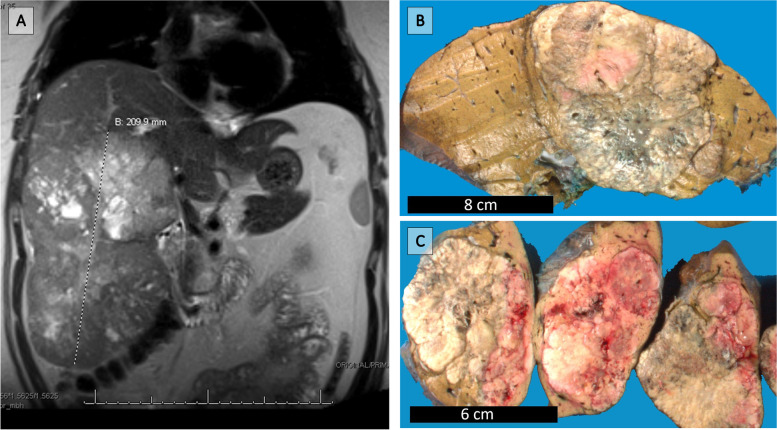
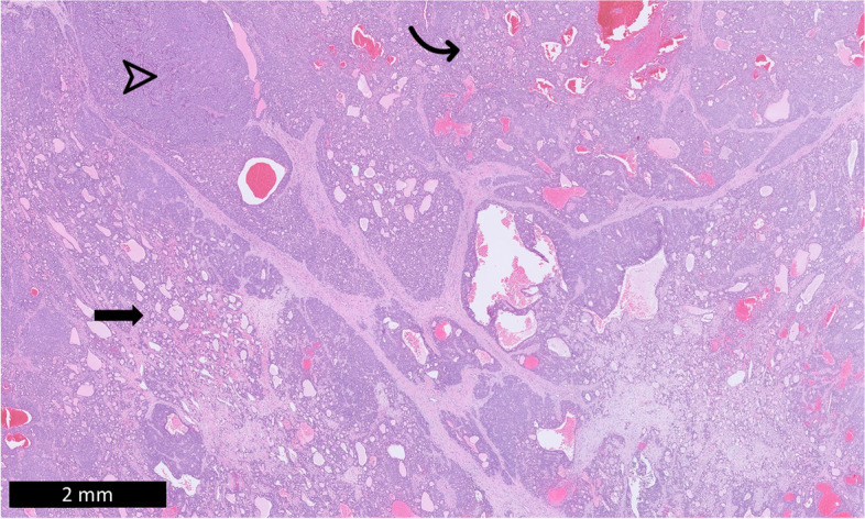
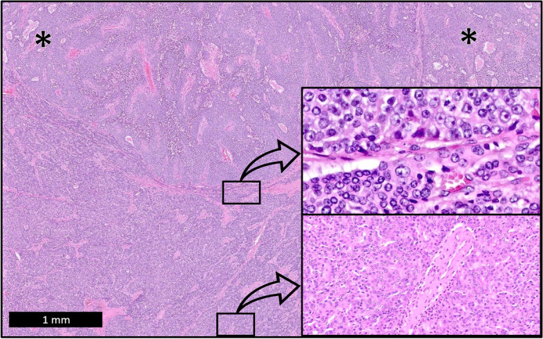
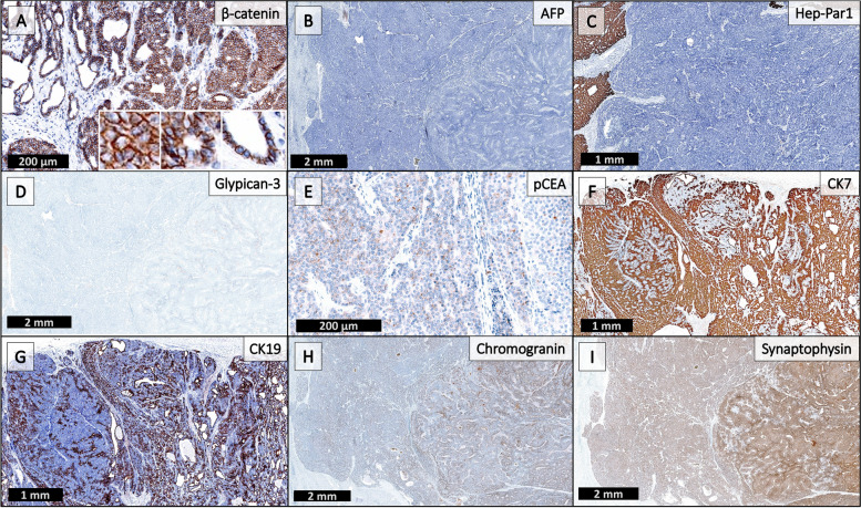
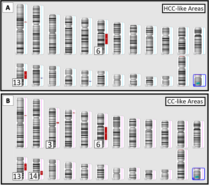
Case presentation: We present an unusual case of a combined hepatocellular-cholangiocarcinoma (cHCC-CC) with neuroendocrine features in a pediatric patient. A 16-year-old presented with abdominal pain and a 21.0 cm mass in the right hepatic lobe with extension into the left lobe. Histology showed a poorly differentiated tumor with a solid, tubuloglandular, and microcystic architecture. Immunohistochemistry results were negative for hepatic markers, positive for markers of biliary differentiation, and positive for neuroendocrine differentiation. The neoplasm was reviewed at several institutions with differing diagnoses. Single nucleotide polymorphism (SNP) chromosomal microarray (CMA) showed large deletions within chromosomes 6q and 13q in both the hepatocellular-like areas and the cholangiocarcinoma-like areas, with additional large deletions in the cholangiocarcinoma-like areas, supporting origin from hepatocellular carcinoma. The final diagnosis was a cHCC-CC with neuroendocrine features.

Conclusions: Diagnosis of cHCC-CCs relies predominately on histomorphology, as per the 2018 International Consensus Group on the nomenclature of cHCC-CC. These findings in this case support that the pathological classification of these lesions be based on molecular data, which could better direct treatment. Further classification of cHCC-CCs and determination of their clinicopathological relevance will require more interobserver consistency and continued molecular profiling of these lesions.

Keywords: Cholangiocarcinoma; Hepatoblastoma; Hepatocellular Carcinoma; Liver; Neuroendocrine; Pediatric; cHCC-CC.


Conflict of interest statement

The authors declare no competing interests.

Figures

Fig. 1
Fig. 1
T2 MRI shows an enlarged liver measuring 29.0 × 22.0 x 10.0 cm with mass lesion (A). Grossly identified are two tan-white to tan-pink, focally hemorrhagic abutting tumors measuring 21.5 cm and 4.5 cm (B and C)
Fig. 2
Fig. 2
The mass demonstrates multiple histologic patterns including tubuloglandular structures (curved arrow), solid sheets and trabeculae (arrowhead), and small spaces lined by biliary-like epithelium and filled with eosinophilic proteinaceous luminal material (arrow). (H&E)
Fig. 3
Fig. 3
Two distinct histological patterns of the tumor are identified, with cholangiocytic differentiation (asterisks) identified in the top lesion (H&E, low power). Small uniform tumor “cancer stem cells” with small basophilic vesicular nuclei, occasional inconspicuous nucleoli, scant pale eosinophilic cytoplasm, indistinct cell borders, and frequent mitoses are seen throughout and are prominent at transitional zones (top inlay; H&E, high power). Tumor cells of similar cytology with more abundant eosinophilic cytoplasm surround an unpaired artery in the HCC-like areas (bottom inlay; H&E, intermediate power)
Fig. 4
Fig. 4
IHC panel. β-catenin (A) shows membranous but not nuclear staining, while alpha-fetoprotein (B) is negative; these findings are not supportive of hepatoblastoma. Negative Hep-Par1 (C), negative Glypican-3 (D), and largely non-canalicular staining pattern of pCEA (E) suggest poorly differentiated tumor if of hepatic origin. CK7 (F) and CK19 (G) show positive staining (strongly positive within the ductular components), highlighting cholangiolar-like differentiation. Focal areas of strongly positive chromogranin (H) and diffusely positive synaptophysin (I) highlight neuroendocrine-like differentiation
Fig. 5
Fig. 5
Chromosomal microarray (CMA) was performed on the histologically HCC-like (A) and histologically CC-like (B) areas. Both areas revealed large deletions (red bars) in chromosomes 6q and 13q and the CC-like component revealed additional large deletions in chromosomes 3p and 14q. The results favor that the neoplastic cells arose from a single clone with genetic heterogeneity causing the tumor’s histologic diversity

References

    1. WHO Classification of Tumours Editorial Board. Digestive System Tumours. 5th ed. Vol. 1. Lyons: International Agency for Research on Cancer; 2019.
    1. Zimmermann A. Hepatoblastoma with cholangioblastic features ('cholangioblastic hepatoblastoma’) and other liver tumors with bimodal differentiation in young patients. Med Pediatr Oncol. 2002;39(5):487–491. doi: 10.1002/mpo.10173. - DOI - PubMed
    1. Nishino H, Hatano E, Seo S, Shibuya S, Anazawa T, Iida T, et al. Histological features of mixed neuroendocrine carcinoma and hepatocellular carcinoma in the liver: a case report and literature review. Clin J Gastroenterol. 2016;9(4):272–279. doi: 10.1007/s12328-016-0669-0. - DOI - PubMed
    1. Joseph NM, Tsokos CG, Umetsu SE, Shain AH, Kelley RK, Onodera C, et al. Genomic profiling of combined hepatocellular-cholangiocarcinoma reveals similar genetics to hepatocellular carcinoma. J Pathol. 2019;248(2):164–178. doi: 10.1002/path.5243. - DOI - PubMed
    1. Fujii H, Zhu XG, Matsumoto T, Inagaki M, Tokusashi Y, Miyokawa N, et al. Genetic classification of combined hepatocellular-cholangiocarcinoma. Hum Pathol. 2000;31(9):1011–1017. doi: 10.1053/hupa.2000.9782. - DOI - PubMed

Publication types

MeSH terms