. 2023 Apr;15(4):1060-1071.
doi: 10.1111/os.13648. Epub 2023 Feb 13.

Long Noncoding RNA PCGEM1 Facilitates Tumor Growth and Metastasis of Osteosarcoma by Sponging miR-433-3p and Targeting OMA1

Affiliations

Long Noncoding RNA PCGEM1 Facilitates Tumor Growth and Metastasis of Osteosarcoma by Sponging miR-433-3p and Targeting OMA1

Jun Li et al. Orthop Surg. 2023 Apr.

Abstract

Objective: Osteosarcoma (OS) is regarded as one of the most common malignant bone tumors, mainly occurring in children and adolescents with high mortality. The dysregulation of lncRNAs is reported to regulate tumor development and be closely related to patient prognosis. Nevertheless, the role of long noncoding RNAs (lncRNAs) prostate-specific transcript 1 (PCGEM1) in OS remains uncharacterized. The current study aimed to explore the role of PCGEM1 in OS.

Methods: Reverse transcription-quantitative polymerase chain reaction (RT-qPCR) was performed to examine the expression of PCGEM1 in OS cell lines. CCK-8, colony formation, Transwell, and western blotting analyses were applied to measure OS cell viability, proliferation, migration, invasion, and epithelial-mesenchymal transition (EMT) after PCGEM1 downregulation. Nuclear-cytoplasmic fractionation, RNA pulldown, RNA immunoprecipitation (RIP), luciferase reporter assays were performed to verify the relationship among PCGEM1, miR-433-3p. and OMA1 in OS. The xenograft tumor models were established to evaluate the effect of PCGEM1 on tumor growth of OS.

Results: In this study, we discovered that PCGEM1 knockdown inhibited cell proliferation, migration, invasion and EMT in OS (P < 0.05). Additionally, PCGEM1 directly bound to miR-433-3p (P < 0.01). OMA1 was confirmed to be a target gene of miR-433-3p (P < 0.05), positively regulated by PCGEM1 but negatively regulated by miR-433-3p. Rescue assays further verified that overexpression of OMA1 reversed the PCGEM1 knockdown-mediated inhibitory effect on the malignant phenotype in OS cells (P < 0.05). Moreover, knockdown of PCGEM1 inhibited tumor growth and metastasis in vivo (P < 0.05).

Conclusions: Overall, PCGEM1 mediated tumor growth and metastasis of OS by sponging miR-433-3p and regulating OMA1, which might provide an innovative strategy for OS diagnosis or treatment.

Keywords: OMA1; Osteosarcoma; PCGEM1; miR-433-3p.


Conflict of interest statement

The authors declare no conflict of interest.

Figures
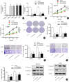
Fig. 1
Fig. 1
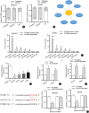
Inhibition of PCGEM1 suppresses cell proliferation, migration, invasion and EMT in OS. (A) RT‐qPCR analysis was used to determine the expression of PCGEM1 in OS cells (MG‐63, U2OS, Saos2 and 143B) and hFOB1.19 cells. (B) The expression of PCGEM1 in MG‐63 and U2OS cells transfected with sh‐PCGEM1#1/2 was detected by RT‐qPCR analysis. (C) The viability of MG‐63 and U2OS cells after inhibition of PCGEM1 was evaluated by CCK‐8 assay. (D) The proliferation of MG‐63 and U2OS cells after knockdown of PCGEM1 was assessed by colony formation assays. (E) The migration and (F) invasion of MG‐63 and U2OS cells after suppression of PCGEM1 were examined by Transwell assays. (G) Western blot analysis was performed to measure the protein levels of E‐cadherin and N‐cadherin in MG‐63 and U2OS cells transfected with sh‐PCGEM1#1/2. *P < 0.05
Fig. 2
Fig. 2
PCGEM1 acts as a molecular sponge for miR‐433‐3p. (A) A nuclear‐cytoplasmic fractionation assay was used to determine the subcellular localization of PCGEM1 in MG‐63 and U2OS cells. (B) The starBase database predicted miRNAs binding to PCGEM1. (C) RNA pulldown assay showed the enrichment of miRNAs from the PCGEM1 probe. (D) RT‐qPCR analysis was performed to determine the expression of miR‐433‐3p in OS cells (MG‐63, U2OS, Saos2 and 143B) and hFOB1.19 cells. (E) The expression of miR‐433‐3p in MG‐63 and U2OS cells transfected with sh‐PCGEM1#1 was measured using RT‐qPCR. (F) The expression of miR‐433‐3p in MG‐63 and U2OS cells transfected with miR‐433‐3p mimics was determined by RT‐qPCR. (G) The binding sequences were predicted by starBase, and a luciferase reporter assay was used to validate the interaction between PCGEM1 and miR‐433‐3p. * P < 0.05, ** P < 0.01
Fig. 3
Fig. 3
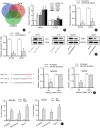
MiR‐433‐3p directly targets OMA1. (A) The Venn diagram shows the potential target genes of miR‐433‐3p predicted by miRanda, PicTar and TargetScan. (B) The mRNA expression of PCCB and OMA1 in OS cells (MG‐63, U2OS, Saos2 and 143B) and hFOB1.19 cells was analyzed by RT‐qPCR. (C‐D) RT‐qPCR and western blot analyses were performed to examine the mRNA and protein levels of OMA1 in MG‐63 and U2OS cells transfected with miR‐433‐3p mimics or sh‐PCGEM1#1. (E) The binding site between miR‐433‐3p and the OMA1 3'UTR was predicted by starBase, and a luciferase reporter assay was conducted to verify the binding of miR‐433‐3p to the OMA1 3'UTR. (F) RIP assay was performed to examine the coexistence of PCGEM1, miR‐433‐3p and OMA1 in RISC. * P < 0.05
Fig. 4
Fig. 4
Upregulation of OMA1 reverses the PCGEM1 knockdown‐induced suppressive effect on the malignant phenotype in OS cells. (A) The overexpression efficiency of pcDNA3.1/OMA1 in MG‐63 and U2OS cells was evaluated by RT‐qPCR and western blot analyses. (B‐C) Cell viability and proliferation were detected by CCK‐8 and colony formation assays in MG‐63 and U2OS cells after transfecting sh‐NC, sh‐PCGEM1#1 or sh‐PCGEM1#1 + OMA1. (D‐E) Cell migration and invasion were tested by Transwell assays in MG‐63 and U2OS cells after transfecting sh‐NC, sh‐PCGEM1#1 or sh‐PCGEM1#1 + OMA1. (F) Western blot analysis was used to determine the E‐cadherin and N‐cadherin levels in MG‐63 and U2OS cells after transfecting sh‐NC, sh‐PCGEM1#1 or sh‐PCGEM1#1 + OMA1. * P < 0.05 vs. the sh‐NC group. # P < 0.05 vs. the sh‐PCGEM1 group
Fig. 5
Fig. 5
Knockdown of PCGEM1 inhibits OS tumor growth and metastasis in vivo. (A) Representative images showing tumor xenografts in nude mice. (B) Tumor volume in nude mice (n = 5). (C) Tumor weight in nude mice (n = 5). (D) The expression of OMA1 and miR‐433‐3p in surgically removed tumor tissues. (E) Representative images and quantification of lung metastasis. * P < 0.05
Fig. 6
Fig. 6
Knockdown of PCGEM1 downregulates Ki67 and OMA1 expression in OS tumor. Positive expression of (A) Ki67 and (B) OMA1 in tumors of nude mice was detected by immunohistochemistry

References

    1. Ritter J, Bielack SS. Osteosarcoma. Ann Oncol. 2010;21(Suppl 7):vii320–5. - PubMed
    1. Sweetnam R. Osteosarcoma. Ann R Coll Surg Engl. 1969;44(1):38–58. - PMC - PubMed
    1. Winkler K, Bielack S, Bieling P. Osteosarcoma. Curr Opin Oncol. 1990;2(3):486–90. - PubMed
    1. Simpson E, Brown HL. Understanding osteosarcomas. Jaapa. 2018;31(8):15–9. - PubMed
    1. Bielack SS, Kempf‐Bielack B, Delling G, Exner GU, Flege S, Helmke K, et al. Prognostic factors in high‐grade osteosarcoma of the extremities or trunk: an analysis of 1,702 patients treated on neoadjuvant cooperative osteosarcoma study group protocols. J Clin Oncol. 2002;20(3):776–90. - PubMed

Substances