. 2023 Mar 20;192(1):106-116.
doi: 10.1093/toxsci/kfad015.

Identification of flucloxacillin-modified hepatocellular proteins: implications in flucloxacillin-induced liver injury

Affiliations

Identification of flucloxacillin-modified hepatocellular proteins: implications in flucloxacillin-induced liver injury

Serat-E Ali et al. Toxicol Sci. .

Abstract

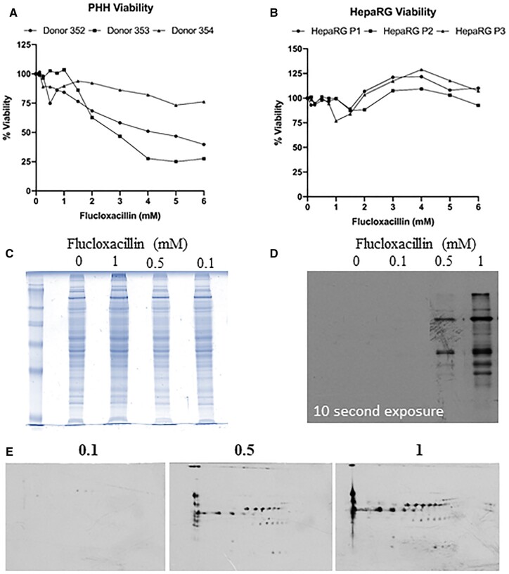
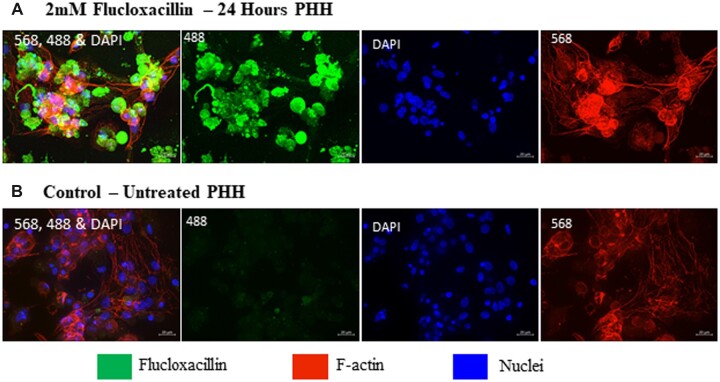
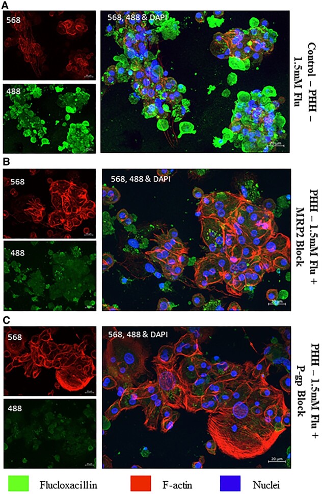
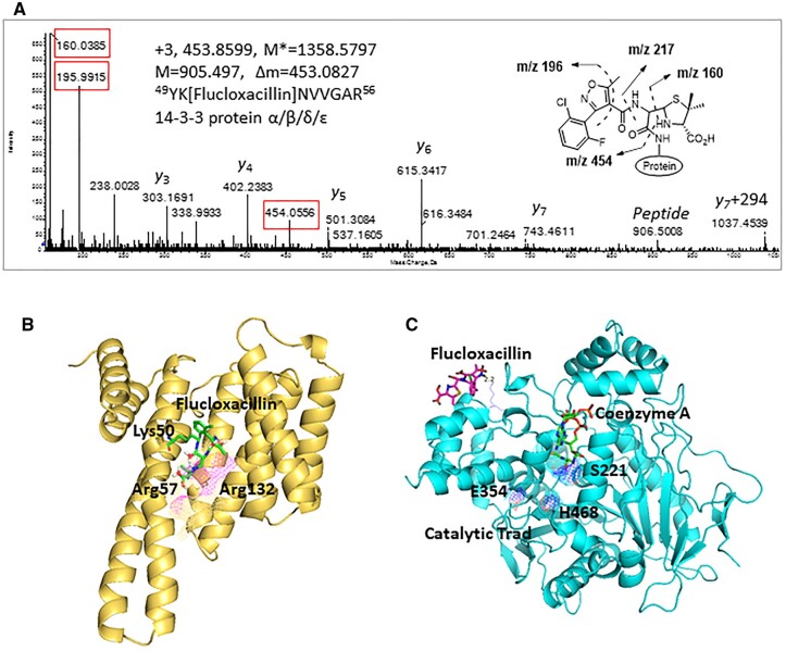
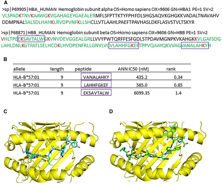
Flucloxacillin is a β-lactam antibiotic associated with a high incidence of drug-induced liver injury. Although expression of HLA-B*57:01 is associated with increased susceptibility, little is known of the pathological mechanisms involved in the induction of the clinical phenotype. Irreversible protein modification is suspected to drive the reaction through the provision of flucloxacillin-modified peptides that are presented to T-cells by the protein encoded by the risk allele. In this study, we have shown that flucloxacillin binds to multiple proteins within human primary hepatocytes, including major hepatocellular proteins (hemoglobin and albumin) and mitochondrial proteins. Inhibition of membrane transporters multidrug resistance-associated protein 2 (MRP2) and P-glycoprotein (P-gp) appeared to reduce the levels of covalent binding. A diverse range of proteins with different functions was found to be targeted by flucloxacillin, including adaptor proteins (14-3-3), proteins with catalytic activities (liver carboxylesterase 1, tRNA-splicing endonuclease subunit Sen2, All-trans-retinol dehydrogenase ADH1B, Glutamate dehydrogenase 1 mitochondrial, Carbamoyl-phosphate synthase [ammonia] mitochondrial), and transporters (hemoglobin, albumin, and UTP-glucose-1-phosphate uridylyltransferase). These flucloxacillin-modified intracellular proteins could provide a potential source of neoantigens for HLA-B*57:01 presentation by hepatocytes. More importantly, covalent binding to critical cellular proteins could be the molecular initiating events that lead to flucloxacillin-induced cholestasis Data are available via ProteomeXchange with identifier PXD038581.

Keywords: covalent binding; flucloxacillin DILI; hepatocytes; membrane transporters.


Figures

Figure 1.
Figure 1.
Flucloxacillin modified proteins were detected in human primary hepatocytes. A, The viability of PHHs decreases after flucloxacillin treatment, with approximately 50% reduction in viability between 2 and 3 mM in 2 out of 3 donors. B HepaRGs show no significant reduction in viability upon flucloxacillin treatment. C, PHHs were treated with increasing concentrations of flucloxacillin for 16 h. Coomassie blue staining shows no change in protein abundances upon flucloxacillin treatment. D, A dose-dependent increase in flucloxacillin protein binding was detected using anti-flucloxacillin antibody. E, Multiple flucloxacillin proteins were detected by 2-dimensional Western blot analysis using anti-flucloxacillin antibody.
Figure 2.
Figure 2.
Qualitative assessment of flucloxacillin binding in PHHs. PHHs were treated with 2 mM flucloxacillin for 24 h to visualize localization and binding of flucloxacillin. Although binding of flucloxacillin (488, green) can be visualized within the cells (A) compared to the untreated control (B), the presence or localization at BC is not clearly defined. 40× magnification, scale bar 20 µm.
Figure 3.
Figure 3.
Inhibition of flucloxacillin efflux in PHH. In order to elucidate the role of membrane transporters in transporting flucloxacillin, PHH were treated with 1.5 mM flucloxacillin (488 green) for 24 h with/without inhibitors of transporters. A, Flucloxacillin extensively forms covalent adducts in PHH. Covalent binding of flucloxacillin in PHH was significantly reduced upon the addition of MRP2 inhibitor (MK571, 30 µM) (B) and P-gp inhibitor (valspodar, 12.5 µM) (C). 40× Magnification, scale bar 20 µm.
Figure 4.
Figure 4.
Covalent binding of flucloxacillin to hepatic proteins. A, A representative MS/MS spectrum shows an adduct was formed on 14-3-3 protein (YK[Flucloxacillin]NVVGAR)—M* = observed flucloxacillin-modified peptide mass, M = theoretical unmodified peptide mass. Characteristic fragment ions from flucloxacillin are highlighted (red boxes). B, Molecular modeling predicts that covalent binding of flucloxacillin to Lys50 on 14-3-3 proteins (PDB code 4E2E) would clash with Arg57 and Arg132 (purple mesh), the key amino acids involved in phosphorylation of binding partner proteins. C, Flucloxacillin (purple) covalently binds to multiple lysine residues on liver carboxylesterase 1, which are distant from the catalytic domain (blue mesh, PDB code 2H7C) and the ligand (coenzyme A, green) binding pocket. Images are illustrated by PyMOL (The PyMOL Molecular Graphics System, Version 1.3 Schrödinger, LLC.).
Figure 5.
Figure 5.
Flucloxacillin predominately targets hemoglobin in human hepatocytes. A, Six out of 11 total lysine residues on hemoglobin subunit α and 7 out of 11 total lysine residues on hemoglobin subunit β were modified by flucloxacillin (modified lysine residues are highlighted in red and peptides sequence are in green). B, Some hemoglobin peptides containing modified lysine residues are also good binders to HLA-B*57:01, a risk allele associated with flucloxacillin-induced liver injury (purple boxes). C, In silicon modeling shows peptide VANALAHKY derived from hemoglobin binds to the binding groove of HLA-B*57:01(PDB code 5vuf, Illing et al., 2018), with P9 (Y) occupying the F pocket. D, The predicted conformation of flucloxacillin-haptenated VANALAHK[flucloxacillin]Y is different from the native peptide, with flucloxacillin molecule reaching out of the binding groove, available for T cell recognition. Images are illustrated by PyMOL (The PyMOL Molecular Graphics System, Version 1.3 Schrödinger, LLC.).

References

    1. Andreatta M., Nielsen M. (2016). Gapped sequence alignment using artificial neural networks: application to the MHC class I system. Bioinformatics 32, 511–517. - PMC - PubMed
    1. Baldo B. A., Pham N. H., Weiner J. (1995). Detection and side-chain specificity of IgE antibodies to flucloxacillin in allergic subjects. J. Mol. Recognit. 8, 171–177. - PubMed
    1. Bencharit S., Edwards C. C., Morton C. L., Howard-Williams E. L., Kuhn P., Potter P. M., Redinbo M. R. (2006). Multisite promiscuity in the processing of endogenous substrates by human carboxylesterase 1. J. Mol. Biol. 363, 201–214. - PMC - PubMed
    1. Burban A., Sharanek A., Hue R., Gay M., Routier S., Guillouzo A., Guguen-Guillouzo C. (2017). Penicillinase-resistant antibiotics induce non-immune-mediated cholestasis through hsp27 activation associated with PKC/P38 and PI3K/AKT signaling pathways. Sci. Rep. 7, 1815. - PMC - PubMed
    1. Daly A. K., Donaldson P. T., Bhatnagar P., Shen Y., Pe'er I., Floratos A., Daly M. J., Goldstein D. B., John S., Nelson M. R., for the DILIGEN study., et al. (2009). HLA-B*5701 genotype is a major determinant of drug-induced liver injury due to flucloxacillin. Nat. Genet. 41, 816–819. - PubMed

Publication types