. 2023 Jan 18;4(2):100181.
doi: 10.1016/j.xhgg.2023.100181. eCollection 2023 Apr 13.

Whole genome sequencing for USH2A-associated disease reveals several pathogenic deep-intronic variants that are amenable to splice correction

Affiliations

Whole genome sequencing for USH2A-associated disease reveals several pathogenic deep-intronic variants that are amenable to splice correction

Janine Reurink et al. HGG Adv. .

Abstract

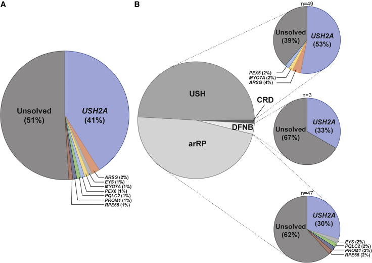
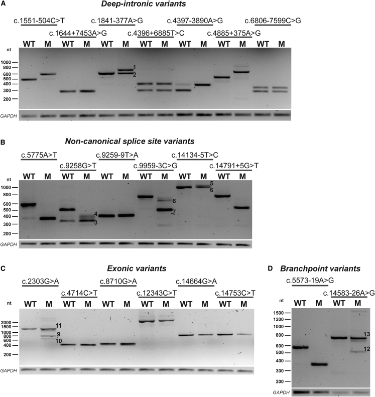
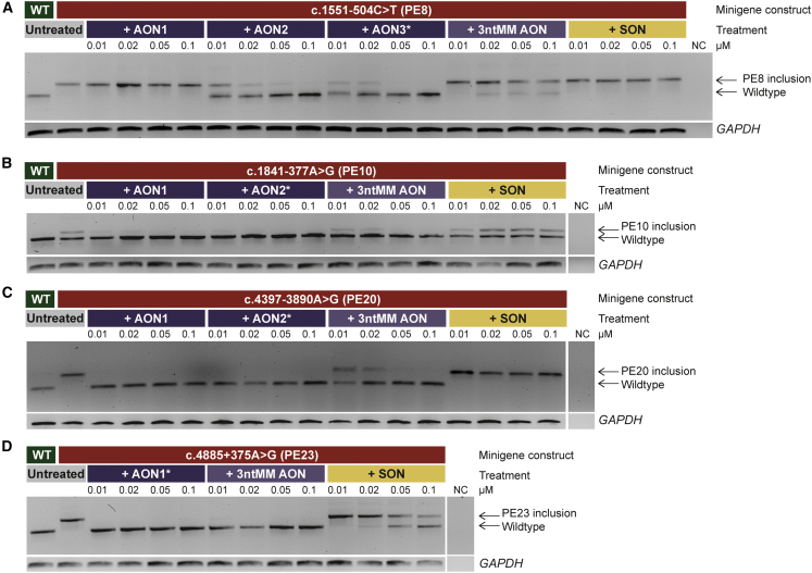
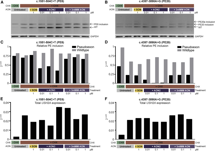
A significant number of individuals with a rare disorder such as Usher syndrome (USH) and (non-)syndromic autosomal recessive retinitis pigmentosa (arRP) remain genetically unexplained. Therefore, we assessed subjects suspected of USH2A-associated disease and no or mono-allelic USH2A variants using whole genome sequencing (WGS) followed by an improved pipeline for variant interpretation to provide a conclusive diagnosis. One hundred subjects were screened using WGS to identify causative variants in USH2A or other USH/arRP-associated genes. In addition to the existing variant interpretation pipeline, a particular focus was put on assessing splice-affecting properties of variants, both in silico and in vitro. Also structural variants were extensively addressed. For variants resulting in pseudoexon inclusion, we designed and evaluated antisense oligonucleotides (AONs) using minigene splice assays and patient-derived photoreceptor precursor cells. Biallelic variants were identified in 49 of 100 subjects, including novel splice-affecting variants and structural variants, in USH2A or arRP/USH-associated genes. Thirteen variants were shown to affect USH2A pre-mRNA splicing, including four deep-intronic USH2A variants resulting in pseudoexon inclusion, which could be corrected upon AON treatment. We have shown that WGS, combined with a thorough variant interpretation pipeline focused on assessing pre-mRNA splicing defects and structural variants, is a powerful method to provide subjects with a rare genetic condition, a (likely) conclusive genetic diagnosis. This is essential for the development of future personalized treatments and for patients to be eligible for such treatments.

Keywords: USH2A; Usher syndrome; antisense oligonucleotides; minigene splice assay; photoreceptor precursor cells; pseudoexon; retinitis pigmentosa; splicing; usherin; whole genome sequencing.


Conflict of interest statement

The authors declare no competing interests.

Figures

Figure 1
Figure 1
Overview of causative genes in a cohort of 100 USH and arRP-affected individuals (A) In 49 of the 100 probands, (likely) biallelic (likely) pathogenic variants were identified in genes associated with autosomal recessive retinitis pigmentosa (arRP) or Usher syndrome (USH). (B) The solved rate per phenotype (arRP, deafness [DFNB] or USH) is shown. The majority (61%) of USH subjects are genetically explained, compared with 38% of arRP subjects and one out of three DFNB affected individuals. The remaining subject (in dark gray) was diagnosed with cone-rod dystrophy (CRD) with hearing loss and remained unsolved.
Figure 2
Figure 2
Minigene splice assays for 21 variants with a potential effect on splicing Representative gel images from minigene splice assays for deep-intronic variants, non-canonical splice site variants, exonic variants, and branchpoint variants. The numbers visualized on the gel correspond to the protein effects described below. (A) Three deep-intronic variants (c.1551-504C>T, c.4397-3890A>G, and c.4885+375A>G32) resulted in PE inclusion in all amplified transcripts. Variant c.1841-377A>G, resulted in the inclusion of a 94 nt PE (1) and remaining conventionally spliced transcript (2). (B) Two non-canonical splice site variants (c.5775A>T and c.14791+5G>T) resulted in the complete skipping of exon 28 or exon 67 in all detected transcripts, whereas variant c.9259-9T>A resulted in a 7-nt extension. Variant c.9258G>T was shown to have a dual effect: partial skipping of exon 46 (3) and alternative partial skipping of exon 46 (4). Likewise, variant c.14134-5T>C resulted in a 47-nt extension upstream of exon 65 (5) and remaining conventionally spliced transcript (6). Variant c.9959-3C>G induced two alternative pre-mRNA splicing events: skipping of complete exon 51 (7) and partial skipping of exon 51 (8) with incomplete penetrance. (C) Variant c.2303G>A had multiple effects, with part of the transcripts lacking the center region of exon 13 (9), part lacking the 3′ side of exon 13 (10) and part being conventionally spliced mRNA (11). (D) Variant c.5573-19A>G resulted in skipping of exon 28 in all transcripts, whereas variant c.14583-26A>G resulted in skipping of exon 67 (12) and conventionally spliced mRNA (13). Transfections were performed in duplicate, GAPDH was used as loading control in all experiments. An overview of the observed effects on pre-mRNA splicing and the consequences on protein level is listed in Table S7. M, variant of interest; nt, nucleotides; WT, wild type.
Figure 3
Figure 3
Antisense oligonucleotide (AON) treatment dilution series for four deep-intronic targets Representative gel images from AON treatment studies in minigenes. (A) Two out of three AONs (AON2 and AON3) were effective in reverting a 118-nt pseudoexon (PE) inclusion in intron 8, caused by variant c.1551-504C>T, for all tested concentrations. A three-nucleotide mismatch antisense oligonucleotide (3ntMM AON) of the most potent AON (AON3, marked with an asterisk) and a scrambled oligonucleotide (SON), had minimal to no effect on reverting PE inclusion. (B) Variant c.1841-377A>G resulted in partial inclusion of a 94-nt PE in intron 10. Both AONs efficiently reverted PE inclusion, while a 3ntMM AON of AON2 had reduced effect and an SON was not effective. (C) Variant c.4397-3890A>G caused inclusion of an 87-nt PE in intron 20. Both AONs efficiently reverted PE inclusion, while a 3ntMM AON of AON2 had reduced effect and an SON was not effective. (D) Variant c.4885+375A>G causes inclusion of a 130-nt PE in intron 23. The AON that could be designed for the region was efficient in reverting PE inclusion. A 3ntMM AON was fully effective as well and an SON had reduced effect. NC, negative control; nt, nucleotides; WT, wild type. Transfections were performed in duplicate, GAPDH was used as loading control in all experiments.
Figure 4
Figure 4
AON treatment of photoreceptor precursor cells (PPCs) differentiated from patient-derived induced pluripotent stem cells (iPSCs) (A) RT-PCR on mRNA of PPC samples that were heterozygous for c.1551-504C>T and subsequent gel electrophoresis showed that ∼50% of transcripts harbor a pseudoexon (PE), while 50% are wild-type (WT) transcripts. Treatment with three different concentrations of antisense oligonucleotide 3 (AON3) against PE8 for 48 h resulted in a reduction, but not complete reversion of PE inclusion. Treatment with a scrambled oligonucleotide (SON) or 3-nucleotide mismatch AON (3ntMM AON) did not result in reduction of PE inclusion. GAPDH was used as loading control. (B) In PPCs heterozygous for c.4397-3890A>G, inclusion of PE20 was fully reverted after treatment with AON2 against PE20, while an SON and 3ntMM AON did not have an effect. (C and D) The effect observed with RT-PCR was confirmed with RT-qPCR. Levels of transcripts containing the PE of interest as well as WT transcript was compared with USH2A exon 13 expression (normalized against GUSB) and confirmed that AON3 against PE8 can partially rescue PE8 inclusion, while AON2 against PE20 has a full effect. (E and F) Expression of USH2A (exon 13) compared with GUSB. USH2A expression was relatively low in all samples; however, lowest in samples not treated with cycloheximide (CHX).

References

    1. Vaché C., Besnard T., le Berre P., García-García G., Baux D., Larrieu L., Abadie C., Blanchet C., Bolz H.J., Millan J., et al. Usher syndrome type 2 caused by activation of an USH2A pseudoexon: implications for diagnosis and therapy. Hum. Mutat. 2012;33:104–108. doi: 10.1002/humu.21634. - DOI - PubMed
    1. Liquori A., Vaché C., Baux D., Blanchet C., Hamel C., Malcolm S., Koenig M., Claustres M., Roux A.F. Whole USH2A gene sequencing identifies several new deep intronic mutations. Hum. Mutat. 2016;37:184–193. doi: 10.1002/humu.22926. - DOI - PubMed
    1. Baux D., Vaché C., Blanchet C., Willems M., Baudoin C., Moclyn M., Faugère V., Touraine R., Isidor B., Dupin-Deguine D., et al. Combined genetic approaches yield a 48% diagnostic rate in a large cohort of French hearing-impaired patients. Sci. Rep. 2017;7:16783. doi: 10.1038/s41598-017-16846-9. - DOI - PMC - PubMed
    1. Carss K.J., Arno G., Erwood M., Stephens J., Sanchis-Juan A., Hull S., Megy K., Grozeva D., Dewhurst E., Malka S., et al. Comprehensive rare variant analysis via whole-genome sequencing to determine the molecular pathology of inherited retinal disease. Am. J. Hum. Genet. 2017;100:75–90. doi: 10.1016/j.ajhg.2016.12.003. - DOI - PMC - PubMed
    1. de Bruijn S.E., Fiorentino A., Ottaviani D., Fanucchi S., Melo U.S., Corral-Serrano J.C., Mulders T., Georgiou M., Rivolta C., Pontikos N., et al. Structural variants create new topological-associated domains and ectopic retinal enhancer-gene contact in dominant retinitis pigmentosa. Am. J. Hum. Genet. 2020;107:802–814. doi: 10.1016/j.ajhg.2020.09.002. - DOI - PMC - PubMed

Publication types

Supplementary concepts