. 2023 Jan 31:10:100477.
doi: 10.1016/j.ejro.2023.100477. eCollection 2023.

Current and upcoming radionuclide therapies in the direction of precision oncology: A narrative review

Affiliations

Current and upcoming radionuclide therapies in the direction of precision oncology: A narrative review

Hina J Shah et al. Eur J Radiol Open. .

Abstract

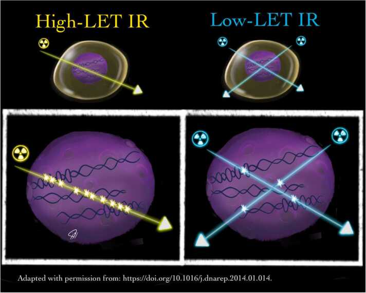
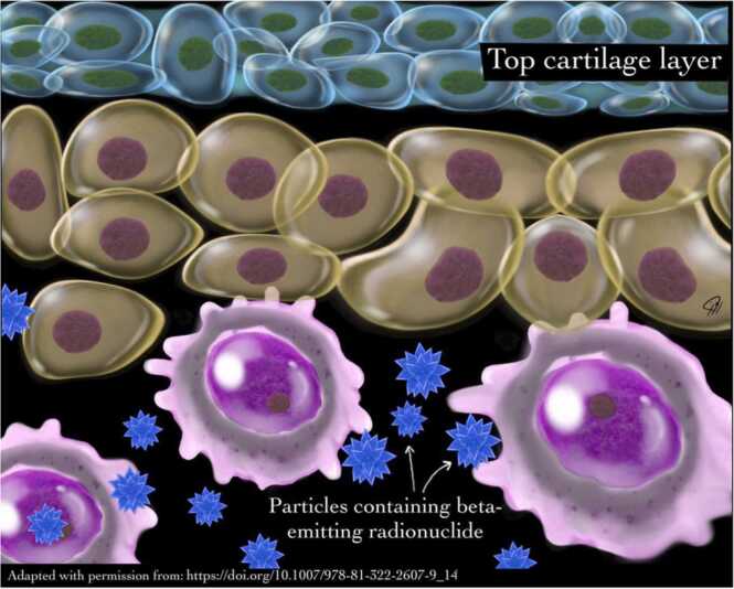
As new molecular tracers are identified to target specific receptors, tissue, and tumor types, opportunities arise for the development of both diagnostic tracers and their therapeutic counterparts, termed "theranostics." While diagnostic tracers utilize positron emitters or gamma-emitting radionuclides, their theranostic counterparts are typically bound to beta and alpha emitters, which can deliver specific and localized radiation to targets with minimal collateral damage to uninvolved surrounding structures. This is an exciting time in molecular imaging and therapy and a step towards personalized and precise medicine in which patients who were either without treatment options or not candidates for other therapies now have expanded options, with tangible data showing improved outcomes. This manuscript explores the current state of theranostics, providing background, treatment specifics, and toxicities, and discusses future potential trends.

Keywords: Cancer imaging; Nuclear medicine; Theranostics.


Conflict of interest statement

The authors declare that they have no known competing financial interests or personal relationships that could have appeared to influence the work reported in this paper.

Figures

Fig. 1
Fig. 1
showing more ionization caused in the path of radionuclide with high linear energy transfer compared to that of radionuclide with low linear energy transfer
Fig. 2
Fig. 2
showing various way in which radionuclide therapy could be delivered. Direct delivery of radionuclide element, using small molecules, peptides, antibodies, nanoconstruct and, microspheres.
Fig. 3
Fig. 3
showing mechanism of action of Ra-223. This radionuclide is administered as an intravenous injection. It binds with hydroxyapatite and is then incorporated into the bony matrix where it causes destruction.
Fig. 4
Fig. 4
60-year-old male with metastatic Prostate cancer Gleason’s score 4 + 5. Non-responder to Hormonal therapy and Docetaxel. (A) Ga-68-PSMA MIP image showing PSMA-avid disease in skeleton. PSA at this time was > 400, He was subsequently treated with 3 cycles of Ac-225-PSMA-617, (B) Ga-68-PSMA MIP image showing near complete resolution of previously seen PSMA-avid disease in skeleton. PSA at this time was 0.1.
Fig. 5
Fig. 5
(A) Ga-68-DOTATATE and (B) F-18-FDG Maximum Intensity Projection (MIP)images of a patient with Grade 1/well-differentiated NET showing multiple areas of somatostatin receptor expressing metastatic disease in image A and negligible FDG uptake in image B. PRRT is suitable in this patient. (C) Ga-68-DOTATATE and (D) F-18-FDG MIP images of a patient with Grade 2/moderately differentiated NET showing multiple areas of somatostatin receptor expressing metastatic disease in the liver in image C and only a few lesions showing concurrent FDG uptake in image D indicating Grade 1/well-differentiated NET. PRRT is suitable in this patient. (E) Ga-68-DOTATATE and (F) F-18-FDG MIP images of a patient with mixed NET showing multiple areas of somatostatin receptor expressing metastatic disease in image E and many lesions showing FDG uptake in image F. One of the area in image F marked with arrow without somatostatin receptor expression in image E indicating that this is high grade/poorly differentiated focus of NET. By using FDG and DOTATATE PET imaging, tumor grade mapping can be done in the whole body.
Fig. 6
Fig. 6
(A) Ga-68-DOTATATE MIP image showing somatostatin receptor expressing disease in liver and upper abdominal lymph nodes. PRRT is a suitable treatment in this patient. (B), (C), (D), (E) Lu-177-DOTATATE planar anterior images after 1,2, 3 and 4 cycles of PRRT respectively, showing decreasing burden of the disease in the liver. Patient received 200 mCi of Lu-177-DOTATATE for each cycle of PRRT. Imaging was performed 18–24 hr after the therapy. (F) Ga-68-DOTATATE MIP post-PRRT image showing partial response to treatment.
Fig. 7
Fig. 7
depicting advantages of somatostatin antagonist over somatostatin analogues.
Fig. 8
Fig. 8
(A)Anterior and (B)Posterior planar images post 30 mCi I-131 showing focal uptake in the thyroid bed. Physiological tracer in the stomach and bowel loops.
Fig. 9
Fig. 9
(A)Anterior and (B)Posterior planar images post 125mCi of I-131 showing focal uptake in the thyroid bed and diffuse heterogeneous uptake in the lung metastatic disease. Physiological tracer in the stomach, bowel loops and urinary bladder.
Fig. 10
Fig. 10
showing different types of intrahepatic tracer distribution which can be seen on Tc-99m-MAA scan after injection of the tracer in the common hepatic artery.
Fig. 11
Fig. 11
(A) Y-90-resin micropheres Maximum Intensity Projection (MIP), axial SPECT, CT and fused SPECT/CT images showing heterogenous tracer uptake in the HCC involving the left lobe of the liver. (B) Y-90-glass microspheres Maximum Intensity Projection (MIP), axial PET, CT and fused PET/CT images showing tracer uptake in the left lobe of the liver. In the right lobe, bland embolization with lipiodol was performed.
Fig. 12
Fig. 12
(A)Anterior and (B)Posterior planar baseline pretreatment 123-I-mIBG images showing uptake in the multiple skeletal sites with Curie score of 23. (C)Anterior and (D)Posterior planar posttreatment 123-I-mIBG images showing uptake in a few skeletal sites with Curie score of 16. Physiological uptake in the myocardium, lungs, liver and urinary bladder.
Fig. 13
Fig. 13
showing mechanism of action of radiation synovectomy

References

    1. Hoefnagel C.A. Radionuclide therapy revisited. Eur. J. Nucl. Med. 1991;18(6):408–431. - PubMed
    1. Troutner D.E. Chemical and physical properties of radionuclides. Int J. Rad. Appl. Instrum. B. 1987;14(3):171–176. - PubMed
    1. Volkert W.A., Goeckeler W.F., Ehrhardt G.J., Ketring A.R. Therapeutic radionuclides: production and decay property considerations. J. Nucl. Med. 1991;32(1):174–185. - PubMed
    1. Zweit J. Radionuclides and carrier molecules for therapy. Phys. Med. Biol. 1996;41(10):1905–1914. - PubMed
    1. Kassis A.I., Adelstein S.J. Radiobiologic principles in radionuclide therapy. J. Nucl. Med. 2005;46(Suppl 1):4S–12S. - PubMed