. 2023 Feb 14;11(2):e0423922.
doi: 10.1128/spectrum.04239-22. Online ahead of print.

Endogenous IL-33 Accelerates Metacestode Growth during Late-Stage Alveolar Echinococcosis

Affiliations

Endogenous IL-33 Accelerates Metacestode Growth during Late-Stage Alveolar Echinococcosis

Brice Autier et al. Microbiol Spectr. .

Abstract

During the course of the infectious disease alveolar echinococcosis (AE), the larval stage of Echinococcus multilocularis develops in the liver, where an initial Th1/Th17 immune response may allow its elimination in resistant individuals. In patients susceptible to infection and disease, the Th2 response initiates later, inducing tolerance to the parasite. The role of interleukin 33 (IL-33), an alarmin released during necrosis and known to drive a Th2 immune response, has not yet been described during AE. Wild-type (WT) and IL-33-/- C57BL/6J mice were infected by peritoneal inoculation with E. multilocularis metacestodes and euthanized 4 months later, and their immune response were analyzed. Immunofluorescence staining and IL-33 enzyme-linked immunosorbent assay (ELISA) were also performed on liver samples from human patients with AE. Overall, metacestode lesions were smaller in IL-33-/- mice than in WT mice. IL-33 was detected in periparasitic tissues, but not in mouse or human serum. In infected mice, endogenous IL-33 modified peritoneal macrophage polarization and cytokine profiles. Th2 cytokine concentrations were positively correlated with parasite mass in WT mice, but not in IL-33-/- mice. In human AE patients, IL-33 concentrations were higher in parasitic tissues than in distant liver parenchyma. The main sources of IL-33 were CD31+ endothelial cells of the neovasculature, present within lymphoid periparasitic infiltrates together with FOXP3+ Tregs. In the murine model, periparasitic IL-33 correlated with accelerated parasite growth putatively through the polarization of M2-like macrophages and release of immunosuppressive cytokines IL-10 and transforming growth factor β1 (TGF-β1). We concluded that IL-33 is a key alarmin in AE that contributes to the tolerogenic effect of systemic Th2 cytokines. IMPORTANCE Infection with the metacestode stage of Echinococcus multilocularis, known as alveolar echinococcosis, is the most severe cestodosis worldwide. However, less than 1% of exposed individuals, in which the immune system is unable to control the parasite, develop the disease. The factors responsible for this interindividual variability are not fully understood. In this in vivo study comparing wild-type and IL-33-/- infected mice, together with data from human clinical samples, we determined that IL-33, an alarmin released following tissue injury and involved in the pathogenesis of cancer and asthma, accelerates the progression of the disease by modulating the periparasitic microenvironment. This suggests that targeting IL-33 could be of interest for the management of patients with AE, and that IL-33 polymorphisms could be responsible for increased susceptibility to AE.

Keywords: Echinococcus multilocularis; IL-1RL1; IL-33; ST2; alveolar echinococcosis; angiogenesis.


Conflict of interest statement

The authors declare no conflict of interest.

Figures

FIG 1
FIG 1
Endogenous interleukin 33 (IL-33) is associated with accelerated course of alveolar echinococcosis (AE). (A) Wet weight of peritoneal metacestodes collected from mice after 4 months of infection. (B) Mouse weight evolution during the course of infection. (C) Quantification of Em14-3-3 gene expression, a parasite viability marker, normalized using the threshold cycle (2–ΔΔCT) method and Echinococcus multilocularis ActII gene. (D) Representative pictures of peritoneal lesions (surrounded by dotted lines) from wild-type (WT) and IL-33−/− mice. Panels A and B: pooled data from 2 independent experiments, with 12 WT and 14 IL-33−/− mice. Panel C: data obtained from one experiment, with 6 WT and 5 IL-33−/− mice. Plots show mean ± standard deviation (SD). Significance was tested using Mann-Whitney U test (A and C) and two-way analysis of variance (ANOVA) for repeated measures with Bonferroni’s post-tests (B). *, P ≤ 0.05; **, P ≤ 0.01; ***, P ≤ 0.001.
FIG 2
FIG 2
Endogenous IL-33 modulates peritoneal cell populations during AE. (A) Composition of peritoneal cell infiltrates at 4 months postinfection. (B) Proportion of CD206+ monocytes/macrophages in peritoneal infiltrates. (C) Quantification of gene expression in the periparasitic tissues, normalized using 2–ΔΔCT method and the Gapdh and Ppid endogenous controls. (D) Linear regression between the weight of lesions and proportions of peritoneal cell populations of WT (blue dots and line) and IL-33−/− mice (orange dots and line). Panels A, B, and D: pooled data from 2 independent experiments, with 5 uninfected (2 WT [close circles] and 3 IL-33−/− [open circles] mice) and 11 infected WT and 14 IL-33−/− mice. Panel C: data obtained from one experiment with 6 WT and 5 IL-33−/− mice. Plots show means ± SD (A, B, and C) and the r2 correlation coefficient (D). Significance was tested using the Kruskal-Wallis test with Dunn’s multiple-comparison post hoc analysis (A), the Mann-Whitney U test (B and C) and the F test (D). *, P ≤ 0.05; **, P ≤ 0.01; ***, P ≤ 0.001.
FIG 3
FIG 3
Endogenous IL-33 modulates concentrations of cytokines in peritoneal lavages during AE. Cytokine concentrations determined by flow cytometry in peritoneal lavages of WT and IL-33−/− mice at 4 months postinfection. Data obtained from one experiment, with 5 uninfected (2 WT [close circles] and 3 IL-33−/− [open circles]) and 6 infected WT and 6 infected IL-33−/− mice. Plots show means ± SD. Significance was tested using the Kruskal-Wallis test with Dunn’s multiple-comparison post hoc analysis. *, P ≤ 0.05; **, P ≤ 0.01; ***, P ≤ 0.001.
FIG 4
FIG 4
Endogenous IL-33 has little impact on cellular and transcription profiles in the liver. (A) Composition of liver cellular infiltrates at 4 months postinfection. Gating strategy was the same as used for peritoneal cells, except that Panel 2 was applied in 1 on 2 experiments. (B) Ratio of CD4+ to CD8+ T cells. (C) Myeloid subpopulations of liver cellular infiltrates. (D) Quantification of gene expression in the liver, normalized using the 2–ΔΔCT method and the Gapdh and Actb endogenous controls. Panels A, B, and D: pooled data from 2 independent experiments, with 5 uninfected (2 WT [close circles] and 3 IL-33−/− [open circles]) and 11 infected WT and 14 infected IL-33−/− mice. Panel C: data obtained from one experiment, with 5 mice in each group. Plots show means ± SD. Significance was tested using the Kruskal-Wallis test with Dunn’s multiple-comparison post hoc analysis. *, P ≤ 0.05; **, P ≤ 0.01; ***, P ≤ 0.001.
FIG 5
FIG 5
Endogenous IL-33 modulates systemic cytokine profiles during AE. (A) Cytokines concentrations determined by flow cytometry in the serum of WT and IL-33−/− mice during the course of infection. Blue and orange symbols indicate statistical significance over time within each group of mice (Kruskal-Wallis with post hoc Dunn’s multiple-comparison test), and black symbols stand for comparison between mice groups at a given time point (Mann-Whitney U test). Concentrations were normalized by logarithmic transformation. Dotted lines show 95% confidence intervals. (B) Heatmaps of pairwise correlation coefficients analysis between serum cytokines concentrations at 4 months postinfection. Dot color and size indicate correlation coefficients and their statistical significance, respectively. Pooled data from 2 independent experiments, with 10 WT and 14 IL-33−/− mice. The parameter “lesions” stands for the weight of the metacestodes 4 months postinfection. *, P ≤ 0.05; **, P ≤ 0.01; ***, P ≤ 0.001.
FIG 6
FIG 6
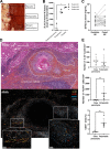
IL-33 is detected in liver alveolar echinococcosis lesions from human patients. (A) Anatomical areas sampled for IL-33 quantification in the liver of patients with AE (n = 3). Squares show the location of biopsy samples. (B) IL-33 concentrations according to the sample site. Quantification after tissue lysis and enzyme-linked immunosorbent assay (ELISA) dosage. Friedman test and post hoc Dunn’s multiple-comparison test (*, P ≤ 0.05). (C) Concentrations of soluble ST2 in sera of 18 patients infected with E. multilocularis, before and after curative surgery. Significance was tested using Wilcoxon matched-pairs signed-rank test. (D) Annotated hematoxylin and eosin staining and immunofluorescence staining of consecutive histological sections, using DAPI (4′,6-diamidino-2-phenylindole), anti-IL-33, anti-CD31, and anti-FoxP3 antibodies, with higher magnifications of structures of interest. Representative picture of biopsy sample slides obtained from 5 patients. (E) Quantification of density of IL-33+ nuclei (top), density of FoxP3+ cells (middle), and proportion of CD31+ area (bottom), in liver parenchyma and periparasitic granuloma, from immunofluorescence staining of human biopsy sample slides (n = 5). Mann-Whitney U test: **, P ≤ 0.01.
FIG 7
FIG 7
Modulation of the host immune response by IL-33 and its impact on parasite growth. (A) Schematic presentation of the putative mechanism of action of IL-33 during alveolar echinococcosis. Step 1: IL-33 is released from neovasculature upon tissue damage by parasite growth. Step 2: IL-33 locally activates ST2+ leukocytes, such as Th2RM, ILC2s, and Tregs, and contributes to polarization of macrophages into the “M2” phenotype. Step 3: ST2+ leukocytes favor parasite growth by orientating immunity toward a tolerogenic phenotype. Blockade of IL-33 for a therapeutic purpose could limit the progression of the disease. (B) Schematic modeling of AE pathophysiology. After a host is contaminated with E. multilocularis eggs, the parasite must pass through 3 immunological events (framed text) to develop a viable metacestode. Once this is finished, immunological factors only impact the parasite’s growth rate, with Th1/Th17 and Th2/Treg responses decreasing and increasing it, respectively. We showed that the alarmin IL-33 locally contributes to Th2 and Treg activity during late-stage AE, thus accelerating the course of the disease.

References

    1. Deplazes P, Rinaldi L, Alvarez Rojas CA, Torgerson PR, Harandi MF, Romig T, Antolova D, Schurer JM, Lahmar S, Cringoli G, Magambo J, Thompson RCA, Jenkins EJ. 2017. Global distribution of alveolar and cystic echinococcosis. Adv Parasitol 95:315–493. doi:10.1016/bs.apar.2016.11.001. - DOI - PubMed
    1. Vuitton DA, McManus DP, Rogan MT, Romig T, Gottstein B, Naidich A, Tuxun T, Wen H, da Silva AM, Vuitton DA, McManus DP, Romig T, Rogan MR, Gottstein B, da Silva AM, Wen H, Naidich A, Tuxun T, Avcioglu A, Boufana B, Budke C, Casulli A, Güven E, Hillenbrand A, Jalousian F, Jemli MH, Knapp J, Laatamna A, Lahmar S, Naidich A, Rogan MT, Sadjjadi SM, Schmidberger J, Amri M, Bellanger A-P, Benazzouz S, Brehm K, Hillenbrand A, Jalousian F, Kachani M, Labsi M, Masala G, da Silva AM, Seyed MS, Soufli I, Touil-Boukoffa C, Wang J, Zeyhle E, Aji T, Akhan O, World Association of Echinococcosis, et al. . 2020. International consensus on terminology to be used in the field of echinococcoses. Parasite 27:41. doi:10.1051/parasite/2020024. - DOI - PMC - PubMed
    1. Kern P, Wen H, Sato N, Vuitton DA, Gruener B, Shao Y, Delabrousse E, Kratzer W, Bresson-Hadni S. 2006. WHO classification of alveolar echinococcosis: principles and application. Parasitol Int 55 Suppl:S283–287. doi:10.1016/j.parint.2005.11.041. - DOI - PubMed
    1. Wen H, Vuitton L, Tuxun T, Li J, Vuitton DA, Zhang W, McManus DP. 2019. Echinococcosis: advances in the 21st century. Clin Microbiol Rev 32:e00075-18. doi:10.1128/CMR.00075-18. - DOI - PMC - PubMed
    1. Vuitton DA, Zhang SL, Yang Y, Godot V, Beurton I, Mantion G, Bresson-Hadni S. 2006. Survival strategy of Echinococcus multilocularis in the human host. Parasitol Int 55:S51–S55. doi:10.1016/j.parint.2005.11.007. - DOI - PubMed