. 2023 Feb;52(1-2):39-51.
doi: 10.1007/s00249-023-01632-5. Epub 2023 Feb 14.

A comparative characterisation of commercially available lipid-polymer nanoparticles formed from model membranes

Affiliations

A comparative characterisation of commercially available lipid-polymer nanoparticles formed from model membranes

Henry Sawczyc et al. Eur Biophys J. 2023 Feb.

Abstract

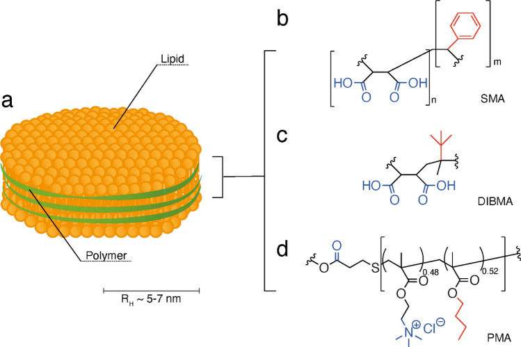
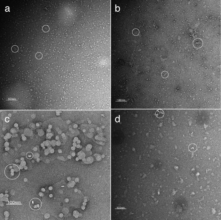
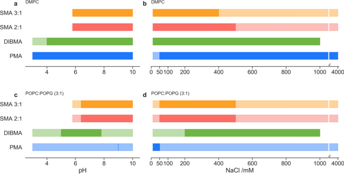
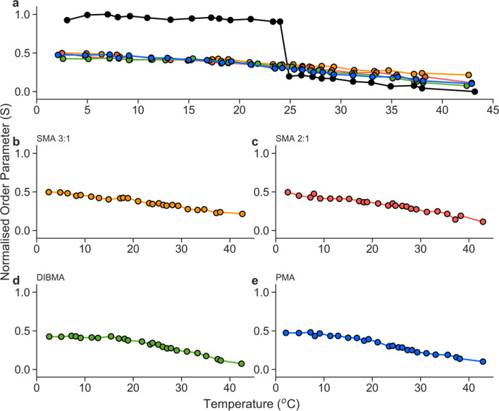
From the discovery of the first membrane-interacting polymer, styrene maleic-acid (SMA), there has been a rapid development of membrane solubilising polymers. These new polymers can solubilise membranes under a wide range of conditions and produce varied sizes of nanoparticles, yet there has been a lack of broad comparison between the common polymer types and solubilising conditions. Here, we present a comparative study on the three most common commercial polymers: SMA 3:1, SMA 2:1, and DIBMA. Additionally, this work presents, for the first time, a comparative characterisation of polymethacrylate copolymer (PMA). Absorbance and dynamic light scattering measurements were used to evaluate solubilisation across key buffer conditions in a simple, adaptable assay format that looked at pH, salinity, and divalent cation concentration. Lipid-polymer nanoparticles formed from SMA variants were found to be the most susceptible to buffer effects, with nanoparticles from either zwitterionic DMPC or POPC:POPG (3:1) bilayers only forming in low to moderate salinity (< 600 mM NaCl) and above pH 6. DIBMA-lipid nanoparticles could be formed above a pH of 5 and were stable in up to 4 M NaCl. Similarly, PMA-lipid nanoparticles were stable in all NaCl concentrations tested (up to 4 M) and a broad pH range (3-10). However, for both DIBMA and PMA nanoparticles there is a severe penalty observed for bilayer solubilisation in non-optimal conditions or when using a charged membrane. Additionally, lipid fluidity of the DMPC-polymer nanoparticles was analysed through cw-EPR, showing no cooperative gel-fluid transition as would be expected for native-like lipid membranes.

Keywords: DIBMA; Lipid-polymer nanoparticles; Lipodisqs; Nanodiscs; PMA; SMALPs.


Conflict of interest statement

The authors wish to declare no conflict of interests.

Figures

Fig. 1
Fig. 1
Diagram of polymer structure used for forming polymer-lipid nanoparticles. a Cartoon representation of polymer-lipid nanoparticles resulting from interaction with polymers (bd), comprised of lipids (shown in yellow) surrounded by a polymer ‘belt’ (shown in green). Multiple polymers have been shown to be capable of forming nanoparticles, such as: b SMA, c DIBMA, and d PMA. The hydrophilic groups are highlighted in blue, and hydrophobic groups highlighted in red
Fig. 2
Fig. 2
Negative stain transmission electron microscopy images of DMPC-polymer nanoparticles: SMA 3:1 (a), SMA 2:1 (b), DIBMA (c), and PMA (d). Nanoparticles were formed by the addition of 1.5% (w/w) polymer to suspended DMPC membranes under optimal conditions (20 mM HEPES, 100 mM NaCl, pH 7.4), followed by size exclusion chromatography to remove excess polymer. White circles highlight abnormal clustering or rouleaux stacking
Fig. 3
Fig. 3
Qualitative range of membrane solubilisation for: DMPC membranes; across a pH range at a constant salt concentration of 100 mM (a); across a NaCl concentration gradient/range (b), and POPC:POPG (3:1); across a pH range (c); across a NaCl concentration gradient/range (d). Ranges are derived from DLS measurements (lighter colour) where nanoparticles were detected (where 2 nm < RH < 10 nm). Darker colour range is indicative of OD350 < 2 after polymer addition, indicating significant membrane solubilisation
Fig. 4
Fig. 4
Qualitative range of membrane solubilisation for: DMPC membranes; across a magnesium concentration from 0 to 25 mM with a constant ionic concentration of 100 mM (a); across a calcium concentration from 0 to 25 mM with a constant ionic concentration of 100 mM (b), and POPC:POPG (3:1); across a magnesium concentration from 0 to 25 mM with a constant ionic concentration of 100 mM (c); across a calcium concentration from 0 to 25 mM with a constant ionic concentration of 100 mM (d). Ranges are derived from DLS measurements (lighter colour) where nanoparticles were detected (where 2 nm < RH < 10 nm). Darker colour range is indicative of OD350 < 2 after polymer addition, indicating significant membrane solubilisation
Fig. 5
Fig. 5
Normalised order parameter (S) of lipid membrane generated from cw-EPR measurement of 1% (molar) 5-PCSL in DMPC-polymer nanoparticles formed by the addition of 1.5% (w/w) polymer to suspended DMPC membranes under optimal conditions (20 mM HEPES, 100 mM NaCl, pH 7.4), followed by size exclusion chromatography to remove excess polymer. DMPC LUVs were generated in the absence of polymer, using freeze–thaw and extrusion methods. a Order parameter of 400 nm DMPC LUVs (black), SMA 3:1 (orange), SMA 2:1(red), DIBMA (green), and PMA (blue) normalised to LUV order parameter. Isolated normalised order parameters of SMA 3:1 (b), SMA 2:1 (c), DIBMA (d), and PMA (e) nanoparticles

References

    1. Anada C, Ikeda K, Egawa A, et al. Temperature- and composition-dependent conformational transitions of amphipathic peptide–phospholipid nanodiscs. J Colloid Interface Sci. 2021;588:522–530. doi: 10.1016/j.jcis.2020.12.090. - DOI - PubMed
    1. Bada Juarez JF, Harper AJ, Judge PJ, et al. From polymer chemistry to structural biology: the development of SMA and related amphipathic polymers for membrane protein extraction and solubilisation. Chem Phys Lipids. 2019;221:167–175. doi: 10.1016/j.chemphyslip.2019.03.008. - DOI - PubMed
    1. Bada Juarez JF, Muñoz-García JC, Inácio dos Reis R, et al. Detergent-free extraction of a functional low-expressing GPCR from a human cell line. Biochim Biophys Acta Biomembr. 2020;1862:183152. doi: 10.1016/j.bbamem.2019.183152. - DOI - PubMed
    1. Bada Juarez JF, Judge PJ, Adam S, et al. Structures of the archaerhodopsin-3 transporter reveal that disordering of internal water networks underpins receptor sensitization. Nat Commun. 2021;12:1–10. doi: 10.1038/s41467-020-20596-0. - DOI - PMC - PubMed
    1. Bakheet TM, Doig AJ. Properties and identification of human protein drug targets. Bioinformatics. 2009;25:451–457. doi: 10.1093/bioinformatics/btp002. - DOI - PubMed

LinkOut - more resources