. 2023 Aug;75(8):1477-1489.
doi: 10.1002/art.42478. Epub 2023 Jun 6.

Interleukin-23 Regulates Inflammatory Osteoclastogenesis via Activation of CLEC5A(+) Osteoclast Precursors

Affiliations

Interleukin-23 Regulates Inflammatory Osteoclastogenesis via Activation of CLEC5A(+) Osteoclast Precursors

Hiroki Furuya et al. Arthritis Rheumatol. 2023 Aug.

Abstract

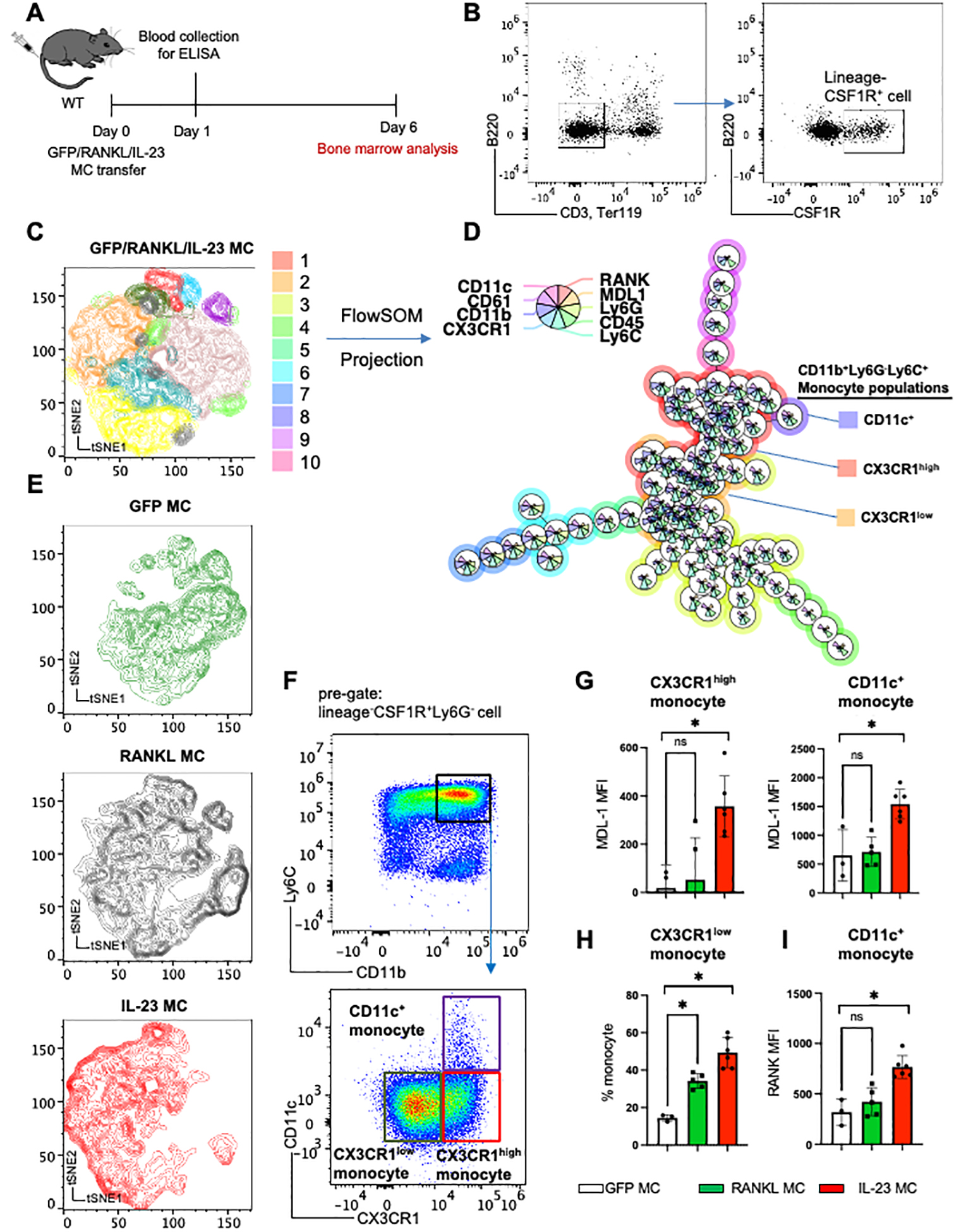
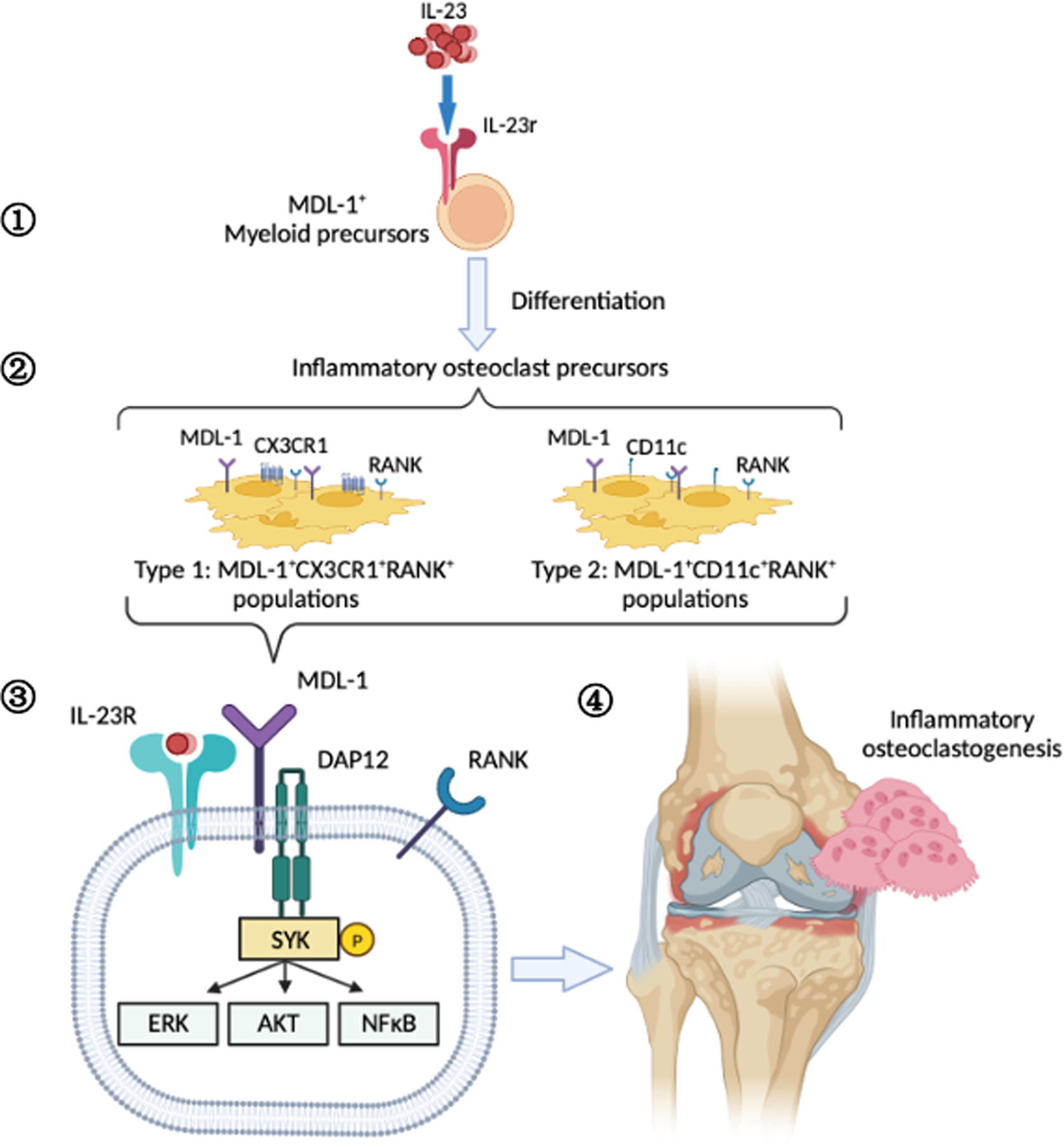
Objective: To investigate the role of interleukin-23 (IL-23) in pathologic bone remodeling in inflammatory arthritis.

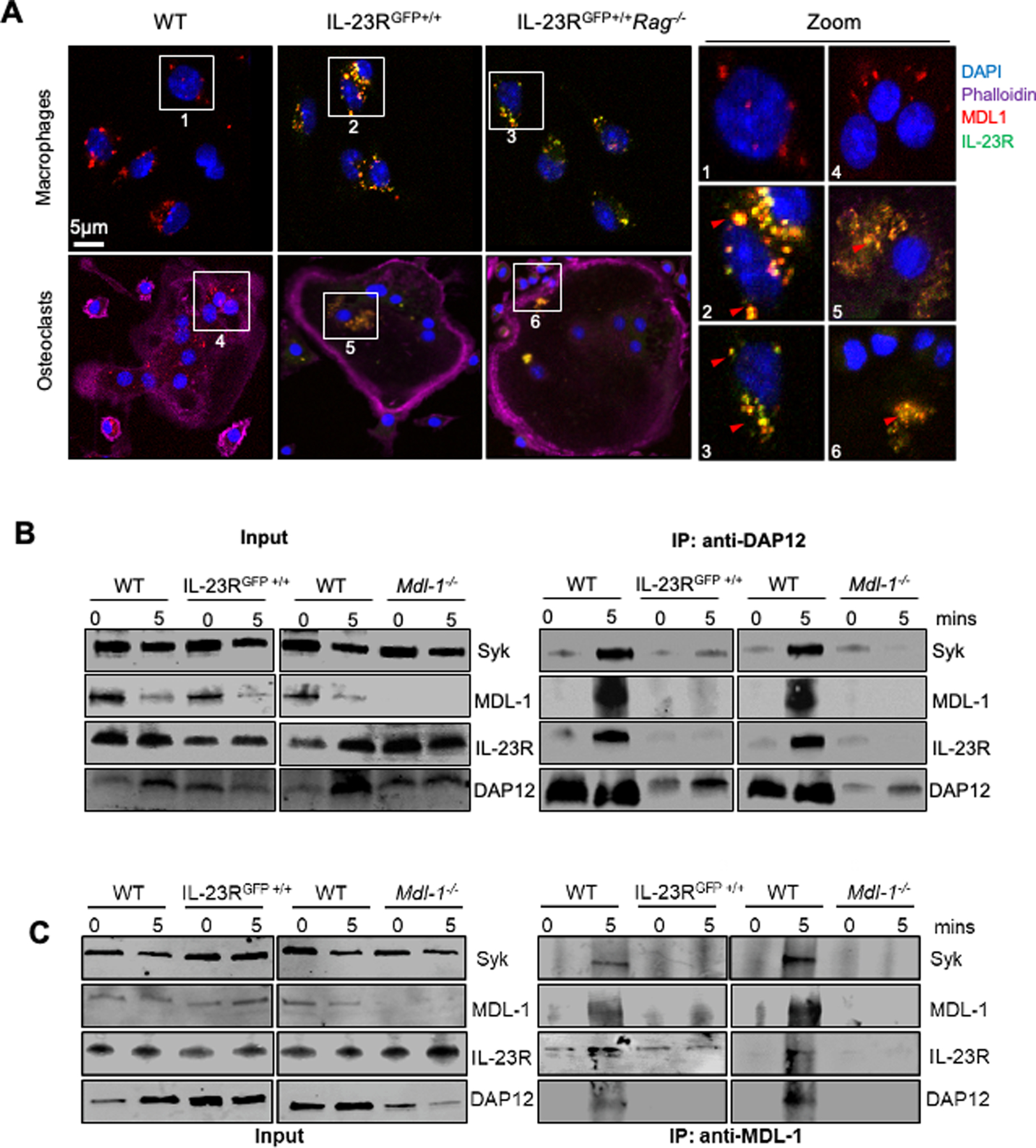
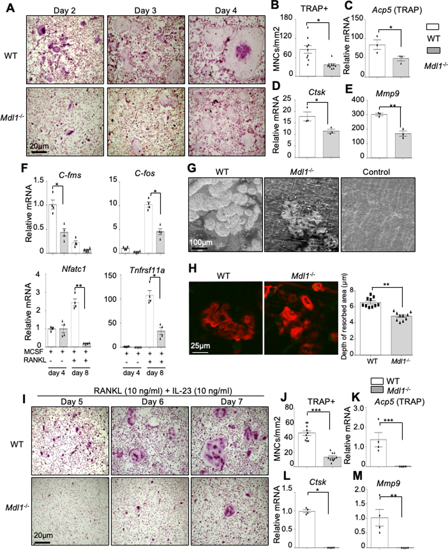
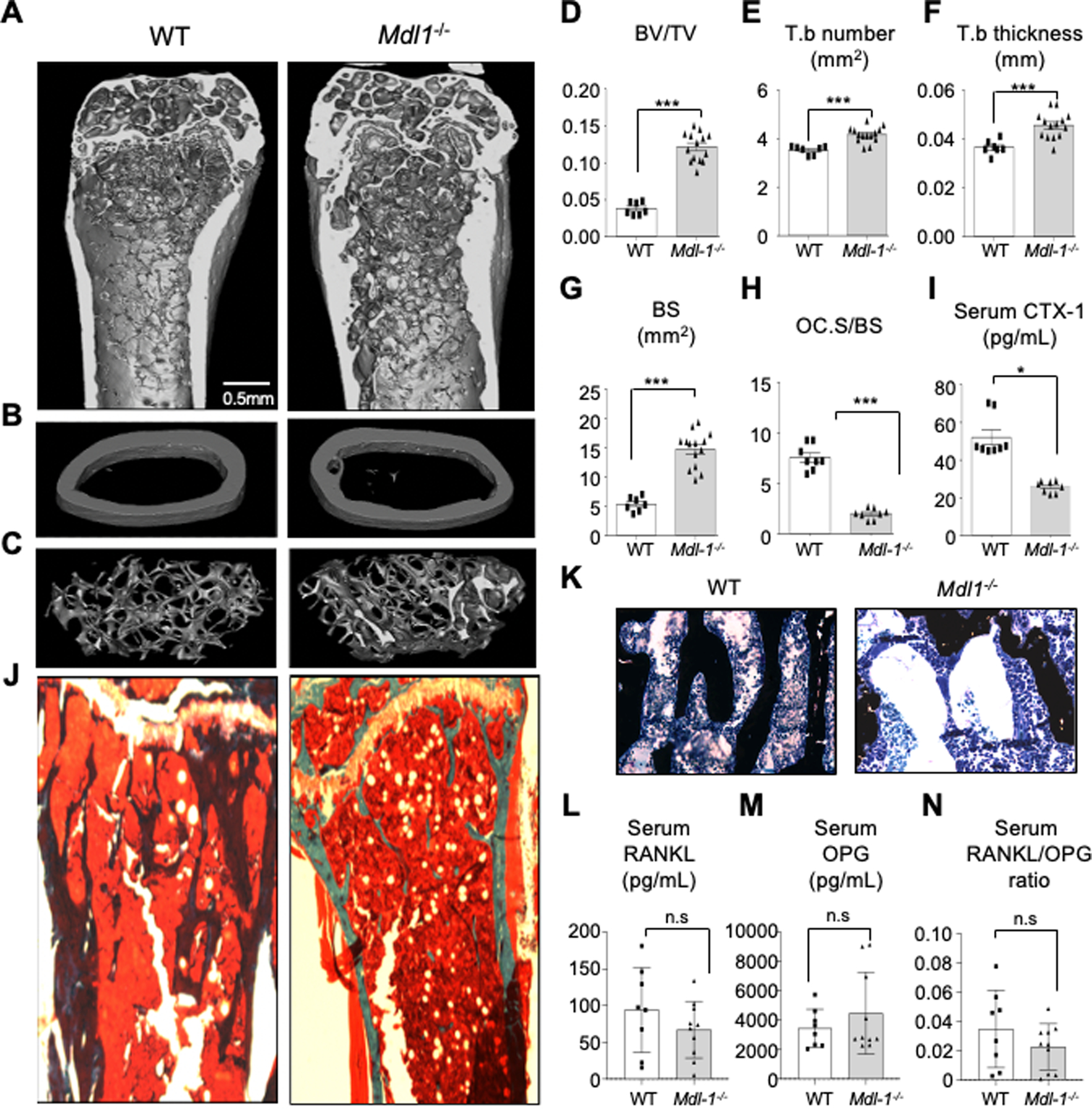
Methods: In this study we investigated the role of IL-23 in osteoclast differentiation and activation using in vivo gene transfer techniques in wild-type and myeloid DNAX-activation protein 12-associating lectin-1 (MDL-1)-deficient mice, and by performing in vitro and in vivo osteoclastogenesis assays using spectral flow cytometry, micro-computed tomography analysis, Western blotting, and immunoprecipitation.

Results: Herein, we show that IL-23 induces the expansion of a myeloid osteoclast precursor population and supports osteoclastogenesis and bone resorption in inflammatory arthritis. Genetic ablation of C-type lectin domain family member 5A, also known as MDL-1, prevents the induction of osteoclast precursors by IL-23 that is associated with bone destruction, as commonly observed in inflammatory arthritis. Moreover, osteoclasts derived from the bone marrow of MDL-1-deficient mice showed impaired osteoclastogenesis, and MDL-1-/- mice had increased bone mineral density.

Conclusion: Our data show that IL-23 signaling regulates the availability of osteoclast precursors in inflammatory arthritis that could be effectively targeted for the treatment of inflammatory bone loss in inflammatory arthritis.


Conflict of interest statement

Disclosures

The authors have no financial conflicts of interest.

Figures

Figure 1:
Figure 1:. IL-23 induces the expansion of MDL-1+ osteoclast precursors.
(A) Schematic presentation of GFP (control)/RANKL/IL-23 MC transfer model in WT mice. (B) Representative figure of integrated flow cytometry data showing the gating strategy of lineageCSF1R+ cell used for unsupervised clustering with (C) tSNE plot and (D) FlowSOM based on expression levels of nine surface molecules, showing 10 distinct clusters. Size of each pie slices show the expression levels of each molecule. (E) Cell clusters post-GFP/RANKL/IL-23 MC gene transfer visualized in tSNE plot. (F) Representative gating strategy of three monocyte populations identified by FlowSOM, and (G) Mean Fluorescence Intensity (MFI) of MDL-1 among CX3CR1high and CD11c+ inflammatory monocytes, (H) percentage of CX3CR1low among CD11b+Ly6GLy6C+monocyte populations, and (I) MFI of RANK among CD11c+ monocyte population after GFP/RANKL/IL-23 MC transfer. Data represent mean ± SEM of single experiment. *P<0.05 by Mann-Whitney.
Figure 2:
Figure 2:. IL-23 activates inflammatory DAP12 signaling in MDL-1+ IL-23R+ double positive cells.
(A) Confocal microscopy images of bone marrow derived macrophages from WT, IL-23RGFP+/+ and IL-23RGFP+/+Rag−/− mice stained with MDL-1 (anti-MDL-1-PE-Cyan), IL-23R (anti-GFP-FITC-Green), nucleus (DAPI-blue), and actin (DyLight 650-Phalloidin) showing MDL-1 and IL-23R co-localization (yellow) in macrophages and osteoclasts. Images are representative of three independent experiments. (B) Immunoprecipitations of DAP12 and/or (C) MDL-1 with total cell lysates of WT, IL-23RGFP+/+ and Mdl-1−/− derived BMMs stimulated with mouse rIL-23 (100 ng/ml) for 0, 5 mins, and immunoblotted with indicated antibodies showing DAP12 co-immunoprecipitation with MDL-1, IL-23R, STAT3, and SYK in WT lysates and lack of binding with DAP12 in either IL-23RGFP+/+ and Mdl-1−/− BMMs.
Figure 3:
Figure 3:. MDL-1 regulates the myeloid PU.1 transcriptional program in RANKL signaling.
(A) TRAP staining of WT bone marrow derived cells cultured in indicated showing increased expression of (B) Mdl-1 and Acp5 during osteoclastogenesis. (C) Alkaline phosphatase (ALP) staining of nodules and (D) Alizarin red staining on 21 day cultures of calvaria-derived cells treated with ascorbic acid (ASC), β-glycerophosphate and dexamethasone (Dex) showing induction of osteoblast maturation and (E) expression of osteoblast differentiation markers independently of Mdl-1. (F-G) Hierarchical cluster analysis of selected genes with > 2-fold changes involved in (F) PU.1 pathway, (G) cell death and survival from microarray analysis of WT (n=9) and Mdl-1−/− (n=9) bone marrow cell stimulated with MCSF for 4 days. (H) Images of live/dead (Calcein/EthD1) staining and (I) fold change of macrophage survival (AlamarBlue assay) showing a lower survival rate of Mdl-1−/− macrophage. (J) Total cell lysates of WT and Mdl-1−/− derived BMMs stimulated with mouse rIL-23 (100 ng/ml) for indicated times, immunoblotted with indicated antibodies and (K) protein band density showing significant differences of Erk, Akt, and NF-κB (p65) phosphorylation in Mdl-1−/− macrophages post-treatment. Data represent mean ± SEM of three independent experiments. *P<0.05; ** P<0.01; *** P<0.001 by Mann-Whitney.
Figure 4:
Figure 4:. MDL-1 deficiency impairs osteoclast differentiation and bone resorption.
(A) TRAP stain of WT (n=4) and Mdl-1−/− (n=4) BMM cultured with MCSF and RANKL for indicated days showing delayed osteoclast maturation and (B) lower total number of TRAP+ multinucleated cells (MNCs) in Mdl-1−/− mice. Images are representative of three independent experiments. (C-E) Gene expression analysis showing reduced expression of osteoclast differentiation markers at day 6 and (F) osteoclast precursor at early (day 4) and late (day 8) time-point in Mdl-1−/− mice. (G-H) WT and Mdl-1−/− bone marrow cells cultured on dentine slices for 18 days with MCSF and RANKL showing (G) reduced dentine erosion surface area and (H) reduced depth of resorbed area by Mdl-1−/− osteoclasts. (I) TRAP stain of WT and Mdl-1−/− BMM cultured with MCSF, RANKL and IL-23 for indicated days showing delayed osteoclast maturation under inflammatory condition and (J) lower total number of TRAP+ MNCs in Mdl-1−/− mice. Images are representative of three independent experiments. (K-M) Gene expression analysis showing reduced expression of osteoclast differentiation markers in Mdl-1−/− mice. Data represent mean ± SEM of two independent experiments. *P<0.05; ** P<0.01; *** P<0.001 by Mann-Whitney test.
Figure 5:
Figure 5:. MDL-1 deficiency impairs osteoclast differentiation and bone loss in vivo.
(A-G) High resolution μCT analysis of 26-week old murine (A) femoral bone, (B) mid femur, (cortical bone) and (C) distal femur (trabecular bone) showing a significantly higher ratio of (D) the bone volume/tissue volume BV/TV, (E) Trabecular number, Tb.N, (F) Trabecular thickness, Tb.Th, (G) and Bone surface, BS (in Mdl-1−/− compared to WT mice. (H) Bone histomorphometry analysis showing lower osteoclast surface over bone surface area (Oc.S/BS), (I) lower type I collagen (CTX-1) serum levels (bone resorption marker) in Mdl-1−/−, (J) Von Kossa-stain with (K) Toluidine blue counterstain showing increased calcium deposits in representative bone images. Serum analysis of (L) RANKL, (M) OPG and (N) ratio of RANKL/OPG levels showing no significant changes between WT and Mdl-1−/− mice. Data represent mean ± SEM of two independent experiments. *P<0.05; ** P<0.01; *** P<0.001 by Mann-Whitney test.
Figure 6:
Figure 6:. Schematic of IL-23 activation of MDL-1+ osteoclast precursors in inflammatory arthritis.
Graphical representation of IL-23 effect on the (1) expansion of MDL-1+IL-23r+ myeloid precursors which (2) differentiate into MDL-1+CX3CR1+RANK+ and MDL-1+CD11c+RANK+ inflammatory osteoclast precursors resulting in (3) enhancement of DAP12-costimulatory signaling, (4) and terminal differentiation of osteoclasts contributing to pathogenicity in inflammatory arthritis.

Similar articles

Cited by

References

    1. Boyle WJ, Simonet WS, Lacey DL. Osteoclast differentiation and activation. Nature.2003;423(6937):337–42. - PubMed
    1. Koga T, Inui M, Inoue K, Kim S, Suematsu A, Kobayashi E, et al. Costimulatory signals mediated by the ITAM motif cooperate with RANKL for bone homeostasis. Nature. 2004;428(6984):758–63. - PubMed
    1. O’Brien W, Fissel BM, Maeda Y, Yan J, Ge X, Gravallese EM, et al. RANK-Independent Osteoclast Formation and Bone Erosion in Inflammatory Arthritis. Arthritis Rheumatol. 2016;68(12):2889–900. - PMC - PubMed
    1. Yao Z, Xing L, Boyce BF. NFkB p100 limits TNF-induced bone resorption in mice by a TRAF3-dependent mechanism. The Journal of Clinical Investigation. 2009;119(10):3024–34. - PMC - PubMed
    1. Dickerson TJ, Suzuki E, Stanecki C, Shin HS, Qui H, Adamopoulos IE. Rheumatoid and pyrophosphate arthritis synovial fibroblasts induce osteoclastogenesis independently of RANKL, TNF and IL-6. Journal of autoimmunity. 2012;39(4):369–76. - PMC - PubMed

Publication types