Review
. 2023 Feb 15;133(4):e165654.
doi: 10.1172/JCI165654.

Molecular pathways that drive diabetic kidney disease

Affiliations
Review

Molecular pathways that drive diabetic kidney disease

Samer Mohandes et al. J Clin Invest. .

Abstract

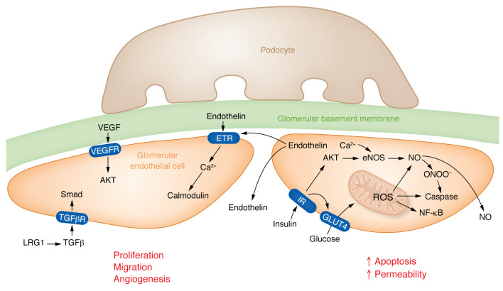
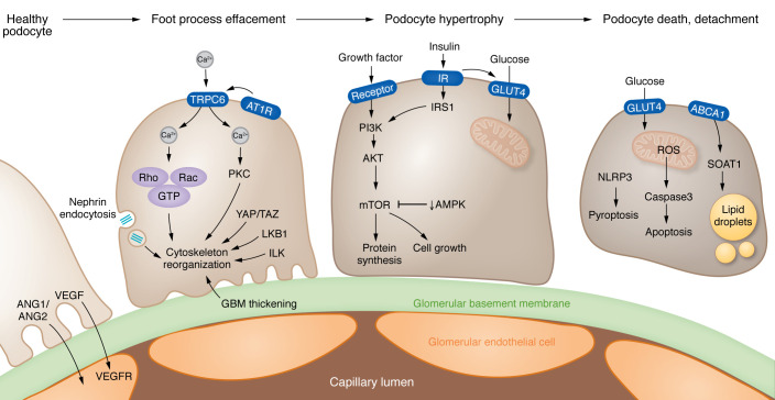
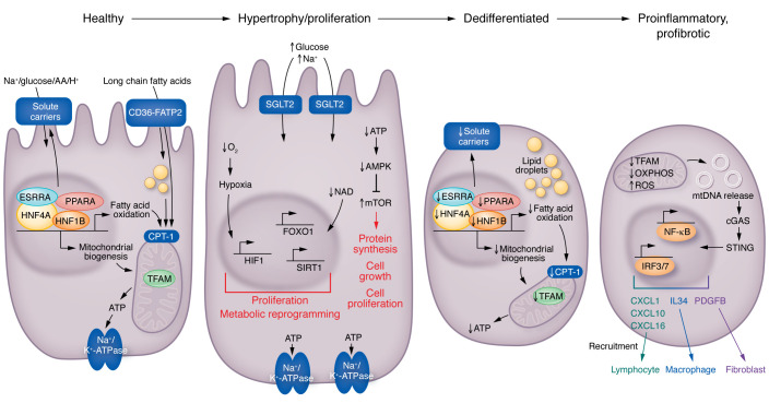
Kidney disease is a major driver of mortality among patients with diabetes and diabetic kidney disease (DKD) is responsible for close to half of all chronic kidney disease cases. DKD usually develops in a genetically susceptible individual as a result of poor metabolic (glycemic) control. Molecular and genetic studies indicate the key role of podocytes and endothelial cells in driving albuminuria and early kidney disease in diabetes. Proximal tubule changes show a strong association with the glomerular filtration rate. Hyperglycemia represents a key cellular stress in the kidney by altering cellular metabolism in endothelial cells and podocytes and by imposing an excess workload requiring energy and oxygen for proximal tubule cells. Changes in metabolism induce early adaptive cellular hypertrophy and reorganization of the actin cytoskeleton. Later, mitochondrial defects contribute to increased oxidative stress and activation of inflammatory pathways, causing progressive kidney function decline and fibrosis. Blockade of the renin-angiotensin system or the sodium-glucose cotransporter is associated with cellular protection and slowing kidney function decline. Newly identified molecular pathways could provide the basis for the development of much-needed novel therapeutics.


Conflict of interest statement

Conflict of interest: The laboratory of KS receives research support from Gilead, GSK, Novo Nordisk, Bayer, Regeneron, Calico, and Novartis. KS is on the advisory board of Jnana Therapeutics, has received consulting fees from Pfizer and Jnana, and holds a provisional patent for Jag1- and Notch-based targeting of chronic kidney disease (patent WO2009035522A1).

Figures

Figure 1
Figure 1. Changes of endothelial cells in diabetes.
(Left) Early DKD is characterized by increased VEGFA expression, VEGFA released by podocytes binds to VEGFR receptors (VEGFR1/2) expressed on glomerular endothelial cells. In addition, VEGFA may also affect the balance between the production of the vasoconstrictive factor endothelin-1 (ET-1) and the vasodilatory factor nitric oxide (NO). By binding to its receptors VEGFR1 and VEGFR2 on GEnCs, VEGFA can stimulate NO production and inhibit ET-1 expression, thus exerting protective effects on the glomerulus. Glomerular endothelial cells release endothelin, which can signal to nearby endothelial cells via endothelin receptors (ETRs). LRG1 potentiates TGFβ signaling to activate Smad pathways in endothelial cells. All 3 signals shown in this cell induce proliferation, migration, and/or angiogenesis in endothelial cells in the DKD glomerulus. (Right) Elevated glucose levels and dysregulated insulin signaling promote increased mitochondrial reactive oxygen species (ROS) levels. Nitric oxide can react with endothelial ROS to release peroxynitrates (ONOO-). Increased oxidative stress will lead to apoptosis of glomerular endothelial cells and enhanced endothelial permeability — changes mostly observed in late DKD. IR, insulin receptor; TGFβR, TGFβ receptor; GLUT4, insulin sensitive glucose transporter 4.
Figure 2
Figure 2. Mechanisms of podocyte dysfunction in DKD.
(Left) Early stages of DKD lead to effacement of podocyte foot processes; multiple pathways contribute to the reorganization of the actin cytoskeleton, including TRPC6-mediated calcium influx activated by angiotensin (ANG) signaling, changes in nephrin endocytosis, and LKB1-associated signaling. In addition, mechanical stress pathways (YAP/TAZ), as well as thickening of the glomerular basement membrane (GBM), can induce integrin and downstream integrin linked kinase (ILK) activation. (Middle) Podocyte hypertrophy also characterizes the early stages of DKD. Growth factor and insulin signaling activates PI3K and mTOR pathways that regulate protein synthesis and cell growth. (Right) Later disease stages are characterized by podocyte death and dedifferentiation. The endoplasmic reticulum enzyme sterol-O-acetyltransferase-1 (SOAT1) facilitates the formation of cholesterol-enriched lipid droplets in podocytes. Dysregulated insulin signaling and high glucose levels trigger oxidative stress and apoptosis. NLRP3-mediated pyroptosis also contributes to podocyte loss. ABCA1, ATP-binding cassette A1; AT1R, angiotensin II receptor type 1; TRPC6, transient receptor potential channel 6.
Figure 3
Figure 3. Changes in kidney proximal tubule cells in diabetes.
Healthy PT cells utilize fatty acids and generate ATP via mitochondrial oxidative phosphorylation to support the Na+/K+ ATP-ase, which then creates a sodium gradient for sodium-mediated glucose, amino acid, or proton reabsorption. Increased tubule glucose presents an increased load for the basal Na+/K+ ATPase and the sodium-mediated glucose cotransporter; more oxygen and ATP are needed to meet this higher metabolic need. The higher AMP/ATP ratio is sensed by AMPK. This excess workload will result in reduced oxygen concentration (relative hypoxia), which is sensed by hypoxia-inducible factor (HIF). HIF activation will induce metabolic reprogramming, and, together with mTOR, will induce tubule cell proliferation and hypertrophy, proliferation, and kidney growth. Later stages of DKD are characterized by dedifferentiation of PT cells. Gene programs associated with mitochondrial biogenesis, fatty acid oxidation, mitochondrial function, and low ATP levels lead to the loss of solute carriers and cellular dedifferentiation. Finally, at later stages of DKD, severe mitochondrial damage will lead to the release of mitochondrial DNA (mtDNA) into the cytosol. Cystosolic mtDNA is then sensed by nucleotide sensing pathways, such as cGAS and stimulator of interferon genes (STING), inducing the activation of IRF3/7 and NF-κB pathways and resulting in transcription of cytokines, growth factors, and downstream immune cell recruitment and fibroblast activation. CPT-1, carnitine palmitoyltransferase 1A; ESRRA, estrogen-related receptor alpha; FATP2, fatty acid transport protein 2; HNF1B, hepatocyte nuclear factor 1B; IRF3/7, inferferon regulatory factor 3/7.

References

    1. Tervaert TW, et al. Pathologic classification of diabetic nephropathy. J Am Soc Nephrol. 2010;21(4):556–563. doi: 10.1681/ASN.2010010010. - DOI - PubMed
    1. Johansen KL, et al. US Renal Data System 2020 annual data report: epidemiology of kidney disease in the United States. Am J Kidney Dis. 2021;77(4 suppl 1):A7–A8. - PMC - PubMed
    1. Thomas MC, et al. Diabetic kidney disease. Nat Rev Dis Primers. 2015;1:15018. doi: 10.1038/nrdp.2015.18. - DOI - PMC - PubMed
    1. Nathan DM, et al. Intensive diabetes treatment and cardiovascular disease in patients with type 1 diabetes. N Engl J Med. 2005;353(25):2643–2653. doi: 10.1056/NEJMoa052187. - DOI - PMC - PubMed
    1. Brosius FC, et al. Mouse models of diabetic nephropathy. J Am Soc Nephrol. 2009;20(12):2503–2512. doi: 10.1681/ASN.2009070721. - DOI - PMC - PubMed

Publication types