. 2023 Feb 15;133(4):e167079.
doi: 10.1172/JCI167079.

Mitochondrial function in macrophages controls cardiac repair after myocardial infarction

Affiliations

Mitochondrial function in macrophages controls cardiac repair after myocardial infarction

David Weissman et al. J Clin Invest. .

Abstract

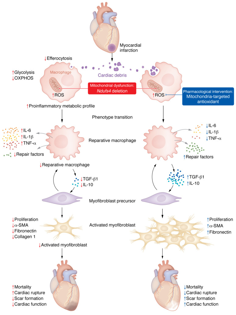
Cardiac healing following acute myocardial infarction (MI) involves the mobilization and activation of immune cells, including macrophages. In the early phase after MI, macrophages adopt a proinflammatory phenotype, while polarizing toward a reparative one in the late stage. Although metabolic reprogramming has been observed during this transition, the mechanistic links to macrophage differentiation are still poorly understood. In this issue of the JCI, Cai, Zhao and colleagues demonstrate that mitochondrial function in macrophages governed the resolution of inflammation and tissue repair by modulating the phagocytic removal of apoptotic cells (so-called efferocytosis) as well as myofibroblast activation. These findings provide important mechanistic insights into the potential relevance of metabolic modulation of macrophage functions following MI, which might lead to alternative therapeutic strategies for MI.


Conflict of interest statement

Conflict of interest: The authors declare that no conflict of interest exists.

Figures

Figure 1
Figure 1. Myeloid-specific deletion of the mitochondrial complex I protein Ndufs4 compromises macrophage function during cardiac repair after MI.
Following acute MI, immune cells, including monocyte-derived macrophages, remove cell debris through efferocytosis but can also amplify the inflammatory response by secreting proinflammatory mediators. Subsequently, proinflammatory macrophages transition from a proinflammatory to a reparative phenotype, a process that associates with metabolic remodeling: shifting from relying on glycolysis (associated with the proinflammatory phenotype) toward mitochondrial OXPHOS (associated with the reparative phenotype). This transition is characterized by increased secretion of antiinflammatory cytokines, chemokines, and repair factors that stimulate the activation and proliferation of cardiac myofibroblasts, which promote tissue repair and scar formation. In this issue of the JCI, Cai, Zhao, and co-authors (20) report that mitochondrial dysfunction in macrophages, induced by Ndufs4 deletion, can cause excessive mitochondrial ROS production, which primes the cells to a greater inflammatory response while impairing efferocytosis. This process, in turn, blunts macrophage polarization, reducing the reparative phenotype, and suppresses the proliferation and activation of myofibroblasts, resulting in poor wound healing and increased mortality. Interestingly, mitochondria-targeted ROS scavenging improved efferocytosis and myofibroblast functions, resulting in reduced infarct rupture and mortality.

Comment on

  • Mitochondrial Dysfunction in Macrophages Promotes Inflammation and Suppresses Repair After Myocardial Infarction

References

    1. Nahrendorf M, et al. The healing myocardium sequentially mobilizes two monocyte subsets with divergent and complementary functions. J Exp Med. 2007;204(12):3037–3047. doi: 10.1084/jem.20070885. - DOI - PMC - PubMed
    1. Heidt T, et al. Differential contribution of monocytes to heart macrophages in steady-state and after myocardial infarction. Circ Res. 2014;115(2):284–295. doi: 10.1161/CIRCRESAHA.115.303567. - DOI - PMC - PubMed
    1. Prabhu SD, Frangogiannis NG. The biological basis for cardiac repair after myocardial infarction: from inflammation to fibrosis. Circ Res. 2016;119(1):91–112. doi: 10.1161/CIRCRESAHA.116.303577. - DOI - PMC - PubMed
    1. Honold L, Nahrendorf M. Resident and monocyte-derived macrophages in cardiovascular disease. Circ Res. 2018;122(1):113–127. doi: 10.1161/CIRCRESAHA.117.311071. - DOI - PMC - PubMed
    1. Dobaczewski M, et al. Transforming growth factor (TGF)-β signaling in cardiac remodeling. J Mol Cell Cardiol. 2011;51(4):600–606. doi: 10.1016/j.yjmcc.2010.10.033. - DOI - PMC - PubMed

Publication types