. 2023 Feb 21;120(8):e2219758120.
doi: 10.1073/pnas.2219758120. Epub 2023 Feb 14.

Split aminoacyl-tRNA synthetases for proximity-induced stop codon suppression

Affiliations

Split aminoacyl-tRNA synthetases for proximity-induced stop codon suppression

Han-Kai Jiang et al. Proc Natl Acad Sci U S A. .

Abstract

Synthetic biology tools for regulating gene expression have many useful biotechnology and therapeutic applications. Most tools developed for this purpose control gene expression at the level of transcription, and relatively few methods are available for regulating gene expression at the translational level. Here, we design and engineer split orthogonal aminoacyl-tRNA synthetases (o-aaRS) as unique tools to control gene translation in bacteria and mammalian cells. Using chemically induced dimerization domains, we developed split o-aaRSs that mediate gene expression by conditionally suppressing stop codons in the presence of the small molecules rapamycin and abscisic acid. By activating o-aaRSs, these molecular switches induce stop codon suppression, and in their absence stop codon suppression is turned off. We demonstrate, in Escherichia coli and in human cells, that split o-aaRSs function as genetically encoded AND gates where stop codon suppression is controlled by two distinct molecular inputs. In addition, we show that split o-aaRSs can be used as versatile biosensors to detect therapeutically relevant protein-protein interactions, including those involved in cancer, and those that mediate severe acute respiratory syndrome-coronavirus-2 infection.

Keywords: genetic code expansion; noncanonical amino acids; pyrrolysyl-tRNA synthetase; stop codon suppression; synthetic biology.


Conflict of interest statement

The authors declare no competing interest.

Figures
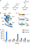
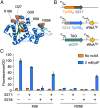
Fig. 1.
Fig. 1.
Development of a split “Ca. M. alvus” PylRS. (A) Structures of ncAAs used in this study. (B) The crystal structure of the PylRS from “Ca. Methanomethylophilus alvus” (PDB: 6ezd). The seven sites at which the enzyme was split into N-terminal and C-terminal fragments are labeled. (C) The three-plasmid system used to identify active split PylRS variants. The N-terminal (PylRSN) and C-terminal (PylRSC) fragments were fused to interacting peptides SYNZIP17 (SZ17) and SYNZIP18 (SZ18), respectively. (D) sfGFP expression in E. coli cells coexpressing various SYNZIP-fused PylRSN and PylRSC fragments in the presence (blue bars) and absence (orange bars) of the PylRS substrate mIF. Data are displayed as the mean ± SEM for three biological replicates.
Fig. 2.
Fig. 2.
Split PylRS must be fused to interacting polypeptides for activity. (A) sfGFP expression in E. coli cells coexpressing PylRSN and PylRSC fragments with and without fusion to SYNZIP peptides. Data were collected in the presence (blue bars) and absence (orange bars) of 2 mM mIF and are displayed as the mean ± SEM for three biological replicates. (B) The three-plasmid system to detect split PylRS activity using a chloramphenicol acetyltransferase reporter. SYNZIP-fused PylRS fragments were coexpressed with a chloramphenicol acetyltransferase gene (CmR) containing an in-frame TAG codon. (C and D) Cells were challenged to grow on media containing mIF and 50 μg/mL chloramphenicol. The PylRS variant split at position Q23 does not require SYNZIP18 for growth (C), whereas, the variant split at position D137 requires both SYNZIP17 (SZ17) and SYNZIP18 (SZ18) (D).
Fig. 3.
Fig. 3.
Developing a split M. jannaschii tyrosyl-tRNA synthetase (MjTyrRS). (A) The crystal structure of the MjTyrRS (PDB: 1j1u). The five sites at which the enzyme was split into N-terminal and C-terminal fragments are labeled. (B) The three-plasmid system used to identify active split MjTyrRS variants. The N-terminal (TyrRSN) and C-terminal (TyrRSC) fragments were fused to interacting peptides SYNZIP17 (SZ17) and SYNZIP18 (SZ18), respectively. MjTyrRS activity was monitored by measuring sfGFP fluorescence in cells expressing a reporter sfGFP gene harboring an in-frame TAG codon. (C) sfGFP expression in E. coli cells coexpressing various SYNZIP-fused TyrRSN and TyrRSC fragments in the presence (blue bars) and absence (orange bars) of the MjTyrRS substrate pIF. Data are displayed as the mean ± SEM for three biological replicates.
Fig. 4.
Fig. 4.
Detecting therapeutically relevant PPIs using split PylRS. (A) The three-plasmid system used to detect the interaction between tBID and Bcl-2 or Mcl-1. The N-terminal PylRS fragment (PylRSN) was fused to tBID or a negative control peptide (Dead BID). The C-terminal PylRS fragment (PylRSC) was fused to Bcl-2 or Mcl-1. PylRS activity was monitored by measuring sfGFP fluorescence in cells expressing a reporter sfGFP gene harboring an in-frame TAG codon. (B and C) sfGFP expression in cells expressing PylRSN-tBID and Bcl-2-PylRSC (B) or Mcl-1-PylRSC (C). (D) A model of the 5-HB from SARS-CoV-2 (based on PDB: 6lxt). The HR1 and HR2 helices are shown in gray and orange, respectively. The 5-HB contains an empty HR2 binding site formed by adjacent HR1 helices. (E) The three-plasmid system used to detect the interaction between the 5-HB and HR2. The 5-HB and an HR2 peptide were fused to PylRSN and PylRSC, respectively. PylRS activity was monitored by measuring sfGFP fluorescence in cells expressing a reporter sfGFP gene harboring an in-frame TAG codon. (F) sfGFP expression in cells coexpressing PylRSN-5-HB and HR2-PylRSC. All data were collected in the presence (blue bars) and absence (orange bars) of 2 mM mIF. Data are displayed as the mean ± SEM for three biological replicates.
Fig. 5.
Fig. 5.
Rapamycin (RAP)-induced stop codon suppression using split MjTyrRS. (A) The N-terminal (TyrRSN) and C-terminal (TyrRSC) MjTyrRS fragments were fused to the RAP-binding proteins FRB and FKBP, respectively. MjTyrRS activity was monitored by measuring sfGFP fluorescence in cells expressing an sfGFP reporter gene harboring an in-frame TAG codon. (B) Robust sfGFP production required the addition of both pIF and RAP to the cell growth media. Data are displayed as the mean ± SEM for three biological replicates.
Fig. 6.
Fig. 6.
Abscisic acid (ABA)-induced stop codon suppression using split PylRS. (A) The three-plasmid system used to detect ABA-induced suppression of TAG and TAA stop codons. The N-terminal (PylRSN) and C-terminal (PylRSC) PylRS fragments were fused to ABI and PYL, respectively. PylRS activity was monitored by measuring sfGFP fluorescence in cells expressing an sfGFP reporter gene harboring an in-frame TAG or TAA codon. (B and C) sfGFP fluorescence in cells expressing an sfGFP reporter harboring an in-frame TAG codon (B) or TAA codon (C). Cells were grown in the presence and absence of 2 mM mIF and 100 μM ABA. (D) The three-plasmid system used to detect ABA-induced stop codon suppression using a chloramphenicol acetyltransferase (CmR) reporter. PYL- and ABI-fused PylRS fragments were coexpressed with a CmR gene containing an in-frame TAG codon. (E) Cells were challenged to grow on media containing 2 mM mIF in the presence and absence of chloramphenicol and ABA. (F) Optimizing the linkers between PylRS fragments and PYL/ABI. All data are displayed as the mean ± SEM of three biological replicates.
Fig. 7.
Fig. 7.
Small-molecule–induced stop codon suppression in HEK293 cells. (A) The two-plasmid system used to measure ABA-induced stop codon suppression in HEK293 cells. The N-terminal (PylRSN) and C-terminal (PylRSC) fragments of PylRS were fused to ABI and PYL, respectively. ABI- and PYL-fused PylRS fragments were expressed from the same promoter using a self-cleaving T2A peptide. Stop codon suppression was determined by measuring SEAP activity in media of cells expressing a SEAP reporter gene harboring a TAG codon at position 41. (B) Relative SEAP activity in cells grown in the presence of BocK and varying concentrations of ABA. Data are displayed as the mean ± SEM for four biological replicates (**P = 0.005, ns = not significant, paired t test). (C) A schematic of the ABA/BocK AND gate.

References

    1. Brophy J. A., Voigt C. A., Principles of genetic circuit design. Nat. Methods 11, 508–520 (2014). - PMC - PubMed
    1. Engstrom M. D., Pfleger B. F., Transcription control engineering and applications in synthetic biology. Synth. Syst. Biotechnol. 2, 176–191 (2017). - PMC - PubMed
    1. Kent R., Dixon N., Contemporary tools for regulating gene expression in bacteria. Trends Biotechnol. 38, 316–333 (2020). - PubMed
    1. Shis D. L., Bennett M. R., Library of synthetic transcriptional AND gates build with split T7 RNA polymerase mutants. Proc. Natl. Acad. Sci. U.S.A. 110, 5028–5033 (2013). - PMC - PubMed
    1. Pu J., Zinkus-Boltz J., Dickinson B. C., Evolution of a split RNA polymerase as a versatile biosensor platform. Nat. Chem. Biol. 13, 432–438 (2017). - PMC - PubMed

Publication types

LinkOut - more resources