. 2023 Apr 19;111(8):1264-1281.e5.
doi: 10.1016/j.neuron.2023.01.017. Epub 2023 Feb 13.

Brief synaptic inhibition persistently interrupts firing of fast-spiking interneurons

Affiliations

Brief synaptic inhibition persistently interrupts firing of fast-spiking interneurons

Simon Chamberland et al. Neuron. .

Abstract

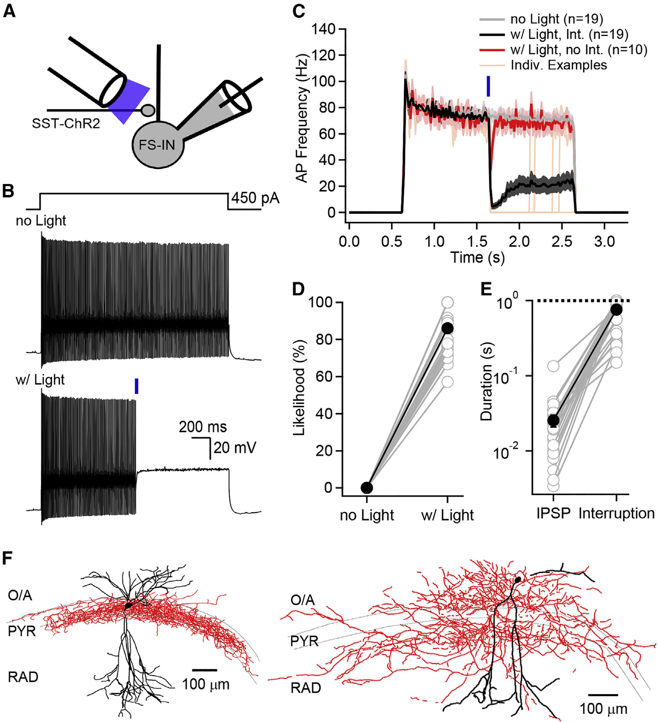
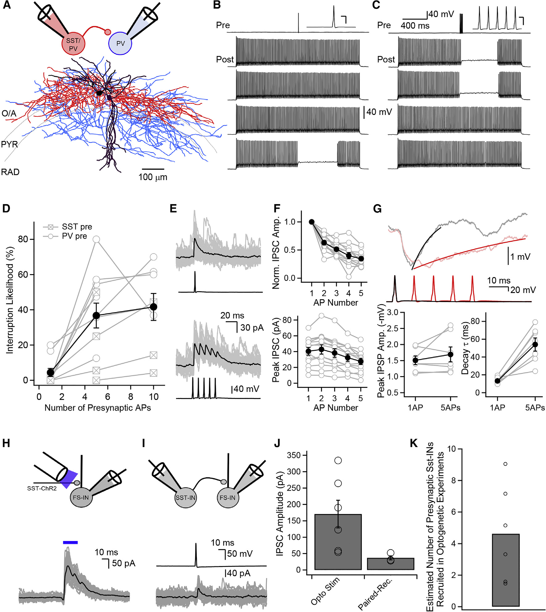
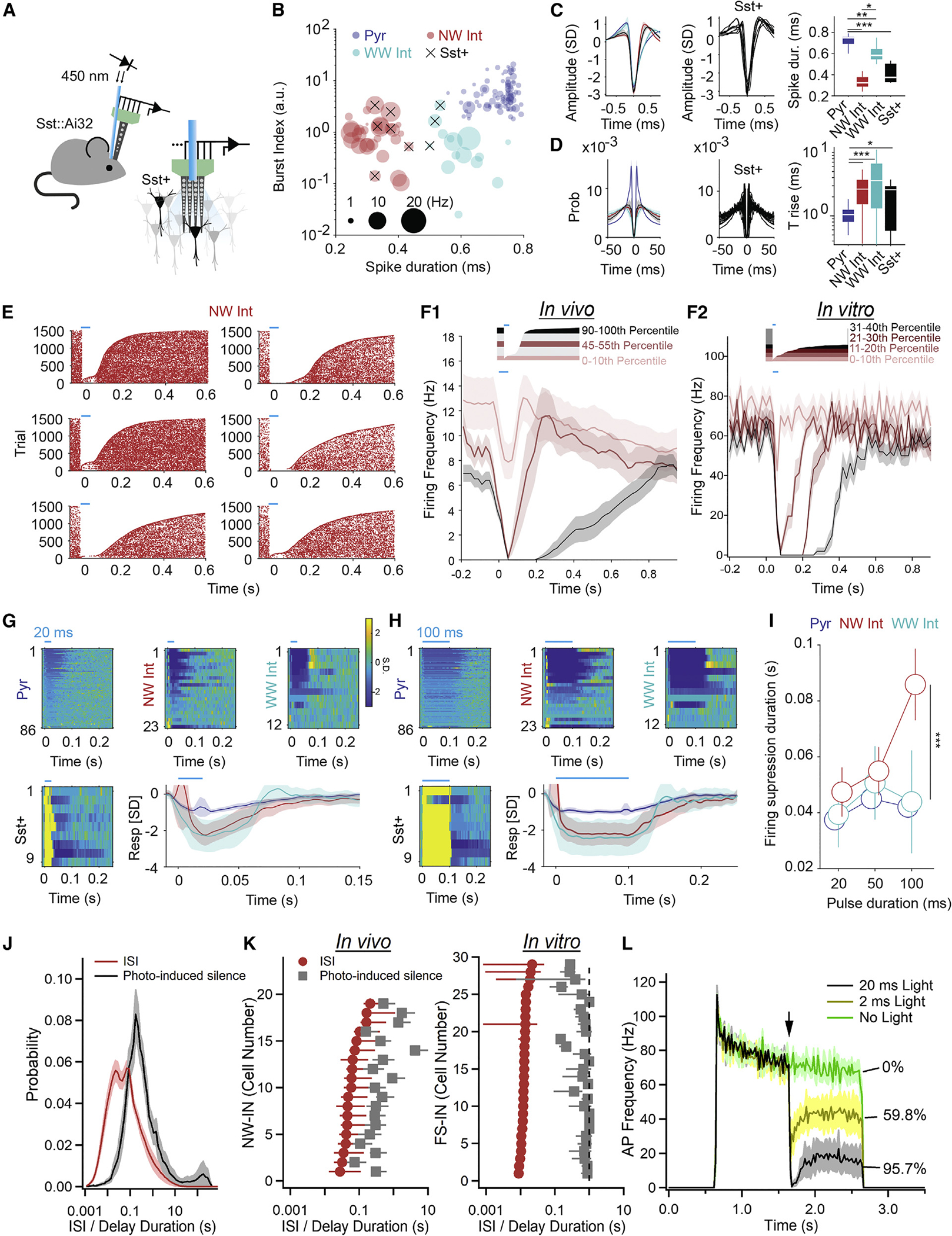
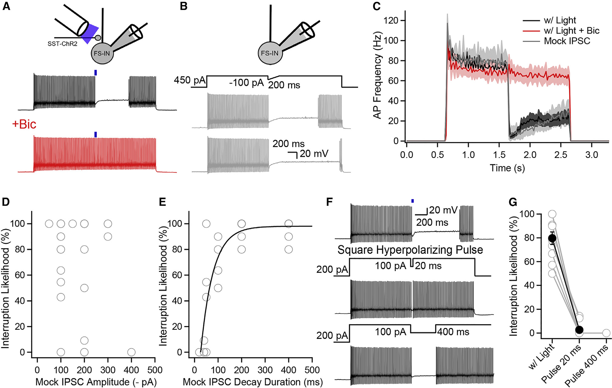
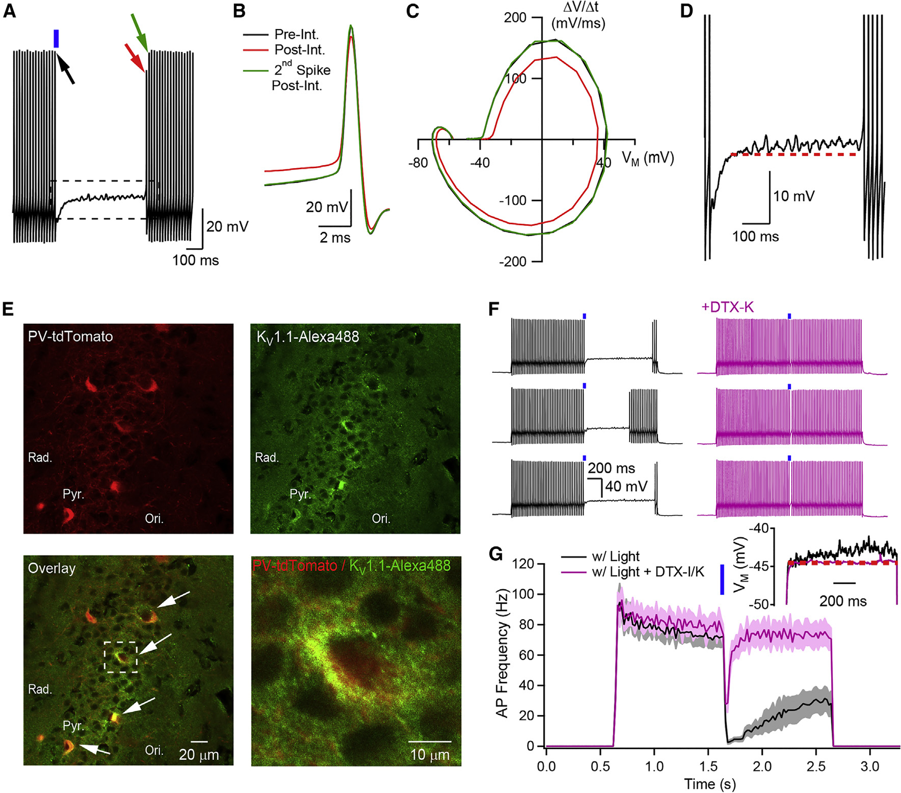
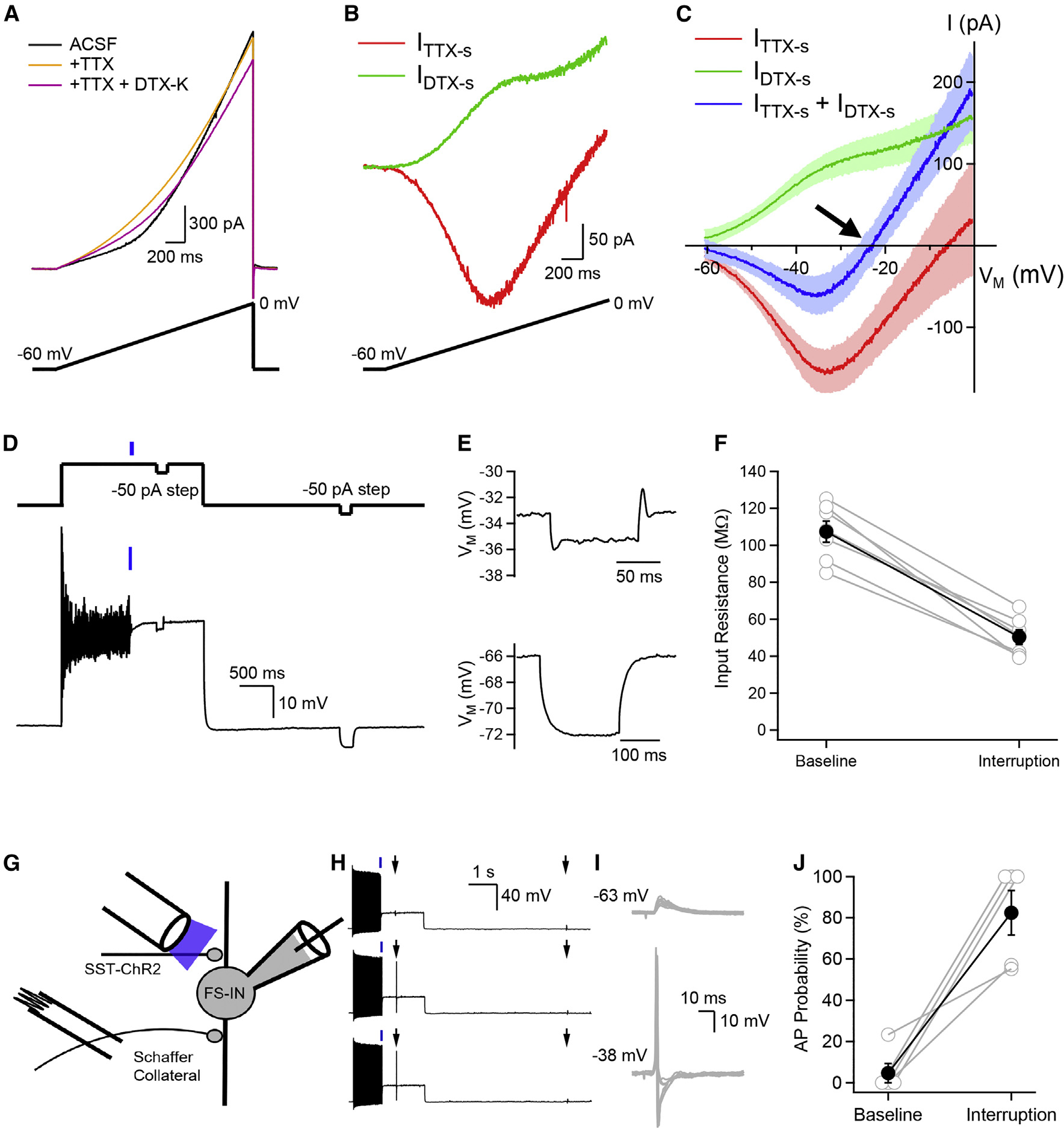
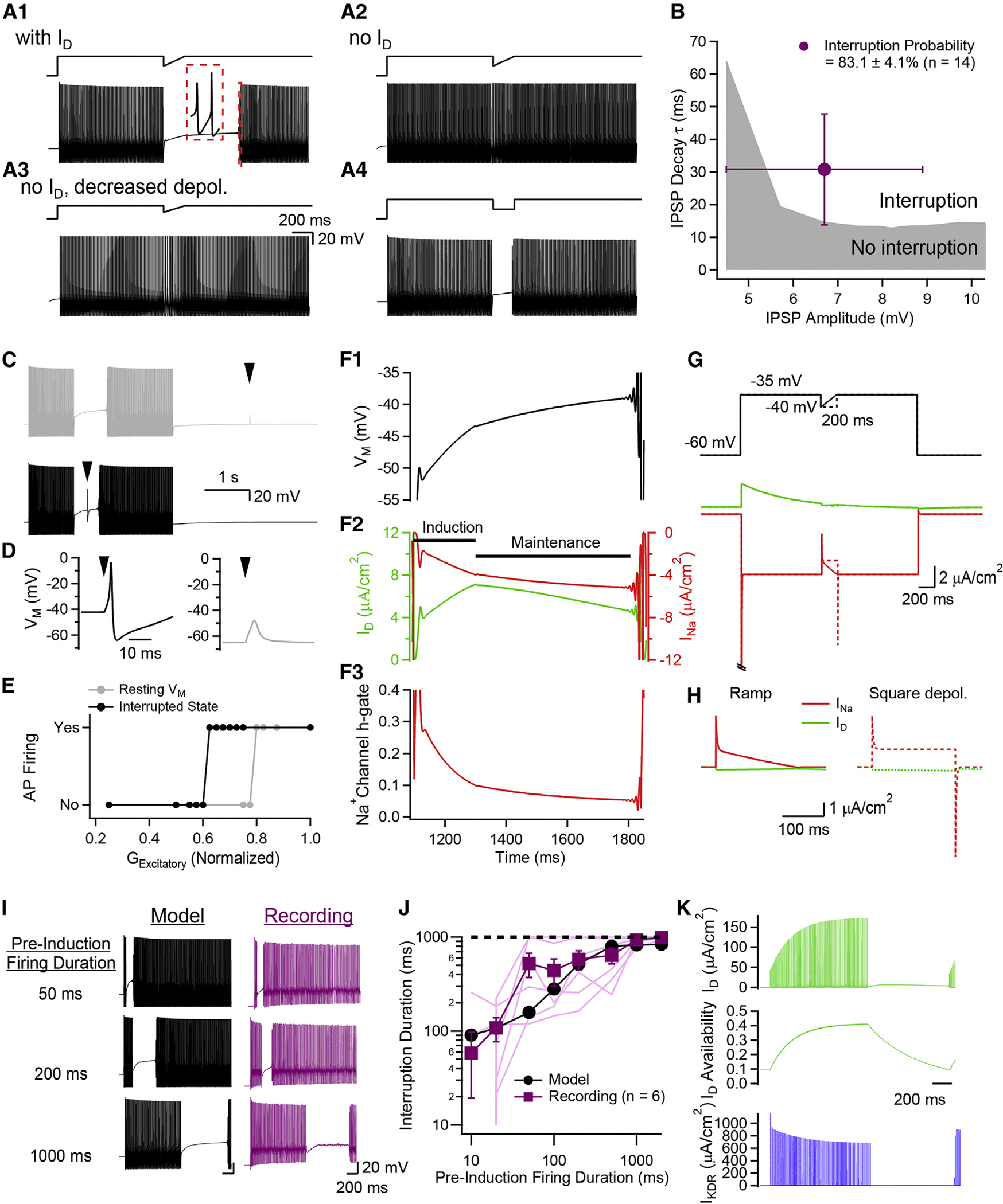
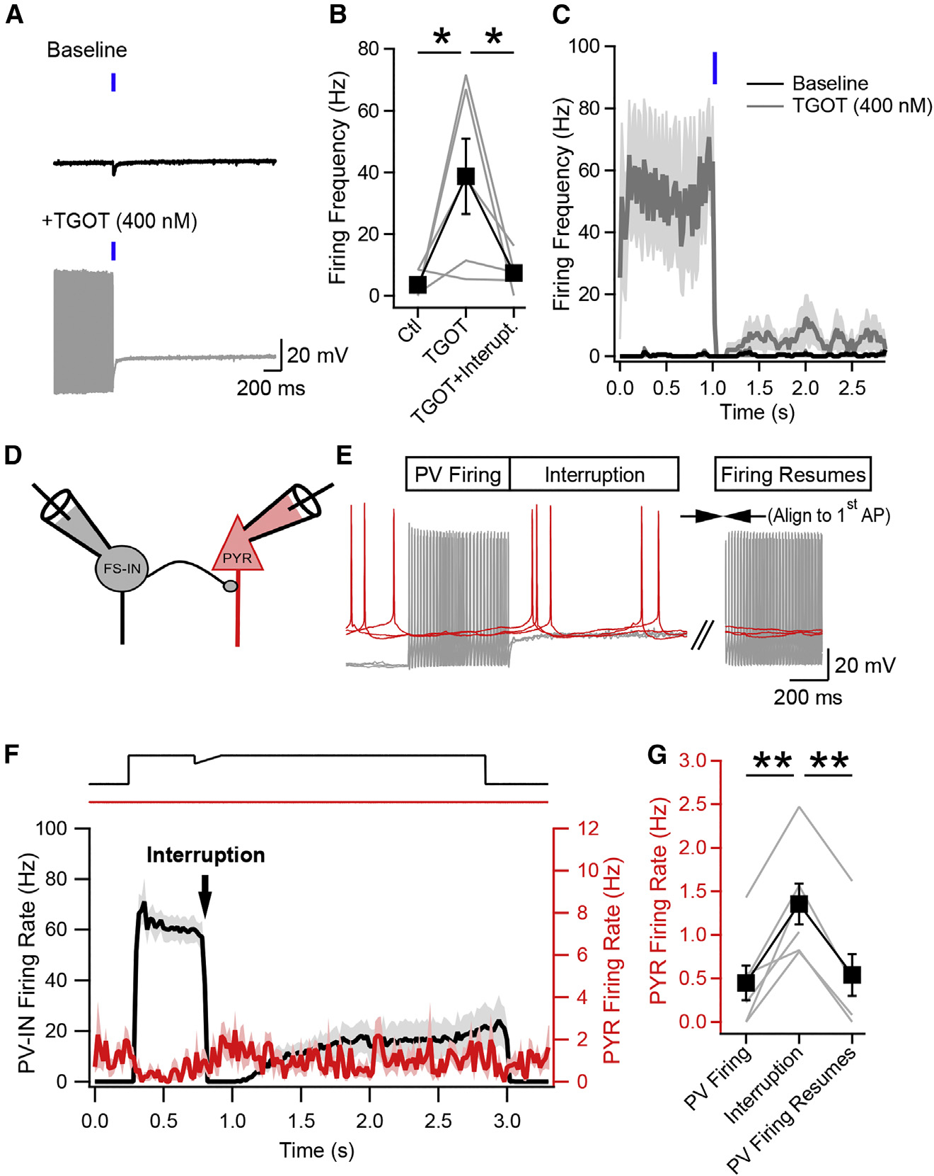
Neurons perform input-output operations that integrate synaptic inputs with intrinsic electrical properties; these operations are generally constrained by the brevity of synaptic events. Here, we report that sustained firing of CA1 hippocampal fast-spiking parvalbumin-expressing interneurons (PV-INs) can be persistently interrupted for several hundred milliseconds following brief GABAAR-mediated inhibition in vitro and in vivo. A single presynaptic neuron could interrupt PV-IN firing, occasionally with a single action potential (AP), and reliably with AP bursts. Experiments and computational modeling reveal that the persistent interruption of firing maintains neurons in a depolarized, quiescent state through a cell-autonomous mechanism. Interrupted PV-INs are strikingly responsive to Schaffer collateral inputs. The persistent interruption of firing provides a disinhibitory circuit mechanism favoring spike generation in CA1 pyramidal cells. Overall, our results demonstrate that neuronal silencing can far outlast brief synaptic inhibition owing to the well-tuned interplay between neurotransmitter release and postsynaptic membrane dynamics, a phenomenon impacting microcircuit function.

Keywords: fast-spiking interneurons; hippocampus; inhibition; persistent activity.


Conflict of interest statement

Declaration of interests The authors declare no competing interests.

Figures

Figure 1.
Figure 1.. Synaptic inhibition persistently interrupts firing of PV-Ins
(A) Recording configuration. (B) PV-INs depolarized with rectangular current waveform. Optogenetic stimulation (blue bar) generated an IPSP followed by a persistent interruption of firing. (C) Summary data, firing frequency vs. time for experiments exemplified in (B), with optogenetic stimulation (black) or without (light gray). Red trace, average of traces with light but when no interruption was induced; orange traces, exemplar trials. (D) Likelihood of observing an interruption. Collective results from PV-INs shown in (C) and 10 additional neurons. (E) Duration of IPSP compared with silent period associated with interruption. The dashed line represents the 1-s duration of the depolarizing step, a cap on the interruption duration. (F) Neurolucida reconstructions of recorded PV-INs. Dendrites black, axon red.
Figure 2.
Figure 2.. A single presynaptic interneuron can interrupt PV-IN firing
(A) Recording configuration and Neurolucida reconstruction of a synaptically connected pair of INs. Dendrites of presynaptic IN shown in black, axon in red. Dendrites of postsynaptic neurons shown in purple, axon in blue. (B) Current-clamp recordings in a pair of INs. A single AP in the presynaptic cell suffices to interrupt postsynaptic firing occasionally. Four consecutive epochs shown. (C) Same pair as in (B), with five APs at 100 Hz. Insets, AP for (B) and (C); calibration: 40 mV vertical, 5 ms horizontal. (D) Interruption likelihood varies with number of presynaptic APs. Five- and ten-AP bursts delivered at 100 Hz. Depolarizing current pulse amplitude in postsynaptic PV-INs, 255 ± 23 pA (n = 11). Presynaptic PV- and SST-INs had similar likelihood to interrupt firing when five APs were evoked (n = 7 and n = 4, respectively; p = 0.11). (E) IPSCs recorded at 0 mV in PV-INs. Black traces, average of 50 consecutive sweeps (gray). (F) Top, normalized IPSC amplitude, measured from trough-to-peak, vs. stimulus number. Bottom, absolute peak amplitudes of the IPSC burst from prestimulus baseline. (G) Top, current-clamp recordings of single- (black) and five-AP- (red) evoked IPSPs (averages of 3 consecutive sweeps). Bottom, with AP repetition, peak IPSP amplitude hardly changed, whereas decay time constant greatly increased. (H–J) IPSCs measured in optogenetic experiments (H), in paired recordings (I); pooled data of IPSC amplitudes (J). (K) Ratio of values in (J) provides estimate of number of optogenetically activated SST-INs synapsing onto a PV-IN.
Figure 3.
Figure 3.. PV-IN silencing persists following optogenetic stimulation in vivo
(A) Recording configuration of multisite silicon probe and optical fiber in CA1. (B) Burst index as a function of spike duration for all neurons sampled (n = 130 units) distinguishes NW-INs (red), CA1-PYRs (blue), WW-INs (teal), and SST-INs (black ×). (C) Average spike waveform for populations identified in (B) (left), including SST-INs (middle), and trough-to-peak spike duration (right). (D) Same as in (C) for firing auto-correlograms and rise time to peak (p < 0.001 for PYR vs. NW-IN). (E) AP raster plots of 6 representative NW-INs during 1,500 trials, ranked by silencing duration induced by 50 ms optogenetic stimulation (blue bars). (F) (F1) Summary graph for all NW-INs sampled, showing the averages of trials across neurons for the lowest, middle, and highest deciles. (F2) Similar analysis performed on in vitro data qualitatively parallels the findings in (F1) (see discussion). (G–I) Optogenetic stimulation for 20 ms (G) or 100 ms (H) in other cell types results in briefer silencing duration than in NW-INs. Warmer colors correspond to higher firing rates (inset). (I) Delay to recovery of spiking as a function of optogenetic stimulation duration. (J) The distributions of inter-spike intervals (ISIs) and optogenetic-induced silence duration for NW-INs are significantly different (Kolmogorov-Smirnov test: p < 0.0001, K = 0.7). (K) Average of ISIs and optogenetic-induced silences for individual neurons, in vivo and in vitro. (L) Effect of varying optogenetic stimulus strength on the apparent interruption duration in vitro. *p < 0.05; **p < 0.01; ***p < 0.001 for all statistical tests.
Figure 4.
Figure 4.. Postsynaptic hyperpolarizations through GABAAR activation or current injection interrupt PV-Ins
(A) Bicuculline (10 μM) abolishes optogenetically induced interruption of firing. (B) Hyperpolarizing current injections (mock IPSCs) reliably interrupt PV-INs. (C) AP frequency vs. time for optogenetically evoked stimulation before (black) and after Bic (red). Data also shown for mock-IPSC-induced interruption (gray). (D) Interruption likelihood vs. mock-IPSC amplitude. (E) Interruption likelihood vs. mock-IPSC duration. Exponential fit shown. (F) Rectangular hyperpolarizing pulse (20 or 400 ms) fails to interrupt firing. (G) Interruption likelihood for paired experiments with optogenetic stimulation or rectangular hyperpolarizing pulses.
Figure 5.
Figure 5.. Kv1.1 is required for interruption of firing
(A) Current-clamp recording from a PV-IN. (B) Zoomed-in data from (A), showing the three APs indicated by arrows. (C) Phase-plane plot for the three APs in (B). (D) During the interruption, the VM undergoes subthreshold oscillations and gradually depolarizes (dashed line, constant VM reference). (E) Immunohistochemistry reveals that Kv1.1 is expressed in PV-INs in regions bordering CA1-PYR layer. White arrows, PV-INs with strong somatic KV1.1 expression. (F) Optogenetically induced interruption before (black) and after (purple) exposure to DTX-K (three consecutive epochs each). (G) AP frequency vs. time for experiments in absence or presence of DTX-K or DTX-I. Inset: DTX-K prevents gradual membrane depolarization.
Figure 6.
Figure 6.. Interplay between Kv1.1-current and Na+-dependent current can support a stable point in VM, a depolarized yet hyperresponsive state
(A) Voltage-clamp recordings from a PV-IN in control (black), in TTX (gold) and with both TTX and DTX-K (purple). (B) Arithmetic subtraction reveals IDTX-s and ITTX-s. (C) Current vs. voltage. IDTX-s and ITTX-s measured in same neurons; shaded areas, standard error. (D) VM dynamics during the firing interruption. (E) VM responses to hyperpolarizing current pulses, during interruption (top) or at resting VM (bottom). (F) Input resistance measured at baseline and during interruption. (G) Experimental design, including fixed amplitude stimulation. (H) Three consecutive sweeps, subthreshold EPSPs at rest become suprathreshold during the interruption. (I) Changes in VM evoked by Schaffer collateral stimulation at resting potential (top) or during the interruption (bottom). (J) AP probability for stimuli delivered at resting VM (baseline) or during interruption.
Figure 7.
Figure 7.. A single-compartment conductance-based model reproduces core features of interruption
(A) Model-generated firing patterns. Inset (A1), first two APs upon firing resumption. (B) An inhibitory conductance in the model reliably interrupted firing. Model parameters comparable to experimentally measured IPSPs reliably generating interruptions (purple cross). Pre-interruption firing duration kept constant (1 s) across experimental and modeling conditions. (C–E) (C) Model neuron is hypersensitive to excitatory inputs during the firing interruption. An excitatory conductance (arrowhead) was introduced at resting VM (gray) or during the interruption (black). (D) At resting VM, the excitatory conductance is subthreshold while the same conductance triggers an AP during interruption. (E) Quantification of excitatory strength required to generate AP, showing increased excitability in interrupted state. (F) (F1) VM during interruption. (F2) ID and INa dynamics during interruption. (F3) Na+ channel inactivation variable (h-gate) during interruption. (G and H) Voltage-clamping the model with VM dynamics known to interrupt neurons (full line) or to resume their firing (dotted line). INa transient triggered by offset of rectangular hyperpolarization. (I) Longer pre-induction bursts are associated with increased duration of interruption in simulations and experiments. Interruptions induced by an inhibitory conductance in model and a mock IPSC in experiments (as in Figure 4B). (J) Interruption duration plateaued as pre-induction firing was prolonged in simulations and experiments (n = 6 neurons; p = 0.18; two-way ANOVA) due to saturating degree of removal of ID inactivation for longer firing episodes. (K) Membrane current dynamics underlying the 1,000 ms pre-induction firing duration (I, bottom).
Figure 8.
Figure 8.. The firing interruption is effective for oxytocin-induced PV-IN firing and disinhibits CA1 pyramidal neurons
(A) Current-clamp recording from a PV-IN at baseline (top) and after TGOT (bottom). 10 traces each overlayed for baseline and TGOT. (B) Pooled data (n = 5) showing effect of TGOT and further optogenetic activation of SST-INs. (C) SST-IN-mediated synaptic inhibition persistently interrupts PV-INs driven to fire by OXTR activation (n = 4). (D and E) Paired recording from a PV-IN (gray) synaptically connected to a CA1-PYR (red); 3 consecutive sweeps during interruption induced by mockIPSC as in (F). (F) AP firing frequency for PV-INs (black) and CA1-PYRs (n = 6 pairs). Shaded areas, standard error. (G) AP frequency recorded in the CA1-PYR for500 ms windows measured during PV-IN firing, at interruption onset (n = 6; **p < 0.01) and following PV-IN firing resumption (n = 4; **p < 0.01; 2 PYRs excluded because resumption of PV-IN firing too rare to reliably assess PYR firing rate).

References

    1. Klausberger T, and Somogyi P (2008). Neuronal diversity and temporal dynamics: the unity of hippocampal circuit operations. Science 321, 53–57. 10.1126/science.1149381. - DOI - PMC - PubMed
    1. Pelkey KA, Chittajallu R, Craig MT, Tricoire L, Wester JC, and McBain CJ (2017). Hippocampal GABAergic inhibitory interneurons. Physiol. Rev. 97, 1619–1747.10.1152/physrev.00007.2017. - DOI - PMC - PubMed
    1. Freund TF, and Buzsáki G (1996). Interneurons of the hippocampus. Hippocampus 6, 347–470. 10.1002/(SICI)1098-1063(1996)6:4&lt;347::AID-HIPO1&gt;3.0.CO;2-I. - DOI - PubMed
    1. Stark E, Eichler R, Roux L, Fujisawa S, Rotstein HG, and Buzsáki G (2013). Inhibition-induced theta resonance in cortical circuits. Neuron 80, 1263–1276. 10.1016/j.neuron.2013.09.033. - DOI - PMC - PubMed
    1. Cardin JA, Carlé n M, Meletis K, Knoblich U, Zhang F, Deisseroth K, Tsai LH, and Moore CI (2009). Driving fast-spiking cells induces gamma rhythm and controls sensory responses. Nature 459, 663–667. 10.1038/nature08002. - DOI - PMC - PubMed

Publication types

MeSH terms

LinkOut - more resources