

Cerebral malaria-using the retina to study the brain
- PMID: 36788363
- PMCID: PMC10397347
- DOI: 10.1038/s41433-023-02432-z
Cerebral malaria-using the retina to study the brain
Abstract
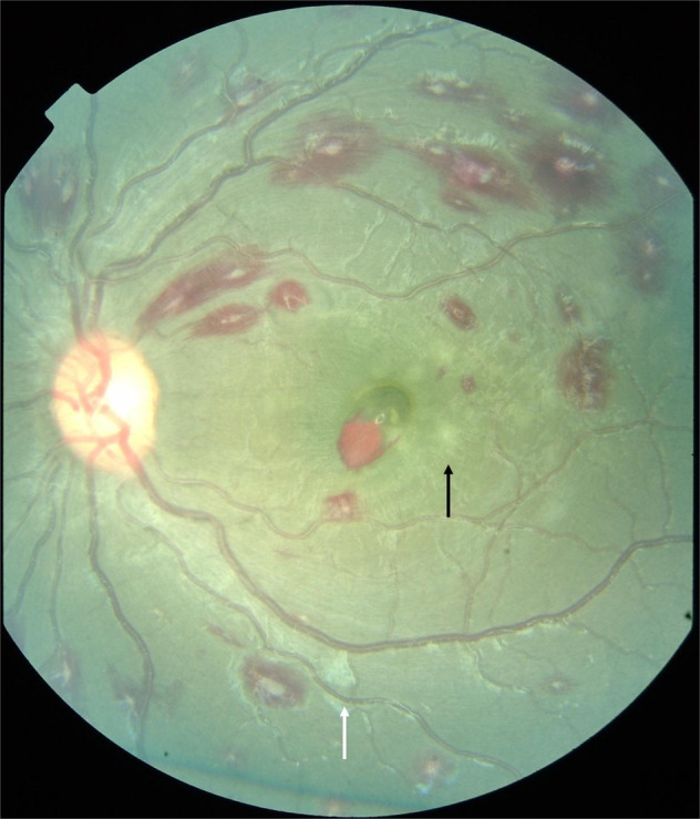
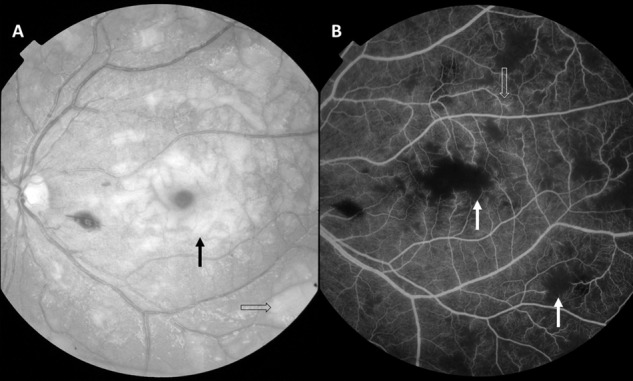
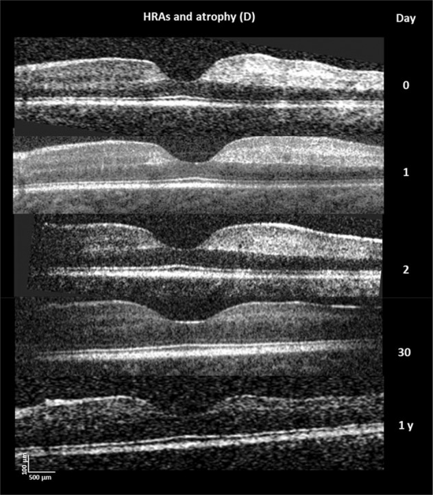
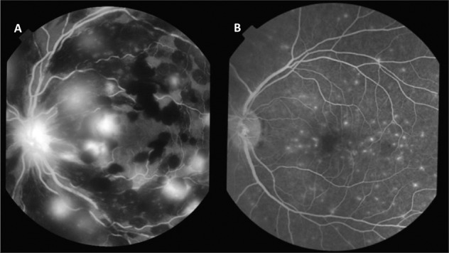
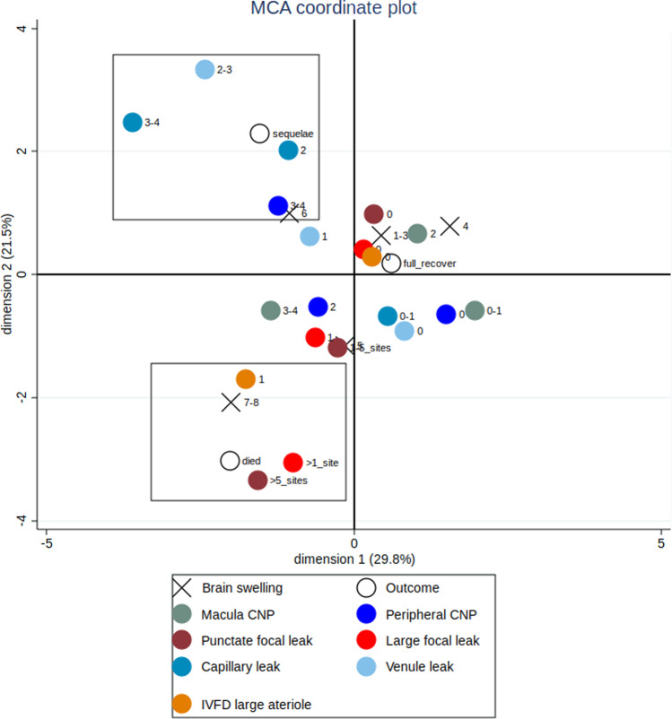
Cerebral malaria (CM) remains a common cause of death of children in Africa with annual mortality of 400 000. Malarial retinopathy is a unique set of fundus signs which has diagnostic and prognostic value in CM. Assessment of malarial retinopathy is now widely utilised in clinical care, and routinely incorporated into clinical studies to refine entry criteria. As a visible part of the central nervous system, the retina provides insights into the pathophysiology of this infectious small-vessel vasculitis with adherent parasitised red blood cells. Fluorescein angiography and optical coherence tomography (OCT) have shown that patchy capillary non-perfusion is common and causes ischaemic changes in the retina in CM. It is likely this is mirrored in the brain and may cause global neurological impairments evident on developmental follow up. Three types of blood-retina barrier breakdown are evident: large focal, punctate, and vessel leak. Punctate and large focal leak (haemorrhage in formation) are associated with severe brain swelling and fatal outcome. Vessel leak and capillary non-perfusion are associated with moderate brain swelling and neurological sequelae. These findings imply that death and neurological sequelae have separate mechanisms and are not a continuum of severity. Each haemorrhage causes a temporary uncontrolled outflow of fluid into the tissue. The rapid accumulation of haemorrhages, as evidenced by multiple focal leaks, is a proposed mechanism of severe brain swelling, and death. Current studies aim to use optic nerve head OCT to identify patients with severe brain swelling, and macula OCT to identify those at risk of neurological sequelae.
摘要: 脑型疟疾 (CM) 仍为非洲儿童死亡的一个常见原因, 每年的死亡人数可达40万人。疟疾性视网膜病变为具有独特眼底体征的一系列改变, 对CM具有诊断和预后价值。在临床工作中, 对于疟疾性视网膜病变的评估已得到广泛应用, 并常规纳入临床研究以完善入选标准。作为中枢神经系统中唯一肉眼可见的组织, 视网膜提供了对这种寄生于红细胞的传染性小血管炎的病理生理学了解的有用信息。荧光素血管造影和相干光断层扫描 (OCT) 显示, 斑片状毛细血管无灌注很常见, 并导致CM患者的视网膜缺血性改变。这些病理性改变可能反映在大脑中的病变, 并导致在发育的随访中出现了明显的的全神经系统损伤。三种类型的血-视网膜屏障破坏是显而易见的: 大面积病灶、点状病灶和血管渗漏。点状和大面积病灶渗漏 (出血形成) 与严重的脑组织肿胀和致命的临床解决有关。血管渗漏和毛细血管无灌注与中度脑组织肿胀和神经系统后遗症有关。这些发现意味着死亡和神经系统后遗症具有不同的机制, 并不是严重后遗症的结局。每一次出血都会导致液体暂时不受控制地流出到组织中。多处病灶渗漏表明, 出血的快速积累, 是严重脑肿胀和死亡的可能机制。目前的研究旨在使用视盘OCT来识别严重脑水肿的患者, 以及使用黄斑OCT来识别那些具有神经系统后遗症风险的患者。.
© 2023. The Author(s).
Conflict of interest statement
The authors declare no competing interests.
Figures
References
-
- WHO. World Malaria Report 2019. 2019 04/12/19. https://www.who.int/publications/i/item/9789241565721.
-
- White N. Malaria. In: Jeremy Farrar PH, T Junghanss, G Kang, D Lalloo, N White, editor. Manson’s Tropical Diseases. 23 ed. London: Elsevier; 2014. p. 532-600.
Publication types
MeSH terms
Grants and funding
LinkOut - more resources
Full Text Sources
Medical
Research Materials
