Randomized Controlled Trial
. 2023 Feb 14;23(1):99.
doi: 10.1186/s12903-023-02803-8.

Evaluation of muco-adhesive tacrolimus patch on caspase-3 induced apoptosis in oral lichen planus: a randomized clinical trial

Affiliations
Randomized Controlled Trial

Evaluation of muco-adhesive tacrolimus patch on caspase-3 induced apoptosis in oral lichen planus: a randomized clinical trial

Suzan S Ibrahim et al. BMC Oral Health. .

Abstract

Background: The study compared the clinical effectiveness of topical Tacrolimus (TAC) in patches or gel with Triamcinolone acetonide (TRI) gel for erosive/atrophic oral lichen planus (OLP) and investigated the influence of these therapies on Caspase-3 expression as a marker of apoptosis.

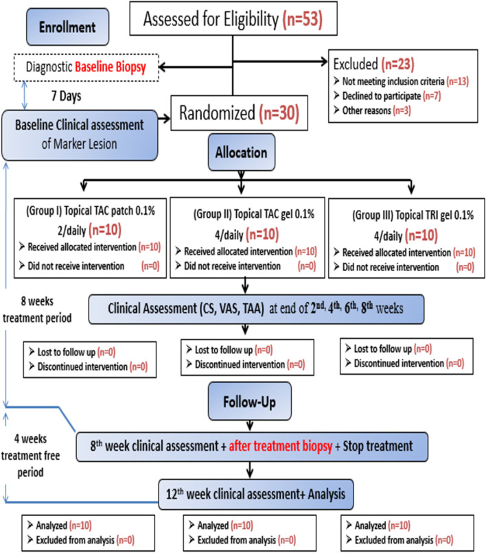
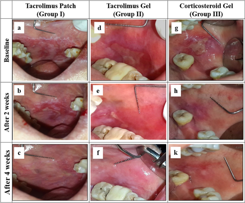
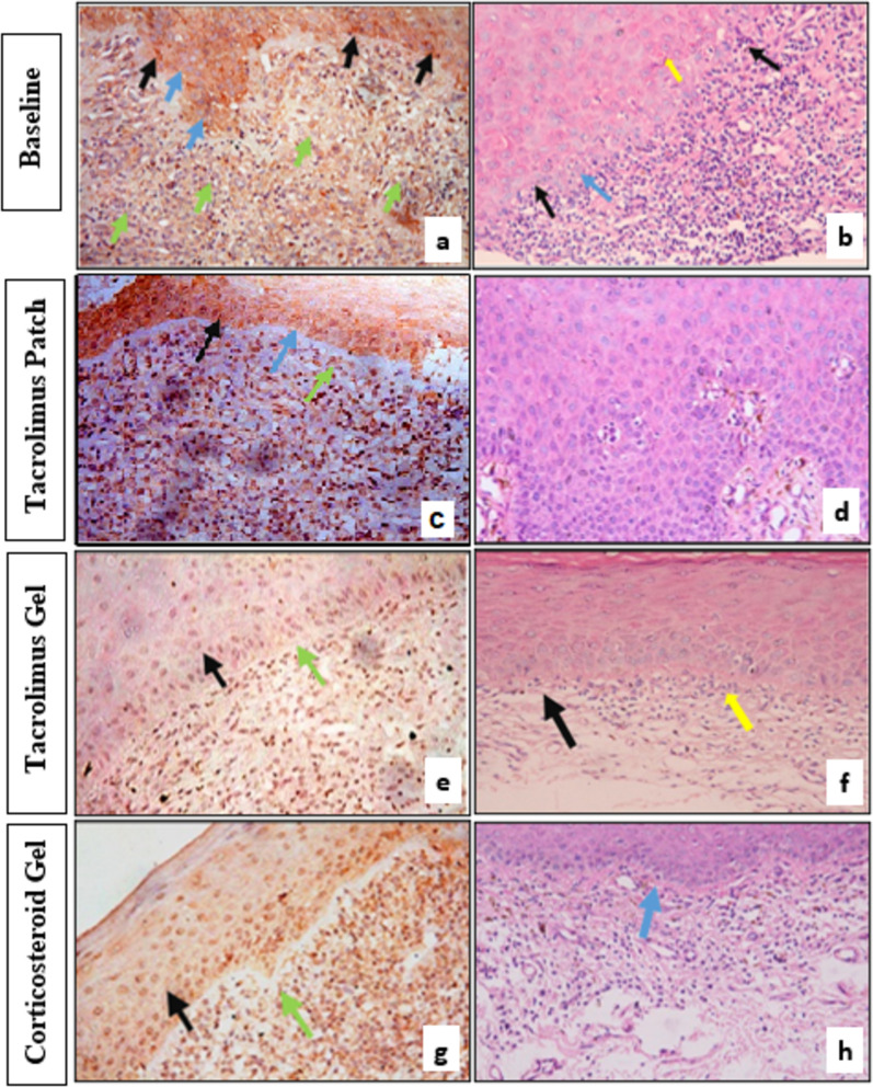
Methods: Thirty patients were randomly assigned into three equal groups to receive either topical TAC 0.1% patch twice daily, topical TAC 0.1% gel, or topical TRI 0.1% gel four times daily for 8 weeks. Each patient's clinical score (CS), visual analogue scale (VAS), and total atrophic area (TAA) of the marker lesion were measured at baseline, 2, 4, and 8 weeks of treatment, as well as after 4 weeks of treatment free period. Caspase-3 expression and lymphocytic counts (LC) were assessed in pre- and post-treatment biopsied stained sections.

Results: TAC patch resulted in a higher reduction in CS [- 14.00 (15.54%)] and VAS [- 70.21 (15.82%)] followed by TAC gel then TRI gel within the first two weeks. The reduction in VAS and TAA were significantly higher in TAC groups compared to TRI gel, although the difference between TAC treatment was not significant and this was observed throughout the treatment and follow-up periods. Caspase-3 expression increased in connective tissue in all groups. It decreased significantly within the epithelium in both TAC groups but increased in TRI gel. (LC) were significantly lowered with the TAC patch compared to other groups. The percentage change in Caspase-3 epithelial expression was significantly correlated to the CS, TAA, and LC.

Conclusion: Both TAC patch and gel significantly decreased pain and lesion size than TRI gel, with a significant reduction in Caspase-3 expression within the epithelium in comparison to the increase seen with TRI gel. The study protocol was registered at www.

Clinicaltrials: gov (NCT05139667) on 01/12/2021.

Keywords: Apoptotic keratinocytes; Caspase-3; Oral lichen planus; Tacrolimus; Triamcinolone acetonide.


Conflict of interest statement

The authors declare that they have no conflict of interest.

Figures

Fig. 1
Fig. 1
Tacrolimus mucoadhesive patch applied to the marker lesion on the buccal mucosa in Group I patient
Fig. 2
Fig. 2
A study CONSORT flow diagram illustrating patients’ enrolment and outcomes assessment
Fig. 3
Fig. 3
Photographs of erosive and atrophic oral lichen planus marker lesions in the buccal mucosa in 3 patients from Group I (a), Group II (d), and Group III (g), at baseline. Reduction in lesion size and severity by topical Tacrolimus patch (b), and slight reduction in ulcer size and extension with topical application of Tacrolimus gel (e), and Triamcinolone Acteonid gel (h), after 2 weeks’ treatment. Complete healing of oral lesions treated with Tacrolimus patch (c), and marked signs of healing of oral lesions from the same patients treated by topical tacrolimus gel (f), and corticosteroid gel (k), after 4 weeks’ treatment
Fig. 4
Fig. 4
a Photomicrograph of OLP specimen at base line showing nuclear and cytoplasmic immunopositivity of some of the epithelial cells (black arrow) while other cells exhibited cytoplasmic immunopositivity and immunonegative nuclei (blue arrow). The connective tissue cells were found to be more frequently immune negative (green arrow). (caspase 3 × 40). b Photomicrograph of OLP specimen at base line showing dead keratinocytes (civatte bodies) (yellow arrow), vacuoles in the basal cell layer (blue arrow). The epithelial connective tissue junction obscured by lymphocytes (black arrow). (H&E ×40). c Photomicrograph of OLP specimen after treatment with tacrolimus patch, showing epithelial cells with cytoplasmic immunopositivity (black arrow), while the majority of epithelial cells nuclei were immunonegative (blue arrow). Some connective tissue cells were immunopostive (green arrow). (Group I) (caspase 3 × 20). d Photomicrograph of one of the OLP specimens after treatment with tacrolimus patch, showing hyperplastic epithelium. Note the bland appearance of the hyperplastic epithelium. Group I (H&E ×40). e Photomicrograph of OLP specimen after treatment with tacrolimus gel, exhibiting mild nuclear and cytoplasmic immunopositivity in some of the epithelial cells (black arrow) and most of the connective tissue cells (green arrow). (Group II) (caspase 3 × 20). f Photomicrograph of OLP specimen after treatment with tacrolimus gel showing relatively scant lymphocytes (yellow arrow), evident epithelial-connective tissue junction (black arrow). Group II (H&E ×20). g Photomicrograph of OLP specimen at base line showing nuclear and cytoplasmic immunopositivity of the epithelial cells (black arrow). Most of the connective tissue demonstrated immunopostive reaction (green arrow). (caspase 3 × 20) (Group III). h Photomicrograph of OLP specimen after treatment showing few lymphocytes, evident epithelial-connective tissue junction notes some basal epithelial cells exhibiting vacuolation (blue arrow). Group III (H&E ×20)

Similar articles

Cited by

References

    1. Li C, Tang X, Zheng X, Ge S, Wen H, Lin X, et al. Global prevalence and incidence estimates of oral lichen planus: a systematic review and meta-analysis. JAMA Dermatol. 2020;156:172–181. doi: 10.1001/JAMADERMATOL.2019.3797. - DOI - PMC - PubMed
    1. Gorouhi F, Davari P, Fazel N. Cutaneous and mucosal lichen planus: a comprehensive review of clinical subtypes, risk factors, diagnosis, and prognosis. Sci World J. 2014 doi: 10.1155/2014/742826. - DOI - PMC - PubMed
    1. Cheng YSL, Gould A, Kurago Z, Fantasia J, Muller S. Diagnosis of oral lichen planus: a position paper of the American Academy of Oral and Maxillofacial Pathology. Oral Surg Oral Med Oral Pathol Oral Radiol. 2016;122:332–354. doi: 10.1016/J.OOOO.2016.05.004. - DOI - PubMed
    1. Adams JM. Ways of dying: multiple pathways to apoptosis. Genes Dev. 2003;17:2481–2495. doi: 10.1101/GAD.1126903. - DOI - PubMed
    1. Thornberry NA, Lazebnik Y. Caspases: enemies within. Science. 1998;281:1312–6. doi: 10.1126/SCIENCE.281.5381.1312. - DOI - PubMed

Publication types

Associated data