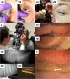
Review of Literature on Intraductal Meibomian Gland Probing with Insights from the Inventor and Developer: Fundamental Concepts and Misconceptions
- PMID: 36789289
- PMCID: PMC9922486
- DOI: 10.2147/OPTH.S390085
Review of Literature on Intraductal Meibomian Gland Probing with Insights from the Inventor and Developer: Fundamental Concepts and Misconceptions
Abstract
Obstructive Meibomian gland dysfunction (MGD) affects millions of patients around the world. Its effective treatment with intraductal meibomian gland probing (MGP), was first reported in 2010. Since then, MGP has provided relief to thousands of patients globally suffering with refractory MGD. The purpose of Meibomian gland probing is restoring the integrity of the gland's central duct by entering the gland through the natural orifice, releasing fixed obstruction thought to be periductal fibrosis, thereby establishing and/or confirming the patency of the duct, and concurrently equilibrating intraductal pressure as well as promoting gland functionality with meibum production. There may or may not be immediate secretion of meibum upon successful restoration of ductal integrity depending on the gland's state of function and degree of atrophy. One double-blind placebo-controlled study has been conducted and, with the accumulated evidence of over 12 other peer reviewed articles in the scientific literature, overwhelmingly indicates that MGP is a safe and effective treatment for the MGD patient refractory to prior standard care and as a first-line treatment. This paper describes relevant fundamental concepts, dispels commonly held misconceptions, and provides an objective review of the current understanding and effectiveness of MGP for the treatment of obstructive MGD. Our analysis will better equip clinicians to draw informed conclusions about both subjective and objective findings reported in MGP studies and researchers to design future robust studies that provide meaningful results.
Keywords: MGD; MGP clinical trial; MGP studies; dry eye; meibomian gland dysfunction; obstructive MGD.
© 2023 Warren and Maskin.
Conflict of interest statement
SLM is a >5% owner of MGD Innovations, Inc. which holds patents on instrumentation and methods for intraductal diagnosis and treatment of meibomian gland disease (MGD). SLM also has patents on the use of jojoba-based treatment options for MGD. He also reports patent royalties from Katena (nos: 9510844, 10159599, and 11110003). NAW is chair of the Not A Dry Eye Foundation. The authors report no other conflicts of interest in this work.
Figures


References
-
- Kheirkhah A, Kobashi H, Girgis J, Jamali A, Ciolino JB, Hamrah P. A randomized, sham-controlled trial of intraductal meibomian gland probing with or without topical antibiotic/steroid for obstructive meibomian gland dysfunction. Ocul Surf. 2020;18(4):852–856. doi: 10.1016/j.jtos.2020.08.008 - DOI - PMC - PubMed
Publication types
LinkOut - more resources
Full Text Sources
Miscellaneous

