. 2023 Feb 15:12:e82015.
doi: 10.7554/eLife.82015.

Structural insights into actin isoforms

Affiliations

Structural insights into actin isoforms

Amandeep S Arora et al. Elife. .

Abstract

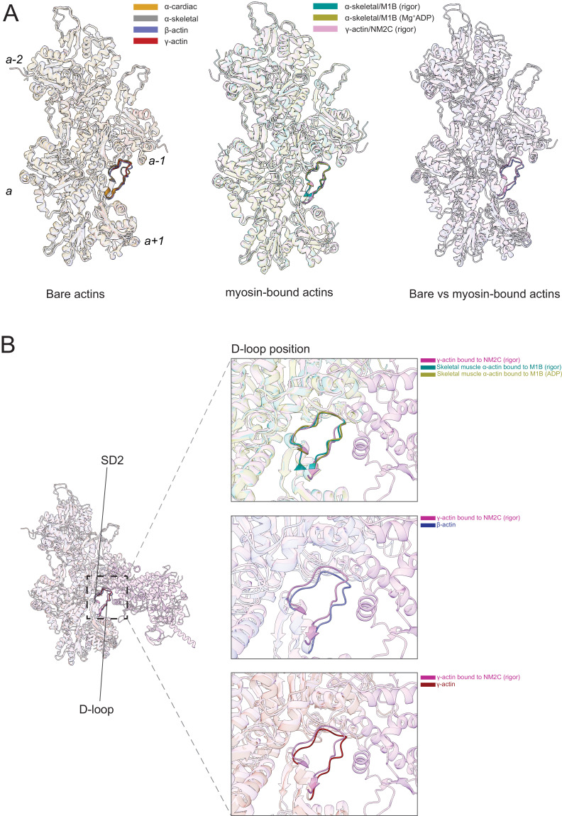
Actin isoforms organize into distinct networks that are essential for the normal function of eukaryotic cells. Despite a high level of sequence and structure conservation, subtle differences in their design principles determine the interaction with myosin motors and actin-binding proteins. Therefore, identifying how the structure of actin isoforms relates to function is important for our understanding of normal cytoskeletal physiology. Here, we report the high-resolution structures of filamentous skeletal muscle α-actin (3.37 Å), cardiac muscle α-actin (3.07 Å), ß-actin (2.99 Å), and γ-actin (3.38 Å) in the Mg2+·ADP state with their native post-translational modifications. The structures revealed isoform-specific conformations of the N-terminus that shift closer to the filament surface upon myosin binding, thereby establishing isoform-specific interfaces. Collectively, the structures of single-isotype, post-translationally modified bare skeletal muscle α-actin, cardiac muscle α-actin, ß-actin, and γ-actin reveal general principles, similarities, and differences between isoforms. They complement the repertoire of known actin structures and allow for a comprehensive understanding of in vitro and in vivo functions of actin isoforms.

Keywords: actin; biochemistry; chemical biology; cytoskeleton; isoforms; macromolecular structure; molecular biophysics; myosin; none; structural biology.

Plain language summary

The protein actin is important for many fundamental processes in biology, from contracting muscle to dividing a cell in two. As actin is involved in such a variety of roles, human cells have slightly different versions of the protein, known as isoforms. For example, alpha-actin is vital for contracting muscle, while beta- and gamma-actin drive cellular processes in non-muscle cells. In order to carry out its various functions, actin interacts with many other proteins inside the cell, such as myosin motors which power muscle contraction. These interactions rely on the precise chain of building blocks, known as amino acids, that make up the actin isoforms; even subtle alterations in this sequence can influence the behavior of the protein. However, it is not clear how differences in the amino acid sequence of the actin isoforms impact actin’s interactions with other proteins. Arora et al. addressed this by studying the structure of four human actin isoforms using a technique called cryo-electron microscopy, where the proteins are flash-frozen and bombarded with electrons. These experiments showed where differences between the amino acid chains of each isoform were located in the protein. Arora et al. then compared their structures with previous work showing the structure of actin bound to myosin. This revealed that the tail-end of the protein (known as the N-terminus) differed in shape between the four isoforms, and this variation may influence how actin binds to others proteins in the cell. These results are an important foundation for further work on actin and how it interacts with other proteins. The structures could help researchers design new tools that can be used to target specific isoforms of actin in different types of laboratory experiments.


Conflict of interest statement

AA, HH, RS, YN, AS, TH, SH, KC No competing interests declared, MB Reviewing editor, eLife

Figures

Figure 1.
Figure 1.. Cryo-electron microscopy (cryo-EM) filament structures of actin isoforms.
(A) Helical reconstruction of skeletal muscle α-actin, (B) cardiac muscle α-actin, (C) β-actin, and (D) γ-actin. Views in (BD) are according to (A). Four individual actin protomers in the filament are shown and denoted with italic numbers. The pointed (−) and barbed (+) ends are indicated. (E) Representative key regions of actin isoforms with corresponding cryo-EM densities in transparent surface representation are shown. The protein backbone and amino acid side chains are shown in licorice and stick representation, respectively. Throughout this work, amino acids are numbered according to the sequence of mature actin isoforms (Figure 1—figure supplement 1C).
Figure 1—figure supplement 1.
Figure 1—figure supplement 1.. Sequence conservation in actin isoforms.
(A) The classical view of the monomer structure of actin. Key regions are indicated. Monomers self-assemble into helical, polarized filaments. The arrangement of individual actin protomers in a filament is shown, and the relative position is denoted with italic numbers. The pointed (−) and barbed (+) end, the nucleotide-binding cleft, and the barbed end groove are indicated. Monomer and filament structure not drawn to scale. (B) ConSurf analysis shows the conservation of amino acid positions in actin isoforms. Variations are shown in cyan color. The protein backbone is shown in licorice representation. (C) Sequence alignment of actin isoforms: rabbit skeletal muscle α-actin (Skα-actin; UniProt ID: P68135), bovine cardiac muscle α-actin (Caα-actin; UniProt ID: Q3ZC07), human smooth muscle α-actin (Smα-actin; UniProt ID: P62736), human β-actin (UniProt ID: P60709), human γ-actin (UniProt ID: P63261), and human smooth muscle γ-actin (Smγ-actin; UniProt ID: Q3ZC07). Conserved amino acids are shown in black, and variable amino acids are highlighted in blue. Amino acids that are absent in mature actin isoforms are shown in gray. Key structural regions and resolved post-translational modifications (PTMs) in our cryo-electron microscopy (cryo-EM) structures are indicated.
Figure 1—figure supplement 2.
Figure 1—figure supplement 2.. Image processing summary for actin isoforms.
(A–D) 2D classes, global map-model Fourier Shell Correlation (FSC) curves, and the local resolution estimated of the experimental maps are shown for skeletal muscle α-actin (A), cardiac muscle α-actin (B), β-actin (C), and γ-actin (D). The gold standard, FSC 0.143 criterion was used to estimate the global resolution of actin isoforms. The local resolution gradient is in Angstrom (Å).
Figure 1—figure supplement 3.
Figure 1—figure supplement 3.. Methylation of H72/H73 in actin isoforms.
(A) The methylated histidine region in the actin protomer is indicated with a rectangle. (B) Superimposition of methylated histidine regions in actin isoform structures. Colors are according to panels C–F. (C) H73Me and neighboring residues in skeletal muscle α-actin. (D) H73Me and neighboring residues in cardiac muscle α-actin. (E) H72Me and neighboring residues in β-actin. (F) H72Me and neighboring residues inγ-actin. Cryo-electron microscopy (Cryo-EM) densities, contoured at ≥2σ, are shown in surface representation in C–F. Panel A is shown in cartoon representation, and panels B–F are shown in stick representation.
Figure 2.
Figure 2.. Conserved nucleotide-binding cleft active site in actin isoforms.
(A–D) Coordination of Mg2+·ADP in the nucleotide-binding cleft of skeletal muscle α-actin (A), cardiac muscle α-actin (B), β-actin (C), and γ-actin (D). Underlines indicate locations of amino acid substitutions between actin isoforms. The protein backbone and side chains are shown in licorice and stick representation, respectively. ADP is shown in cyan-colored stick representation. Electron densities for key amino acids in the nucleotide-binding cleft active site of actin isoforms are shown. Schematic representations of key interactions in the nucleotide-binding cleft active sites of the respective actin isoforms are shown in the right panel. The schematics are not drawn to scale.
Figure 2—figure supplement 1.
Figure 2—figure supplement 1.. Coordination of Mg2+·ADP in the nucleotide-binding cleft of actin isoforms.
(A) Superimposition of the nucleotide-binding cleft active site of actin isoforms. Key amino acids are indicated. ADP is shown in cyan-colored stick representation, and Mg2+ is shown as green sphere. Underlines indicate locations of amino acid substitutions between actin isoforms. The protein backbone and side chains are shown in licorice and stick representation, respectively. (B) Comparison of the position of ADP and Mg2+ in the nucleotide-binding cleft active site of actin isoforms. Colors are according to (A).
Figure 3.
Figure 3.. Similarities and differences between actin isoforms.
(A) Sequence variations at the N-terminus located in SD1 of actin isoforms. (B) Sequence variations in SD1 of actin isoforms. (C) Sequence variations in SD3 of actin isoforms. (D) Sequence variations in SD4 of actin isoforms. SD2 is conserved between actin isoforms. The identical and nonidentical amino acids at sites of substitutions within the actin protomer across isoforms are shown for skeletal muscle α-actin (orange), cardiac muscle α-actin (yellow), β-actin (purple), and γ-actin (teal) as spheres. Note that the first three amino acids of skeletal muscle α-actin and the first amino acid of cardiac muscle α-actin are unresolved in our structures. The protein backbone is shown in licorice representation, and the substituted amino acids are shown in spheres representation.
Figure 3—figure supplement 1.
Figure 3—figure supplement 1.. Amino acid variations along the longitudinal and transverse axis of actin isoforms.
(A–D) Variable amino acid distributions along the longitudinal and transverse axis of actin isoforms. Nonconserved amino acids are shown along with the longitudinal (purple axis) and transverse axis (yellow) for actin subdomains SD1 (A), SD3 (B), and SD4 (C). (D) Distribution of all variable amino acids in SD1, SD3, and SD4 for all actin isoforms. For clarity, only amino acids close to the transverse and longitudinal axis are labeled.
Figure 4.
Figure 4.. Comparative structural analysis of the longitudinal interprotomer interface.
(A–D) Key residues at the interprotomer interface of skeletal muscle α-actin (A), cardiac muscle α-actin (B), β-actin (C), and γ-actin (D). Individual protomers in actin isoforms are oriented according to Figure 4—figure supplement 1A. Underlines indicate locations of amino acid substitutions between actin isoforms. The protein backbone and side chains are shown in licorice and stick representation, respectively.
Figure 4—figure supplement 1.
Figure 4—figure supplement 1.. Transverse and longitudinal interprotomer interfaces in actin isoforms.
(A) Analyzed transverse interprotomer interface (yellow rod) in actin isoforms. (B) Analyzed longitudinal interprotomer interface (lilac rod) in actin isoforms. (C) Substituted amino acids in the longitudinal interprotomer interface of skeletal muscle α-actin and β-actin. Colors of the protomers are according to the Figure 4. (D) Substituted amino acid V286/I287 in the transverse interprotomer interface of skeletal muscle α-actin and β-actin. The protein backbone and amino acid side chains are shown in licorice and stick representation.
Figure 5.
Figure 5.. Comparative structural analysis of the transverse interprotomer interface.
(A–D) Key residues at the interprotomer interface of skeletal muscle α-actin (A), cardiac muscle α-actin (B), β-actin (C), and γ-actin (D). Individual protomers in actin isoforms are oriented according to Figure 4—figure supplement 1B. Underlines indicate locations of amino acid substitutions between actin isoforms. The protein backbone and amino acid side chains are shown in licorice and stick representation, respectively.
Figure 6.
Figure 6.. The actomyosin interface.
(A) Superimposition of bare actin isoform structures in the Mg2+·ADP state (left), superimposition of myosin-bound actin isoforms structures (middle), and overlap of bare versus myosin-bound actin structures (right) are shown. (B) Zoomed-in view of the actomyosin interface at the D-loop region. For clarity, only D-loops involved in the binding of myosins are highlighted in the respective dark colors. The offset between the structures in the lower two panels is caused by a conformational change of SD2 in myosin-bound compared to bare actin structures.
Figure 6—figure supplement 1.
Figure 6—figure supplement 1.. The actomyosin interface of the cardiac thin filament compared to bare cardiac muscle α-actin.
Superimposition of the structures of cardiac myosin-2 bound to the thin filament and bare cardiac muscle α-actin.
Figure 7.
Figure 7.. The N-terminus of actin interacts with loop-2 of myosins.
(A) Close-up view of the N-termini of bare β-actin (blue), bare γ-actin (red), myosin-bound actin isoforms M1B (green, lime), and NM2C (pink). The N-termini are indicated with an asterisk. (B) A cluster of positively charged amino acids in the loop-2 of myosins is in close proximity to amino acids D1Ac to D3 in the N-terminus of bare β-actin (blue) and, (C) a cluster of positively charged amino acids in the loop-2 of myosins is in close proximity to amino acids E1Ac to E3 in the N-terminus of bare γ-actin (red).
Author response image 1.

Update of

References

    1. Abe A, Saeki K, Yasunaga T, Wakabayashi T. Acetylation at the N-terminus of actin strengthens weak interaction between actin and myosin. Biochemical and Biophysical Research Communications. 2000;268:14–19. doi: 10.1006/bbrc.1999.2069. - DOI - PubMed
    1. Adams PD, Afonine PV, Bunkóczi G, Chen VB, Davis IW, Echols N, Headd JJ, Hung L-W, Kapral GJ, Grosse-Kunstleve RW, McCoy AJ, Moriarty NW, Oeffner R, Read RJ, Richardson DC, Richardson JS, Terwilliger TC, Zwart PH. PHENIX: a comprehensive python-based system for macromolecular structure solution. Acta Crystallographica. Section D, Biological Crystallography. 2010;66:213–221. doi: 10.1107/S0907444909052925. - DOI - PMC - PubMed
    1. Ali R, Zahm JA, Rosen MK. Bound nucleotide can control the dynamic architecture of monomeric actin. Nature Structural & Molecular Biology. 2022;29:320–328. doi: 10.1038/s41594-022-00743-5. - DOI - PMC - PubMed
    1. Arnesen T, Marmorstein R, Dominguez R. Actin’s N-terminal acetyltransferase uncovered. Cytoskeleton. 2018;75:318–322. doi: 10.1002/cm.21455. - DOI - PMC - PubMed
    1. Baranwal S, Naydenov NG, Harris G, Dugina V, Morgan KG, Chaponnier C, Ivanov AI. Nonredundant roles of cytoplasmic β- and γ-actin isoforms in regulation of epithelial apical junctions. Molecular Biology of the Cell. 2012;23:3542–3553. doi: 10.1091/mbc.E12-02-0162. - DOI - PMC - PubMed

Publication types

MeSH terms