. 2023 Feb;169(2):001298.
doi: 10.1099/mic.0.001298.

Characterization of a TatA/TatB binding site on the TatC component of the Escherichia coli twin arginine translocase

Affiliations

Characterization of a TatA/TatB binding site on the TatC component of the Escherichia coli twin arginine translocase

Emmanuele Severi et al. Microbiology (Reading). 2023 Feb.

Abstract

The twin arginine transport (Tat) pathway exports folded proteins across the cytoplasmic membranes of prokaryotes and the thylakoid membranes of chloroplasts. In Escherichia coli and other Gram-negative bacteria, the Tat machinery comprises TatA, TatB and TatC components. A Tat receptor complex, formed from all three proteins, binds Tat substrates, which triggers receptor organization and recruitment of further TatA molecules to form the active Tat translocon. The polytopic membrane protein TatC forms the core of the Tat receptor and harbours two binding sites for the sequence-related TatA and TatB proteins. A 'polar' cluster binding site, formed by TatC transmembrane helices (TMH) 5 and 6 is occupied by TatB in the resting receptor and exchanges for TatA during receptor activation. The second binding site, lying further along TMH6, is occupied by TatA in the resting state, but its functional relevance is unclear. Here we have probed the role of this second binding site through a programme of random and targeted mutagenesis. Characterization of three stably produced TatC variants, P221R, M222R and L225P, each of which is inactive for protein transport, demonstrated that the substitutions did not affect assembly of the Tat receptor. Moreover, the substitutions that we analysed did not abolish TatA or TatB binding to either binding site. Using targeted mutagenesis we introduced bulky substitutions into the TatA binding site. Molecular dynamics simulations and crosslinking analysis indicated that TatA binding at this site was substantially reduced by these amino acid changes, but TatC retained function. While it is not clear whether TatA binding at the TMH6 site is essential for Tat activity, the isolation of inactivating substitutions indicates that this region of the protein has a critical function.

Keywords: MD simulations; Tat pathway; TatC; mutagenesis; protein transport; twin arginine signal peptide.


Conflict of interest statement

The authors declare no conflict of interests

Figures

Fig. 1.
Fig. 1.
(a) Structures of TatA (blue), TatB (orange) and TatC (green). The transmembrane helices (TMHs) and amphipathic helices (APHs) of TatA and TatB are indicated, together with the TMH numbering in TatC. (b) A model for the resting state of the TatABC receptor complex, showing the interactions of the transmembrane helices, with the constituent subunits coloured as in (a). Polar cluster interactions between TatC residues T208 and Q215 (green) and TatB residue E8 (orange).
Fig. 2.
Fig. 2.
Mutagenesis of TatC TMH6. (a) Amino acid sequence of TatC TMH6 and flanking regions. Inactivating substitutions falling in this region of TatC that have been identified previously [15, 41, 57] are indicated by red font. Residues boxed in grey have been shown by cysteine-substitution and crosslinking (M205, L206 [26]) or co-evolution analysis, molecular simulations and mutagenesis [15] to form the TatA/TatB TMH5 binding site. Residues shown boxed in yellow are part of the TMH6 binding site [26]. (b) Structural model of TatA bound at the TMH6 binding site. (c) Phenotypic characterization of TatC single amino acid TMH6 variants. Spot tests of strain DADE (ΔtatABCD, ΔtatE) carrying pTAT1d derivatives producing TatA, TatB and the indicated amino acid TMH6 variant in TatC. Strains were re-grown from overnight cultures in liquid medium to an OD600 of 1, decimally diluted and spotted (10 μl) on LB medium with or without added 2 % SDS, and finally incubated overnight aerobically at 37 °C.
Fig. 3.
Fig. 3.
Membrane accumulation levels of TatC single amino acid TMH6 variants expressed from a low-copy vector. Western blots of membrane fractions from MΔBC (as MC4100, ΔtatBC) cultures harbouring derivatives of the low-copy construct pC*BC101FLAG expressing TatB and the indicated amino acid TMH6 variant in C-terminally FLAG-tagged TatC. Crude membrane fractions were prepared from exponentially growing cultures as described in the Methods, blotted and probed with an anti-FLAG monoclonal antibody. An equal amount of total protein was loaded in each lane.
Fig. 4.
Fig. 4.
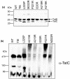
BN-PAGE analysis of Tat complexes containing TatC TMH6 variants. (a) Crude membranes from strain DADE (ΔtatABCD, ΔtatE) producing the indicated TatC variants alongside wild-type TatA and TatB from plasmid pTAT1d were solubilized by addition of 2 % (w/v) digitonin and analysed by (a) SDS-PAGE and Western blotting with a mix of anti-TatB and anti-TatC antibodies to assess protein levels; and (b) BN-PAGE (4–16% Bis-Tris Native PAGE gels; 20 µg solubilized membrane per lane) followed by Western blotting with an anti-TatC antibody as indicated.
Fig. 5.
Fig. 5.
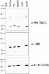
Co-purification of TatB and variant TatC with the Tat substrate SufI. Strain DADE-P (ΔtatABCD, ΔtatE, pcnB1) producing the indicated TatCHIS variants alongside wild-type TatB and the Tat substrate SufIFLAG from plasmid pFATBCHIS-sufI FLAG were inoculated from overnight cultures at a starting OD600 of 0.05 and grown for 3 h in the presence of 1 mM IPTG after which membrane fractions were produced as described in the Methods. Membranes were solubilized by addition of 2 % (w/v) digitonin and incubated with Ni-NTA-magnetic beads to separate TatCHIS-containing complexes. Affinity-bound complexes were eluted by the thermal treatment of the beads and analysed on SDS-PAGE with anti-His (for TatC), anti-TatB and anti-FLAG antibodies. ‘tatC’: DADE-P harbouring pFATΔA-sufI FLAG expressing WT TatC without a His-tag [8]; ‘ΔtatC’: DADE-P carrying a pFATBCHIS-sufI FLAG derivative with a PCR-induced frameshift within the TMH6 coding region of tatC HIS, used here as a further negative control.
Fig. 6.
Fig. 6.
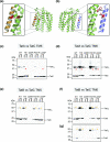
In vivo disulphide cross-linking between TatC harbouring TMH6 substitutions and TatA or TatB. (a and b) Diagnostic crosslinks between TatA/TatB and TatC can be used to probe occupancy of (a) the TMH5 site (L9C substituted TatA or TatB and M205C TatC) and (b) the TMH6 site (L9C substituted TatA or TatB and F213C TatC). (c–g) Membranes from strain DADE (ΔtatABCD, ΔtatE) producing the indicated TatC variants and either the M205C or F213C substitutions alongside L9C variants of either TatA or TatB from the low-copy plasmid pTAT101cysless were analysed by anti-TatC Western blotting after exposure of whole cells to either 1.8 mM CuP (oxidizing, ‘ox’) or 10 mM DTT (reducing, ‘red’). Crosslinks are shown between (c) TatA[L9C] and TatC[M205C]; (d) TatA[L9C] and TatC[M213C]; (e) TatB[L9C] and TatC[M205C]; (f) TatB[L9C] and TatC[F213C]. (g) Is a reload of the membranes from (f) with reduced exposure to prevent signal saturation. In each case red asterisks mark the positions of TatA–TatC crosslinks and yellow asterisks the TatB–TatC crosslinks.
Fig. 7.
Fig. 7.
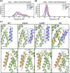
Molecular simulations of the interactions of TatA with TatC TMH6 for the WT and P221R, L225P and TIE mutants. (a) Distances between TatA and TatC TMH6. (b) Distances between TatB and TatC TMH5. The centre of mass of the transmembrane helices was used to calculate the distances. (c) Snapshots of the MD simulations showing the displacement of TatA (blue) and TatB (orange) from the TatC (green) TMH6 and TMH5 binding sites, respectively. The three TatC mutants are shown in comparison with the WT.

References

    1. Palmer T, Stansfeld PJ. Targeting of proteins to the twin-arginine translocation pathway. Mol Microbiol. 2020;113:861–871. doi: 10.1111/mmi.14461. - DOI - PMC - PubMed
    1. Theg SM. Chloroplast transport and import. Photosynth Res. 2018;138:261–262. doi: 10.1007/s11120-018-0593-x. - DOI - PubMed
    1. Berks BC. The twin-arginine protein translocation pathway. Annu Rev Biochem. 2015;84:843–864. doi: 10.1146/annurev-biochem-060614-034251. - DOI - PubMed
    1. Cline K. Mechanistic aspects of folded protein transport by the Twin Arginine Translocase (Tat) J Biol Chem. 2015;290:16530–16538. doi: 10.1074/jbc.R114.626820. - DOI - PMC - PubMed
    1. De Geyter J, Smets D, Karamanou S, Economou A. Inner membrane translocases and insertases. Subcell Biochem. 2019;92:337–366. doi: 10.1007/978-3-030-18768-2_11. - DOI - PubMed

Publication types

MeSH terms

LinkOut - more resources