

Asymmetry of Peripapillary Retinal Blood Vessel and Retinal Nerve Fiber Layer Thickness Between Healthy Right and Left Eyes
- PMID: 36790798
- PMCID: PMC9940773
- DOI: 10.1167/iovs.64.2.17
Asymmetry of Peripapillary Retinal Blood Vessel and Retinal Nerve Fiber Layer Thickness Between Healthy Right and Left Eyes
Abstract
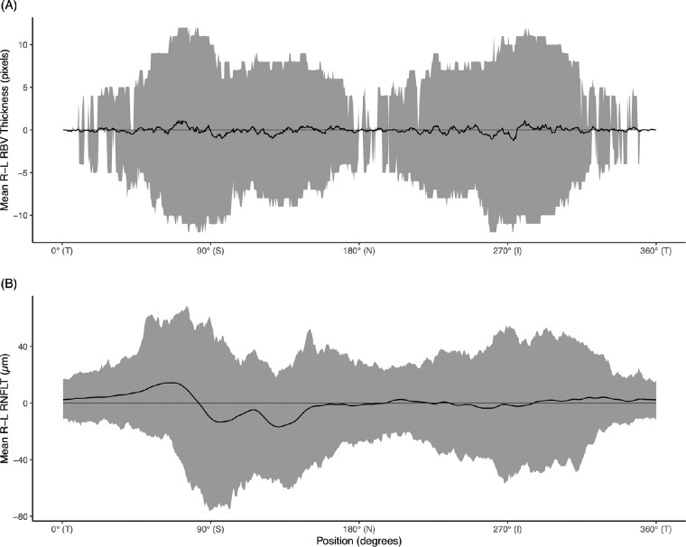
Purpose: The purpose of this study was to determine if there is asymmetry in retinal blood vessel (RBV) position and thickness between right and left eyes (R-L) and evaluate whether R-L asymmetry in RBV thickness is related to R-L asymmetry of retinal nerve fiber layer thickness (RNFLT).
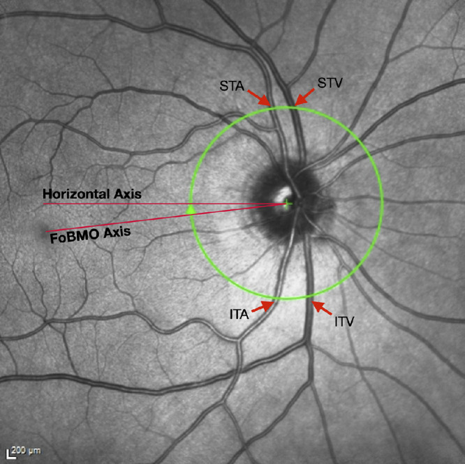
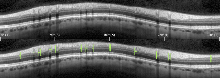
Methods: We analyzed peripapillary circle scan optical coherence tomography (OCT) examinations from healthy White subjects to measure RNFLT and RBV thickness and position relative to the fovea to Bruch's membrane opening axis, for all visible RBV. The R-L asymmetries of RNFLT and RBV thickness were computed for each A-scan. Four major vessels (superior temporal artery [STA] and superior temporal vein [STV], inferior temporal artery [ITA], and vein [ITV]) were identified using infrared images.
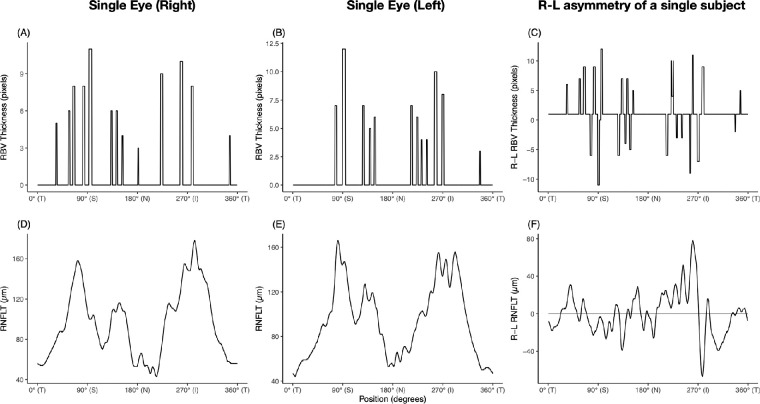
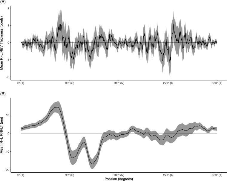
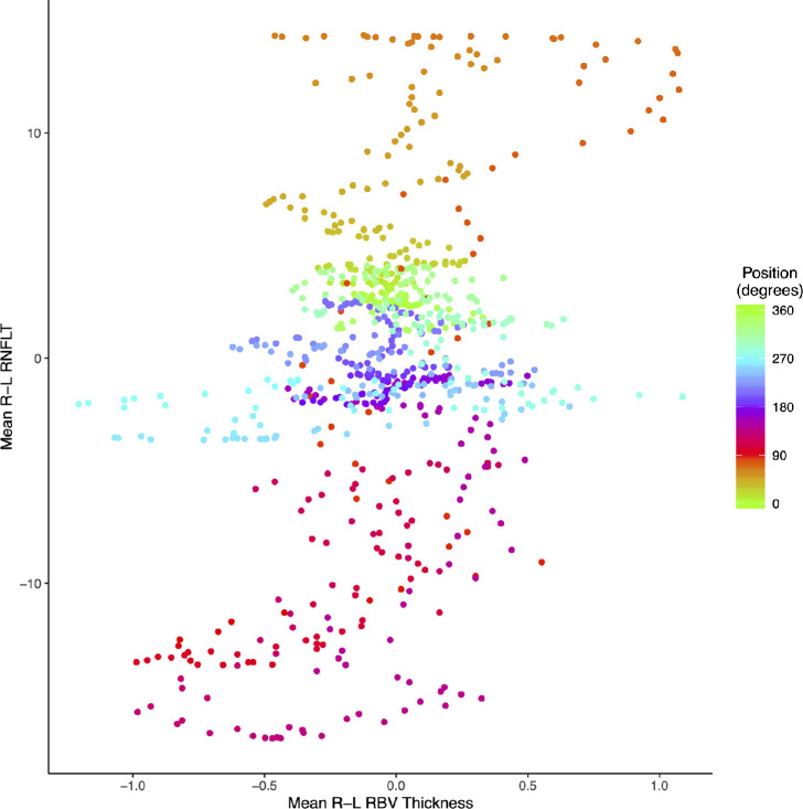
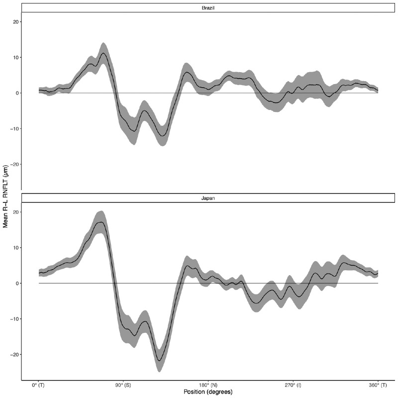
Results: We included 219 individuals. The mean (standard deviation) number of RBV measured per eye was 15.0 (SD = 2.2). The position of the STV and STA was more superior in left eyes than in right eyes, by 2.4 degrees and 3.7 degrees, respectively (P < 0.01). There was no region with significant R-L asymmetry in RBV thickness. RNFLT was thicker in right eyes in the temporal superior region and thicker in left eyes in the superior and nasal superior regions, with the asymmetry profile resembling in a "W" shape. This shape was also present in post hoc analyses in two different populations. The R-L asymmetries of RBV and RNFLT at each A-scan were not significantly associated (P = 0.37).
Conclusions: There is little R-L asymmetry in RBV, and it is not related to RNFLT asymmetry. This study suggests that R-L RNFLT asymmetry is due to factors other than RBV.
Conflict of interest statement
Disclosure:
Figures
Similar articles
-
Correlations Between Retinal Nerve Fiber Layer Thickness and Axial Length, Peripapillary Retinal Tilt, Optic Disc Size, and Retinal Artery Position in Healthy Eyes.J Glaucoma. 2017 Jan;26(1):34-40. doi: 10.1097/IJG.0000000000000550. J Glaucoma. 2017. PMID: 27753756
-
Asymmetry analysis of the retinal nerve fiber layer thickness in normal eyes using optical coherence tomography.Korean J Ophthalmol. 2005 Dec;19(4):281-7. doi: 10.3341/kjo.2005.19.4.281. Korean J Ophthalmol. 2005. PMID: 16491818
-
Bruch's Membrane Opening Minimum Rim Width and Retinal Nerve Fiber Layer Thickness in a Normal White Population: A Multicenter Study.Ophthalmology. 2015 Sep;122(9):1786-94. doi: 10.1016/j.ophtha.2015.06.001. Epub 2015 Jul 18. Ophthalmology. 2015. PMID: 26198806 Free PMC article.
-
Evaluation of Bruch's membrane opening-minimum rim width and retinal nerve fiber layer thickness in adults with anisometropic amblyopia.Photodiagnosis Photodyn Ther. 2020 Dec;32:102023. doi: 10.1016/j.pdpdt.2020.102023. Epub 2020 Sep 23. Photodiagnosis Photodyn Ther. 2020. PMID: 32979546
-
Lateral thinking - Interocular symmetry and asymmetry in neurovascular patterning, in health and disease.Prog Retin Eye Res. 2017 Jul;59:131-157. doi: 10.1016/j.preteyeres.2017.04.003. Epub 2017 Apr 28. Prog Retin Eye Res. 2017. PMID: 28457789 Review.
Cited by
-
Interocular Asymmetry of OCT Retinal Nerve Fiber Layer Values in a Normative Population: The Framingham Heart Study.Transl Vis Sci Technol. 2025 Jun 2;14(6):7. doi: 10.1167/tvst.14.6.7. Transl Vis Sci Technol. 2025. PMID: 40459525 Free PMC article.
-
Retina as a potential biomarker in schizophrenia spectrum disorders: a systematic review and meta-analysis of optical coherence tomography and electroretinography.Mol Psychiatry. 2024 Feb;29(2):464-482. doi: 10.1038/s41380-023-02340-4. Epub 2023 Dec 11. Mol Psychiatry. 2024. PMID: 38081943 Free PMC article.
References
-
- Chen TC, Hoguet A, Junk AK, et al. .. Spectral-domain OCT: Helping the clinician diagnose glaucoma: A report by the American academy of ophthalmology. Ophthalmology. 2018; 125(11): 1817–1827. - PubMed
-
- Park JJ, Oh DR, Hong SP, Lee KW.. Asymmetry analysis of the retinal nerve fiber layer thickness in normal eyes using optical coherence tomography. Korean J Ophthalmol KJO. 2005; 19(4): 281–287. - PubMed
-
- Huynh SC, Wang XY, Burlutsky G, Mitchell P.. Symmetry of optical coherence tomography retinal measurements in young children. Am J Ophthalmol. 2007; 143(3): 518–520. - PubMed
-
- Qian J, Wang W, Zhang X, et al. .. Optical coherence tomography measurements of retinal nerve fiber layer thickness in Chinese children and teenagers. J Glaucoma. 2011; 20(8): 509–513. - PubMed
MeSH terms
Grants and funding
LinkOut - more resources
Full Text Sources
