. 2023 Feb 21;120(8):e2216479120.
doi: 10.1073/pnas.2216479120. Epub 2023 Feb 15.

ALK fusion NSCLC oncogenes promote survival and inhibit NK cell responses via SERPINB4 expression

Affiliations

ALK fusion NSCLC oncogenes promote survival and inhibit NK cell responses via SERPINB4 expression

Tzu-Po Chuang et al. Proc Natl Acad Sci U S A. .

Abstract

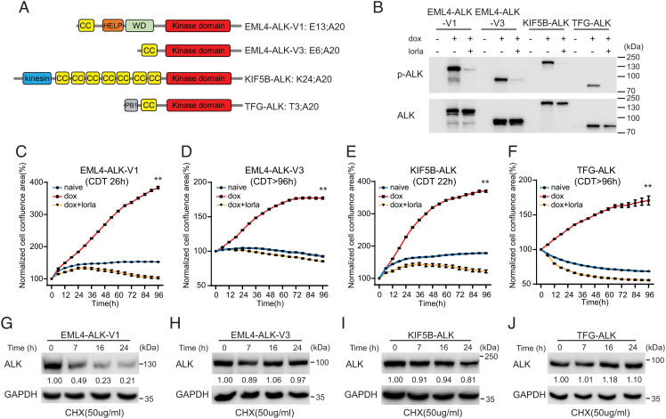
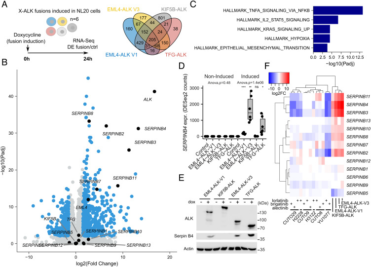
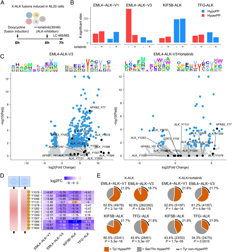
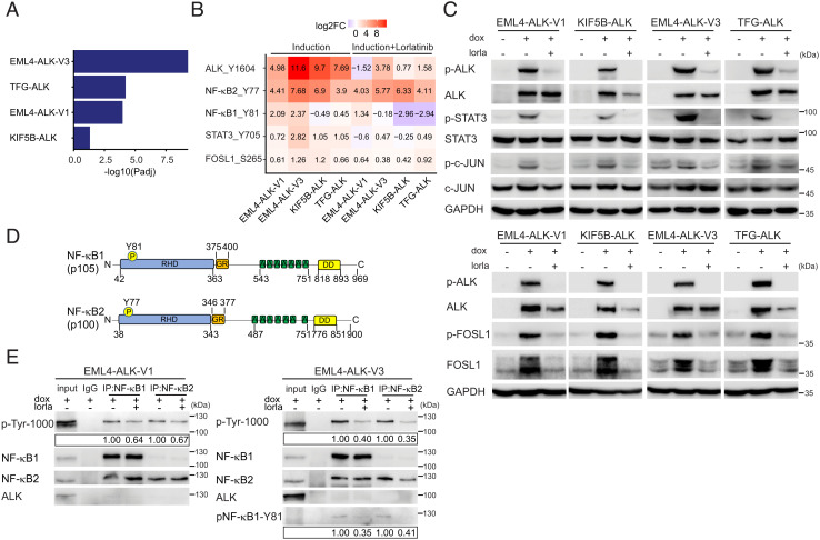
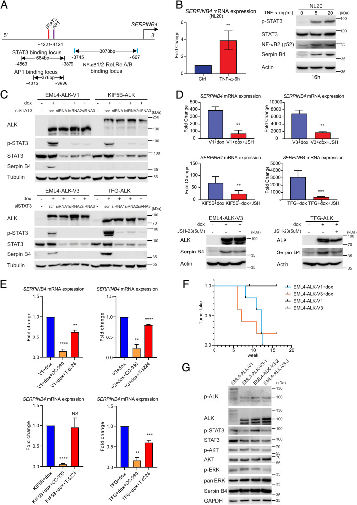
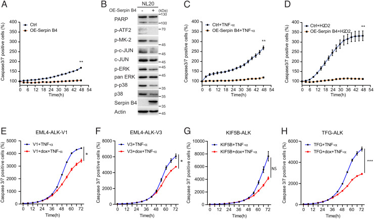
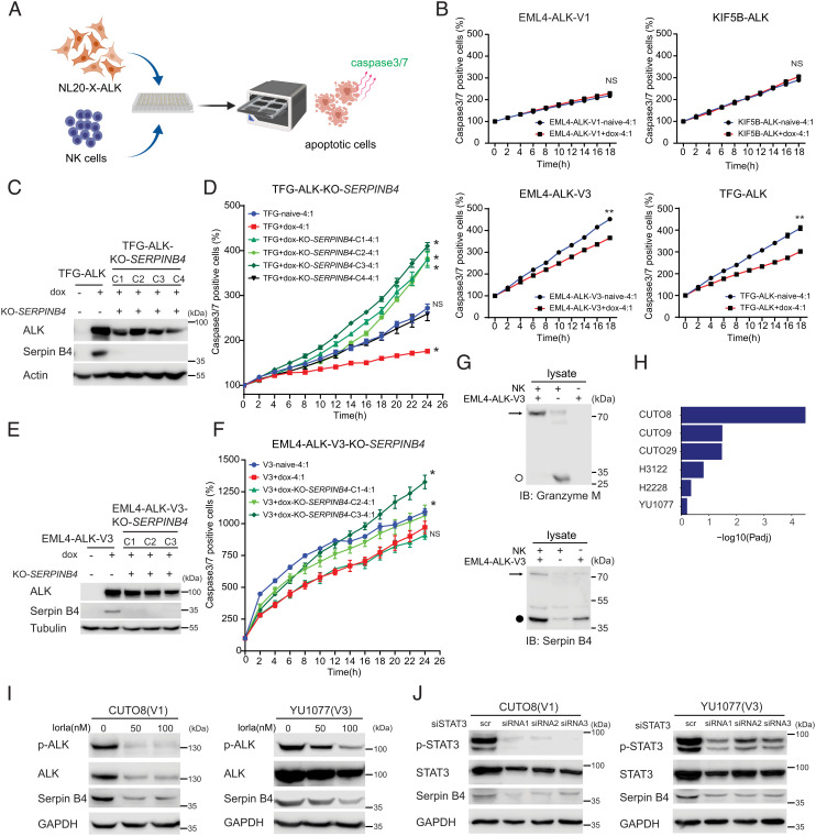
Anaplastic lymphoma kinase (ALK) fusion variants in Non-Small Cell Lung Cancer (NSCLC) consist of numerous dimerizing fusion partners. Retrospective investigations suggest that treatment benefit in response to ALK tyrosine kinase inhibitors (TKIs) differs dependent on the fusion variant present in the patient tumor. Therefore, understanding the oncogenic signaling networks driven by different ALK fusion variants is important. To do this, we developed controlled inducible cell models expressing either Echinoderm Microtubule Associated Protein Like 4 (EML4)-ALK-V1, EML4-ALK-V3, Kinesin Family Member 5B (KIF5B)-ALK, or TRK-fused gene (TFG)-ALK and investigated their transcriptomic and proteomic responses to ALK activity modulation together with patient-derived ALK-positive NSCLC cell lines. This allowed identification of both common and isoform-specific responses downstream of these four ALK fusions. An inflammatory signature that included upregulation of the Serpin B4 serine protease inhibitor was observed in both ALK fusion inducible and patient-derived cells. We show that Signal transducer and activator of transcription 3 (STAT3), Nuclear Factor Kappa B (NF-κB) and Activator protein 1 (AP1) are major transcriptional regulators of SERPINB4 downstream of ALK fusions. Upregulation of SERPINB4 promotes survival and inhibits natural killer cell-mediated cytotoxicity, which has potential for therapeutic impact targeting the immune response together with ALK TKIs in NSCLC.

Keywords: ALK fusion; NGS; anaplastic lymphoma kinase; non-small cell lung cancer; phosphoprofiling.


Conflict of interest statement

The authors declare no competing interest.

Figures

Fig. 1.
Fig. 1.
Characterization of NL20 cells expressing ALK NSCLC fusions. (A) Schematic representation of ALK fusion proteins investigated in this study. The breakpoint located at exon 20 in ALK is identical in all four ALK fusions. EML4-ALK-V1 (E13; A20) and V3 (E6; A20) harbor exons 1 to 13 and 1 to 6 of EML4, respectively; KIF5B-ALK (K24; A20) and TFG-ALK (T3; A20) contain exons 1 to 24 of KIF5B and exons 1 to 3 of TFG, respectively, fused to exon 20 of ALK. Protein domains are indicated as: coiled coil (CC, yellow), hydrophobic motif in EML proteins (HELP) (orange), tryptophan-aspartic acid repeat domain (WD) (green), ALK kinase domain (red), kinesin (blue), and PB1 (gray). (B) ALK fusions were induced in NL20-ALK cells with doxycycline (dox) in the presence or absence of lorlatinib (lorla, 30 nM), and lysates immunoblotted with pY1278-ALK and pan-ALK as indicated. PhosphoY1278-ALK (p-ALK) was employed as readout of ALK inhibition. (CF) NL20-ALK cells were treated with DMSO (blue), doxycycline (red), or doxycycline plus lorlatinib 30 nM (orange). Cell confluence was monitored using an IncuCyte® Live Cell Analysis system. Data points represent mean ± SEM of normalized cell confluence conducted in triplicate. Cell doubling time of individual cell lines is indicated (**< 0.01, two-tailed paired t test). One typical experiment of three independent experiments is shown. (GJ) NL20-ALK stable cell lines were treated with cycloheximide (CHX) for 7, 16 and 24 h, and resulting lysates immunoblotted for ALK and Glyceraldehyde-3-Phosphate Dehydrogenase (GAPDH). Quantification of ALK fusion protein levels compared to GAPDH control is depicted below each lane (n = 3, a representative result is shown).
Fig. 2.
Fig. 2.
Transcriptomic response to ALK fusion expression in NL20 cells. (A) ALK fusions were independently induced in NL20 cells, and RNA-seq-based DE with control cells was determined 24 h after fusion induction with doxycycline (dox). Venn diagram showing the number of DE genes (log2FC +/−1.5 at 1% False Discovery Rate (FDR), using a hyperbolic threshold) for each fusion as indicated. n = 6, RNA-seq analysis from three independent clones per variant performed in duplicate. See SI Appendix, Table S1 for detailed results. (B) Volcano plot showing DE response to EML4-ALK-V3 fusion induction. DE genes indicated in blue with genes discussed in main text indicated and labeled in black. (C) Hallmark GSEA results in 205 common genes that were DE for all four ALK fusions. GSEA was performed using Fisher’s exact test. Bars represent adjusted P-values (−log10 scale) for enriched gene sets at 5% FDR. (D) Box plot showing SERPINB4 expression for all four non-induced and dox-induced ALK fusions and control cells as indicated. ANOVA performed independently on non-induced and induced conditions. Asterisks indicating significance when comparing fusions to Ctrl cells (Student’s t test). NS, non-significant; *< 0.05; **< 0.01; ***< 0.001. (E) Immunoblot validation of Serpin B4 protein expression before and after fusion induction as indicated. (F) Heatmap showing DE log2FC values (color scale at top left) of all 13 SERPINB genes for the four ALK fusions as well as six ALK-positive patient-derived cell lines (CUTO29, H2228, CUTO9, H3122, CUTO8, YU1077) treated with three different ALK inhibitors as indicated. Dendrograms indicate hierarchical clustering results. RNA-seq data generated from H2228 and H3122 as well as from the recent patient-derived CUTO8, 9, 29, and YU1077 cells (24).
Fig. 3.
Fig. 3.
Lorlatinib-dependent phosphoproteomic response to ALK fusion induction. (A) DP between the four different ALK fusions and control NL20 cells was determined 7 h after fusion induction with doxycycline (dox). Cells were treated with lorlatinib 6 h after fusion induction as indicated. (B) Bar plot indicating the number of hyper- and hypophosphorylated sites (log2FC +/−1.5 at 5% FDR, using a hyperbolic threshold) in control and lorlatinib treatment conditions as indicated. (C) Volcano plots showing DP response in control (Left) and lorlatinib-treated (Right) EML4-ALK-V3 cells. DP sites indicated in blue with sites discussed in main text indicated and labeled in black. Sequence logo plots showing position-specific enrichment ±5 amino acids centered around the phosphorylation site for hypo- and hyperphosphorylated sites shown on top. (D) Heatmap showing DP (logFC values) between lorlatinib-treated and untreated fusions for all ALK tyrosine sites. Position in ALK indicated (Left). (E) Pie charts showing the proportion of tyrosine motifs in hyperphosphorylated sites in cells expressing ALK fusions in the presence or absence of lorlatinib. P-values calculated using Fisher’s exact test, estimating likelihood of overrepresentation of tyrosines in hyperphosphorylated sites.
Fig. 4.
Fig. 4.
Activation of STAT3, AP1 and NF-κB downstream of NSCLC ALK fusions. (A) Gene set enrichment for 27 inflammatory response genes (35) in DE genes in ALK fusions as indicated. GSEA was performed using Fisher’s exact test. Bars represent Padj values (−log10 scale). (B) Heatmap of DP logFC values (comparing each fusion to control cells) at ALK-Y1604, NF-κB2-Y77, NF-κB1-Y81, STAT3-Y705, and FOSL1-S265, in the presence (Right) or absence (Left) of lorlatinib. (C) ALK fusions were induced in NL20-ALK cells with doxycycline (dox) in the presence or absence of lorlatinib (lorla, 30 nM), and lysates immunoblotted with pY1278-ALK, pan-ALK, pY705-STAT3, pan-STAT3, pS265-FOSL1, FOSL1, pS63-c-JUN, pan-c-JUN, and GAPDH as indicated. Phospho-ALK (pY1278-ALK) was employed as readout of ALK inhibition. (D) Schematic representation of NF-κB1 and NF-κB2 proteins and phosphotyrosine sites identified in this study. Protein functional domains are indicated as: Rel-homology domain (RHD, blue), glycine-rich region (GR, orange), ankyrin repeat (A, green), and death domain (DD, yellow). (E) NF-κB1/NF-κB2 were immunoprecipitated from EML4-ALK-V1 or EML4-ALK-V3 expressing NL20 cells, in the presence or absence of lorlatinb (lorla), with NF-κB-specific antibodies as indicated, followed by immunoblotting with anti-p-Tyr-1000, NF-κB1, NF-κB2, ALK, and phosphor-specific NF-κB-Y81. Blots are representative of three different experiments. Quantification of phosphotyrosine levels is shown in box. n = 3, a representative result is shown.
Fig. 5.
Fig. 5.
NSCLC ALK fusions drive SERPINB4 expression via STAT3 and NF-κB. (A) Schematic representation of the SERPINB4 promoter region. Red bars show predicted STAT3 binding motifs, and purple and blue bars indicate predicted AP1 and NF-κB binding sites, respectively. (B) TNF-α induces SERPINB4 expression in NL20 cells. NL20 cells were treated with TNF-α for 6 h followed by qRT-PCR on total RNA. (Left) Fold change SERPINB4 mRNA expression normalized to untreated controls (Ctrl), (n = 3; **< 0.01; two-tailed paired t test). (Right) NL20 cells were treated with TNF-α (0, 5, 20 ng/mL) for 16 h, and lysates subjected to immunoblotting analysis with pY705-STAT3, pan-STAT3, NF-κB2, Serpin B4, and Actin antibodies as indicated. (C) NL20-ALK cells were transfected with either control siRNA (scr) or siRNAs (1 to 3) targeting STAT3 upon doxycycline (dox) addition. Resulting cell lysates were immunoblotted with pan-ALK, pY705-STAT3, pan-STAT3, Serpin B4, and Tubulin antibodies as indicated. (D) NL20-ALK cells treated with JSH-23 (5 µM) for 24 h upon doxycycline (dox) addition followed by either qPCR or immunoblotting analysis. Fold change SERPINB4 mRNA expression was normalized to untreated (no dox) controls and data presented as mean ± SD (n = 3; **P < 0.01; two-tailed paired t test). JSH-23-treated and control cell lysates were subjected to immunoblotting analysis with ALK, Serpin B4, and Actin antibodies as indicated. (E) NL20-ALK cells treated with CC-930 (10 µM) or T-5224 (10 µM) for 24 h upon doxycycline (dox) addition followed by qPCR analysis. Fold change SERPINB4 mRNA expression was normalized to untreated controls and data presented as mean ± SD (n = 3; NS, non-significant; **< 0.01; ***< 0.001; ****< 0.0001; two-tailed paired t test. (F) Doxycycline-induced NL20-EML4-ALK-V1 or NL20-EML4-ALK-V3 cells (2.5 × 106 per injection) were injected into female athymic nude mice in the presence or absence of doxycycline (dox, 625 mg/kg delivered in feed, n = 5) as indicated. Animals in each treatment group were maintained for 15 wk and tumor development monitored. Data are presented as tumor incidence over time. Kaplan–Meier analysis with Mantel–Cox log-rank test and Bonferroni-corrected threshold. = 0.0044 for V1 vs. V1+dox, = 0.0128 for V3 vs. V3+dox and = 0.610 for V1+dox vs. V3+dox. (G) Tumors from EML4-ALK-V1 (one tumor sample) and EML4-ALK-V3 (three tumor samples) were harvested, and homogenized cell lysates subjected to immunoblotting analysis with pY1278-ALK, pan-ALK, pY705-STAT3, pan-STAT3, pS473-AKT, pan-Akt, pT202/Y204-ERK1/2, pan-ERK, Serpin B4, and GAPDH antibodies as indicated.
Fig. 6.
Fig. 6.
Serpin B4 expression protects NL20 cells against apoptosis. (A) NL20 cells were transfected with either pcDNA3-SERPINB4 or pcDNA3 (Ctrl) for 24 h, after which cells were replated in 96-well plates and caspase-3/7 positive cell percentages were calculated and normalized using an IncuCyte® Live Cell Analysis system. Each data point represents mean ± SEM of normalized percentage conducted in triplicate (**< 0.01; two-tailed paired t test). One typical experiment of three independent experiments is shown. (B) Lysates of NL20 cells transfected with either pcDNA3-SERPINB4 or pcDNA3 as control were collected after 48 h and subjected to immunoblotting with poly-ADP ribose polymerase (PARP), p-ATF2, p-MK-2, p-c-JUN, c-JUN, p-ERK1/2, pan-ERK, p-p38, p38, Serpin B4, and Actin antibodies as indicated. (C and D) NL20 cells were transfected with either pcDNA3-SERPINB4 or pcDNA3 (Ctrl) for 24 h followed by TNF-α (100 ng/mL) or H2O2 (0.05 M) treatment. Caspase-3/7 positive cell percentages were calculated and normalized using an IncuCyte® Live Cell Analysis system. Each data point represents mean ± SEM of normalized percentage, conducted in triplicate (**< 0.01; two-tailed paired t test). One typical experiment of three independent experiments is shown. (EH) ALK fusion cell lines, EML4-ALK-V1 (E), EML4-ALK-V3 (F), KIF5B-ALK (G), and TFG-ALK (H) in the presence or absence of doxycycline (dox), were treated with TNF-α (100 ng/mL). Caspase-3/7 positive cell percentages were calculated and normalized using an IncuCyte® Live Cell Analysis system. Each data point represents mean ± SEM of normalized percentage conducted in triplicate (NS, non-significant; *< 0.05; ***< 0.001; two-tailed paired t test). One typical experiment of three independent experiments is shown.
Fig. 7.
Fig. 7.
Serpin B4 expression rescues ALK expressing NL20 cells from NK cell-mediated cytotoxicity. (A) Schematic outline of the NK cell and NL20-ALK cell co-culture assay employed. NL20-ALK cells were primed with doxycycline for 2 d, prior to incubation with NK cells. Incucyte® Caspase-3/7 Dye was used as an indicator for cells undergoing apoptosis. Apoptotic signals (green fluorescence) were analyzed by an IncuCyte® Live Cell Analysis system. (B) NL20-ALK cells primed with doxycycline for 2 d were incubated with NK cells at an effector to target cell ratio (E:T) of 4:1. Caspase-3/7 positive cell percentages were calculated and normalized to 0 h using an IncuCyte® Live Cell Analysis system. Each data point represents mean ± SEM of normalized percentage conducted in triplicate (NS, non-significant; **< 0.01; two-tailed paired t test). One typical experiment of three independent experiments is shown. (C) Immunoblotting analysis of Serpin B4 protein expression in parental NL20-TFG-ALK and four independent SERPINB4 CRISPR/CRISPR associated protein 9 (Cas9)-engineered KO clones (C1 to C4). Cell lysates were immunoblotted with ALK, Serpin B4, and Actin antibodies as indicated. (DSERPINB4 KO NL20-TFG-ALK (C1 to C4) and parental control cells were primed with doxycycline (dox) for 2 d prior to co-culturing with NK cells and an E:T ratio of 4:1 and the percentage of caspase-3/7 positive cells calculated. Each data point represents mean ± SEM of normalized percentage conducted in triplicate (NS, non-significant; *< 0.01; two-tailed paired t test). One typical experiment of three independent experiments is shown. (E) Immunoblotting analysis of Serpin B4 protein expression in parental NL20-EML4-ALK-V3 and three independent SERPINB4 CRISPR/Cas9-engineered KO clones (C1 to C3). Cell lysates were immunoblotted with ALK, Serpin B4, and Tubulin antibodies as indicated. (FSERPINB4 KO NL20-EML4-ALK-V3 (C1 to C3) and parental control cells were primed with doxycycline (dox) for 2 d prior to co-culturing with NK cells and an E:T ratio of 4:1 and the percentage of caspase-3/7 positive cells calculated. Each data point represents mean ± SEM of normalized percentage conducted in triplicate (NS, non-significant; *< 0.01; two-tailed paired t test). One typical experiment of three independent experiments is shown. (G) Immunoblotting analysis of co-incubated NK-92 and NL20-EML4-ALK-V3 lysates using Granzyme M and Serpin B4 antibodies indicates expression of Granzyme M (○, 27.5 kDa) in NK-92 cell lysates and Serpin B4 (●, 45 kDa) in NL20-EML4-ALK-V3 cell lysates. The formation of a Serpin B4 and Granzyme M complex (72.5 kDa) in the co-incubated lysates is indicated with an arrow. (H) Gene set enrichment for 27 inflammatory response genes (35) in DE genes in patient-derived cell lines treated with lorlatinib as indicated. GSEA was performed using Fisher’s exact test. Bars represent Padj values (–log10 scale). (I) CUTO8 and YU1077 were treated with lorlatinib (50 or 100 nM), and lysates were immunoblotted with pY1278-ALK, pan-ALK, Serpin B4, and GAPDH as indicated. Phospho-ALK was employed as readout of ALK inhibition. (J) CUTO8 and YU1077 cells were transfected with either control siRNA (scr) or siRNAs (1 to 3) targeting STAT3. Cell lysates were immunoblotted with pY705-STAT3, pan-STAT3, Serpin B4, and GAPDH antibodies as indicated.

References

    1. Rikova K., et al. , Global survey of phosphotyrosine signaling identifies oncogenic kinases in lung cancer. Cell 131, 1190–1203 (2007). - PubMed
    1. Soda M., et al. , Identification of the transforming EML4-ALK fusion gene in non-small-cell lung cancer. Nature 448, 561–566 (2007). - PubMed
    1. Ou S. I., Zhu V. W., Nagasaka M., Catalog of 5’ fusion partners in ALK-positive NSCLC Circa 2020. JTO Clin. Res. Rep. 1, 100015 (2020). - PMC - PubMed
    1. Lin J. J., et al. , Impact of EML4-ALK variant on resistance mechanisms and clinical outcomes in ALK-positive lung cancer. J. Clin. Oncol. 36, 1199–1206 (2018). - PMC - PubMed
    1. Zhang S. S., Nagasaka M., Zhu V. W., Ou S. I., Going beneath the tip of the iceberg. Identifying and understanding EML4-ALK variants and TP53 mutations to optimize treatment of ALK fusion positive (ALK+) NSCLC. Lung Cancer 158, 126–136 (2021). - PubMed

Publication types

MeSH terms