. 2023 Feb 21;120(8):e2213090120.
doi: 10.1073/pnas.2213090120. Epub 2023 Feb 15.

Gatekeeper mutations activate FGF receptor tyrosine kinases by destabilizing the autoinhibited state

Affiliations

Gatekeeper mutations activate FGF receptor tyrosine kinases by destabilizing the autoinhibited state

Alida Besch et al. Proc Natl Acad Sci U S A. .

Abstract

Many types of human cancers are being treated with small molecule ATP-competitive inhibitors targeting the kinase domain of receptor tyrosine kinases. Despite initial successful remission, long-term treatment almost inevitably leads to the emergence of drug resistance mutations at the gatekeeper residue hindering the access of the inhibitor to a hydrophobic pocket at the back of the ATP-binding cleft. In addition to reducing drug efficacy, gatekeeper mutations elevate the intrinsic activity of the tyrosine kinase domain leading to more aggressive types of cancer. However, the mechanism of gain-of-function by gatekeeper mutations is poorly understood. Here, we characterized fibroblast growth factor receptor (FGFR) tyrosine kinases harboring two distinct gatekeeper mutations using kinase activity assays, NMR spectroscopy, bioinformatic analyses, and MD simulations. Our data show that gatekeeper mutations destabilize the autoinhibitory conformation of the DFG motif locally and of the kinase globally, suggesting they impart gain-of-function by facilitating the kinase's ability to populate the active state.

Keywords: NMR spectroscopy; molecular dynamics; receptor tyrosine kinases.


Conflict of interest statement

The authors declare no competing interest.

Figures

Fig. 1.
Fig. 1.
Distribution of gatekeeper mutations within tyrosine kinases from the kinase mutations and drug response database (16). Heatmap of gatekeeper mutations, where the native gatekeeper residue is displayed on the Y-axis and the residue it is mutated to is shown on the X-axis. The greatest occurrence of mutations is colored in red and stems from the same mutation observed in different tyrosine kinases and in multiple tumor samples; white boxes correspond to no observed mutations. Amino acid residues are ordered from hydrophobic to hydrophilic in the Left to Right and Bottom to Top directions.
Fig. 2.
Fig. 2.
Gatekeeper mutants display increased phosphorylation activity in vitro. (A) Cartoon representation of FGFR2K structure (PDB ID: 2PVF) highlighting the catalytic and regulatory spines (C- and R-spines). Left: Whole view of the kinase structure with the nonhydrolyzable nucleotide AMP-PCP (gray), gatekeeper residue (blue), and hydrophobic R- and C-spine residues (gray) represented in sticks. The surface of gatekeeper and hydrophobic spine residues are also displayed in blue and gray, respectively. Mg2+ atoms are shown in green spheres. Right: expanded view of the gatekeeper and hydrophobic spine residues represented as in the Left panel. (B) Phosphorylation assays of wild-type FGFR2K and its gatekeeper mutants evaluated using native gel electrophoresis (Top) and IB analyses with an antibody specific for A-loop phosphorylated FGFR at both Y656 and Y657 (Bottom). (C) Quantification of phosphorylation at A-loop tyrosines (Y656/Y657) from the IB data in panel (B) for FGFR2K and gatekeeper mutants. Intensities for each respective kinase sample are normalized to a value of 1 for the 20-min time point.
Fig. 3.
Fig. 3.
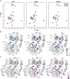
ATP binding increases conformational heterogeneity of gatekeeper mutants. (A) 1H/13C HMQC spectra of WT0Y, V564I0Y, and V564E0Y in the absence (apo state; black) and presence of ATP (20 mM) and Mg2+ (40 mM; purple). Residues highlighted with a box indicate example residues with notable differences between WT0Y and the gatekeeper mutants. (B) Results from CPMG relaxation dispersion experiments and intensity retention calculations plotted on the homology model of autoinhibited FGFR2K. The difference between CPMG relaxation dispersion experiments performed at two frequencies, ΔR2, was calculated as in Eq. 1 and plotted on the homology model of autoinhibited FGFR2K. Significant ΔR2 values are shown from 20 s−1 (blue) to 2 s−1 (light blue). ΔR2 values less than 2 s−1 are shown in white. Peaks with intensity retention (IR) values less than 0.25 from panel (A) are displayed in purple. Hydrophobic R-spine residues are displayed in green; residue labels in bold correspond to the gatekeeper residue and hydrophobic R-spine residues. Note that some residues displayed missing peaks in the CPMG experiment for WT0Y (L647, L675) and V564E0Y (L675) and could not be analyzed; these residues are not displayed in the respective plots. (C) IR values for HMQC spectra from panel (A) plotted on the homology model of autoinhibited FGFR2K. IR values were calculated as the intensities of ATP and Mg2+-bound states divided by the intensities of the apo states. IR values plotted range from 0 (purple) to 1 (white). Hydrophobic R-spine residues are displayed in green; residue labels in bold correspond to the gatekeeper residue and hydrophobic R-spine residues.
Fig. 4.
Fig. 4.
Gatekeeper mutations of FGFR2K enhance the ATP-binding affinity. (A) 1H/13C methyl HMQC spectra of L483 (β1 strand) at three concentrations of ATP using samples of WT0Y (Left), V564I0Y (Middle), and V564E0Y (Right). Full titrations for all peaks are displayed in the supporting information. Note that the L617 peak (22.2 13C ppm and 0.25 1H ppm) was removed from the spectra for clarity. (B). Chemical shift perturbation as a function of ATP concentrations (absolute value shown). 13C chemical shifts were scaled by a factor of 0.25 to account for the 1H chemical shift range. Binding affinity was quantified using a global nonlinear least-square fit to assigned residues experiencing chemical shift perturbations in fast exchange. Individual residues correspond to the symbols as represented below. WT0Y: I548Cδ1, V516, I548Hδ1, L483, V516, L572, L560, L483; V564I0Y: L496, L483, V516, V516, V516, L560, L572; and V564E0Y: V516, L483, I623Cδ1, L483, L560, L603, L603.
Fig. 5.
Fig. 5.
MD simulations reveal disruption of the hydrophobic R-spine within the autoinhibited state. (A and C) The percentage of R-spine formed calculated from MD stimulations every 5 psec starting from the active (A) and autoinhibited states (C) for unphosphorylated wild-type FGFR2K (WT; gray), V564I (blue), V564I (pink), and phosphorylated wild-type FGFR2K (pWT; green). Each bar represents one replicate of 1-μs sampling time. (B and D) Screenshots of representative structures for the gatekeeper (position 564) and hydrophobic R-spine residues (M538, L550, H624, F645) sampled during MD simulations starting from the active (B) and autoinhibited states (D). In each panel, the hydrophobic R-spine residues correspond to the order displayed in the Right panel. Gatekeeper and R-spine residues are shown in sticks and a surface representation.
Fig. 6.
Fig. 6.
Classification of PDB structures and contacts distinguishing active and autoinhibited conformations. (A) Left: Approach to classify FGFR kinase structures using a PCA analysis of the minimum residue distance matrix for conserved residues among the four FGFR isoforms. Right: Crystal structures are plotted on the first two PCs showing two distinct clusters highlighted as autoinhibited and active states. Representative structures are highlighted from each cluster where 3KY2 and 1FGK represent unphosphorylated wild-type FGFR kinases and 3GQI and 2PVF represent phosphorylated wild-type FGFR kinases. 6PNXA and 6PNXB correspond to the substrate-acting and enzyme-acting kinases, respectively, in the A-loop transphosphorylation asymmetric dimer structure of FGFR3 kinase (32). A complete list of PDBs and where they cluster is displayed in SI Appendix, Table S1. (B) Left: Scaled t-value between active and inactive clusters of each distance–distance pair is plotted against the minimum residue distance. Red lines represent the cutoff region used to extract significant autoinhibited and active contacts. Right: Autoinhibited and active contacts are plotted on their representative structure and contacts are highlighted in yellow dotted lines. Key regions in the kinase are colored as follows: αC helix (blue), A-loop (red), and catalytic loop (green).
Fig. 7.
Fig. 7.
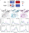
Gatekeeper mutants destabilize the autoinhibited state around the DFG motif and DFG+1 and DFG+2 residues. (A) Heatmap of active or autoinhibited contacts that are formed (red) and broken (blue) during MD simulations. The two heatmap columns refer to simulations starting from the autoinhibited state (Left) or active state (Right). Residue pairs displayed for autoinhibited and active contacts represent the three greatest differences between wild-type and gatekeeper mutants from simulations initiated from the autoinhibited state structure (PDB ID: 3KY2). (B) Representative snapshots of MD simulations of wild-type FGFR2K (gray), V564I (light blue), and V564E (pink) that display contacts formed and disrupted starting from the autoinhibited state. Select residues are displayed that correspond to those identified in panel (A) and those of the DFG motif and gatekeeper position. Black dotted lines represent distances 4.5 Å or less for autoinhibited contacts for the wild-type FGFR2 snapshot and active contacts for the gatekeeper mutation snapshots. (C and D). Density plots corresponding to the minimum residue distance of the indicated autoinhibited (C) or active contact (D) for MD stimulations starting from the autoinhibited conformation. Distances were calculated for each 5 psec of the MD stimulation. Colors of the density plots are displayed within the Right panel of (C).
Fig. 8.
Fig. 8.
Mechanism of gatekeeper mutation activation occurring through the hydrophobic R-spine and DFG motif. (A) F645 chi1 dihedral angle calculated for the autoinhibited (DFGin, inactive) and active (DFGin, active) clusters displayed in Fig. 6A. Two PDBs (4UXQ, 4QRC) within the autoinhibited cluster were excluded from the plot, since they were bound by type II inhibitors and in the DFGout conformation. (B) The percentage of F645 in the autoinhibited conformation (i.e., chi1 angle > 0°) from MD stimulations starting from the autoinhibited state for unphosphorylated wild-type FGFR2K (WT; gray), V564I (blue), V564E (pink), and phosphorylated wild-type FGFR2K (pWT; green). The dihedral angle was calculated every 5 psec of the MD simulation. Each bar in the graph corresponds to one replicate of 1-μs sampling time. (C) Model depicting mutations at the gatekeeper residue (in cyan) disrupting the hydrophobic R-spine (in yellow) of the autoinhibited conformation and influencing the conformation of the DFG phenylalanine (F645). The αC-helix is depicted in gray, the substrate binding site is depicted as a purple oval, and the N- and C-lobes of the kinase are shown as gray circles. The A-loop text in the substrate-binding site forms an autoinhibited interaction, while the A-loop text outside depicts the active conformation of the loop.

Similar articles

Cited by

References

    1. Lemmon M. A., Schlessinger J., Cell signaling by receptor tyrosine kinases. Cell 141, 1117–1134 (2010). - PMC - PubMed
    1. Pon J. R., Marra M. A., Driver and passenger mutations in cancer. Annu. Rev. Pathol. 10, 25–50 (2015). - PubMed
    1. Bailey M. H., et al. , Comprehensive characterization of cancer driver genes and mutations. Cell 174, 1034–1035 (2018). - PMC - PubMed
    1. Sanchez-Vega F., et al. , Oncogenic signaling pathways in the cancer genome atlas. Cell 173, 321–337.e310 (2018). - PMC - PubMed
    1. Torkamani A., Schork N. J., Prediction of cancer driver mutations in protein kinases. Cancer Res. 68, 1675–1682 (2008). - PubMed

Publication types

MeSH terms

Substances