. 2023 Mar 2;127(8):1689-1703.
doi: 10.1021/acs.jpcb.2c07124. Epub 2023 Feb 15.

Inferring Pathways of Oxidative Folding from Prefolding Free Energy Landscapes of Disulfide-Rich Toxins

Affiliations

Inferring Pathways of Oxidative Folding from Prefolding Free Energy Landscapes of Disulfide-Rich Toxins

Rachael A Mansbach et al. J Phys Chem B. .

Abstract

Short, cysteine-rich peptides can exist in stable or metastable structural ensembles due to the number of possible patterns of formation of their disulfide bonds. One interesting subset of this peptide group is the conotoxins, which are produced by aquatic snails in the family Conidae. The μ conotoxins, which are antagonists and blockers of the voltage-gated sodium channel, exist in a folding spectrum: on one end of the spectrum are more hirudin-like folders, which form disulfide bonds and then reshuffle them, leading to an ensemble of kinetically trapped isomers, and on the other end are more BPTI-like folders, which form the native disulfide bonds one by one in a particular order, leading to a preponderance of conformations existing in a single stable state. In this Article, we employ the composite diffusion map approach to study the unified free energy surface of prefolding μ-conotoxin equilibrium. We identify the two most important nonlinear collective modes of the unified folding landscape and demonstrate that in the absence of their disulfides, the conotoxins can be thought of as largely disordered polymers. A small increase in the number of hydrophobic residues in the protein shifts the free energy landscape toward hydrophobically collapsed coil conformations responsible for cysteine proximity in hirudin-like folders, compared to semiextended coil conformations with more distal cysteines in BPTI-like folders. Overall, this work sheds important light on the folding processes and free energy landscapes of cysteine-rich peptides and demonstrates the extent to which sequence and length contribute to these landscapes.


Conflict of interest statement

The authors declare no competing financial interest.

Figures

Figure 1
Figure 1
Folding continuum of the conotoxins described in this work. The Sequential Bond Formation (left) scheme follows a mechanism in which the unfolded peptide exists in a relatively disordered state until two cysteine residues come into proximity with each other and form a native bond. Following this, the native bonds are sequentially formed until the peptide is folded. In contrast, the Collapse and Reshuffle (right) scheme follows a mechanism in which the peptide first undergoes hydrophobic collapse, which puts many cysteine residues into proximity, so non-native bonds form quickly and then undergo reshuffling to achieve the native fold. Locations of conotoxins along the continuum are derived from this work and from Paul George et al.
Figure 2
Figure 2
Multiple sequence alignment of the μ conotoxin sequences considered in this work. Label on the left; sequences in the center, with dashes representing gaps, and sequence length in the rightmost column. In the top row, dashes represent a column with at least one gap, asterisks represent the six cysteines that are forced to align (columns highlighted in red), and periods represent noncysteine sequence alignments with no gap (highlighted in yellow). The alignment was performed with Clustal Omega and modified slightly by hand to enforce cysteines alignment in all columns.
Figure 3
Figure 3
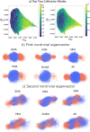
Collective modes identified by the composite diffusion map. In (a), we illustrate the configurations projected into the top two collective modes colored by re2e, end to end length, (left panel) and κ2, relative shape anisotropy (right panel). In (b) and (c), we visualize the two modes through demonstrating the configurations that contribute most strongly to each one. Red corresponds to configurations contributing strongly to one extremum of the mode (low ψ value) while blue corresponds to configurations contributing strongly to the other extremum (high ψ value).
Figure 4
Figure 4
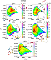
We present the two-dimensional free energy surfaces projected into the two top collective modes of the system using the Pyemma package. Black Xs demonstrate the location of representative frames from disulfide-connected simulations in the native state, projected into the FES using the Nystrom extension., Representative conformations produced with NGLview. We demonstrate two KIIIA conformations from one basin to show that there is some helical content but the basin is not solely comprised of helical conformers. Note that PIIIA’s secondary minimum is occupied by the native state conformers.
Figure 5
Figure 5
Diffusion distances rdiff between free energy minima and average native-state projection into diffusion map coordinates. Each point is calculated as the (a) two-dimensional and (b) one-dimensional distances between the approximate location of the minima identified from the one-dimensional FES fits and the average value of the projected native states calculated from the Nystrom extension, both calculated in terms of the top two eigenmodes of the diffusion map. Error bars represent standard error propagation of the standard deviations reported in Table 4, while the shaded area is calculated from the distance between the lower and upper ends of the ranges shown in Table 3.
Figure 6
Figure 6
Differential population of transient native bonds in free energy landscapes. In this figure, we indicate the portions of the free energy surfaces where cysteines are in close proximity (within a cutoff of 5 Å) and thus bonds might in principle be able to form between them. Black triangles indicate a close proximity between one set of cysteines, and cyan indicates a close proximity between two.
Figure 7
Figure 7
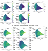
Total contacts. In this plot, we color the two top collective modes by (a) the total number of contacts and (b) the fraction of those total contacts that are native. We observe that, in general, the areas with lower numbers of total contacts have a higher fraction that are native, corresponding to more extended states.
Figure 8
Figure 8
Secondary structure propensities of the μ-conotoxins. Average secondary structure per residue. Secondary structure of reference NMR structure shown above the corresponding residues in each respective plot.
Figure 9
Figure 9
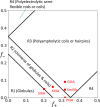
Sequence charge analysis. We plot the fraction of negative charges in each sequence versus the fraction of positive charges in each sequence for the conotoxins (labeled red points) and show where they fall within predefined regions of an approximate phase diagram for intrinsically disordered proteins.

References

    1. Lewis R. J.; Garcia M. L. Therapeutic potential of venom peptides. Nat. Rev. Drug Discovery 2003, 2, 790–802. 10.1038/nrd1197. - DOI - PubMed
    1. Brodie E. D. Toxins and venoms. Curr. Biol. 2009, 19, R931–R935. 10.1016/j.cub.2009.08.011. - DOI - PubMed
    1. Luo L.; Li B.; Wang S.; Wu F.; Wang X.; Liang P.; Ombati R.; Chen J.; Lu X.; Cui J.; et al. Centipedes subdue giant prey by blocking KCNQ channels. Proc. Natl. Acad. Sci. U. S. A. 2018, 115, 1646–1651. 10.1073/pnas.1714760115. - DOI - PMC - PubMed
    1. Luo S.; Zhangsun D.; Harvey P. J.; Kaas Q.; Wu Y.; Zhu X.; Hu Y.; Li X.; Tsetlin V. I.; Christensen S.; et al. Cloning, synthesis, and characterization of αO-conotoxin GeXIVA, a potent α9α10 nicotinic acetylcholine receptor antagonist. Proc. Natl. Acad. Sci. U. S. A. 2015, 112, E4026–E4035. 10.1073/pnas.1503617112. - DOI - PMC - PubMed
    1. Dang B. Chemical synthesis and structure determination of venom toxins. Chin. Chem. Lett. 2019, 30, 1369–1373. 10.1016/j.cclet.2019.03.021. - DOI

Publication types