. 2023 Apr 1;324(4):E347-E357.
doi: 10.1152/ajpendo.00221.2022. Epub 2023 Feb 15.

Thrombospondin-1, CD47, and SIRPα display cell-specific molecular signatures in human islets and pancreata

Affiliations

Thrombospondin-1, CD47, and SIRPα display cell-specific molecular signatures in human islets and pancreata

Neslihan Erdem et al. Am J Physiol Endocrinol Metab. .

Abstract

Thrombospondin-1 (TSP1) is a secreted protein minimally expressed in health but increased in disease and age. TSP1 binds to the cell membrane receptor CD47, which itself engages signal regulatory protein α (SIRPα), and the latter creates a checkpoint for immune activation. Individuals with cancer administered checkpoint-blocking molecules developed insulin-dependent diabetes. Relevant to this, CD47 blocking antibodies and SIRPα fusion proteins are in clinical trials. We characterized the molecular signature of TSP1, CD47, and SIRPα in human islets and pancreata. Fresh islets and pancreatic tissue from nondiabetic individuals were obtained. The expression of THBS1, CD47, and SIRPA was determined using single-cell mRNA sequencing, immunofluorescence microscopy, Western blot, and flow cytometry. Islets were exposed to diabetes-affiliated inflammatory cytokines and changes in protein expression were determined. CD47 mRNA was expressed in all islet cell types. THBS1 mRNA was restricted primarily to endothelial and mesenchymal cells, whereas SIRPA mRNA was found mostly in macrophages. Immunofluorescence staining showed CD47 protein expressed by β cells and present in the exocrine pancreas. TSP1 and SIRPα proteins were not seen in islets or the exocrine pancreas. Western blot and flow cytometry confirmed immunofluorescent expression patterns. Importantly, human islets produced substantial quantities of secreted TSP1. Human pancreatic exocrine and endocrine tissue expressed CD47, whereas fresh islets displayed cell surface CD47 and secreted TSP1 at baseline and in inflammation. These findings suggest unexpected effects on islets from agents that intersect TSP1-CD47-SIRPα.NEW & NOTEWORTHY CD47 is a cell surface receptor with two primary ligands, soluble thrombospondin-1 (TSP1) and cell surface signal regulatory protein alpha (SIRPα). Both interactions provide checkpoints for immune cell activity. We determined that fresh human islets display CD47 and secrete TSP1. However, human islet endocrine cells lack SIRPα. These gene signatures are likely important given the increasing use of CD47 and SIRPα blocking molecules in individuals with cancer.

Keywords: CD47; SIRPα; islets; thrombospondin-1; type 1 diabetes.


Conflict of interest statement

No conflicts of interest, financial or otherwise, are declared by the authors.

Figures

None
Graphical abstract
Figure 1.
Figure 1.
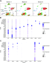
Single-cell expression of CD47, THBS1, and SIRPA mRNA in human islets. UMAP plots of cell clusters of each donor islet preparation. Graphs display donor cells, color-coded for each islet cell population (1,745 cells for donor 1, 4,334 cells for donor 2, and 5,441 cells for donor 3) (A). DotPlots show relative gene expression for each cell population shown on the y-axis [islet marker genes (B); CD47, THBS1, SIRPA, CD274, PDCD1, CTLA4, B2M, and HLA-DRA (C)]. DotPlot graphs display the combined data from all three donors. Dot size indicates the percentage of cells with detectable transcript and color indicates the average expression of each gene by z score. The UMAP and DotPlot graphs were generated using Seurat and RStudio (2022.02.2 Build 485 version). UMAP, uniform manifold approximation and projection.
Figure 2.
Figure 2.
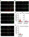
CD47 protein is expressed in the endocrine and exocrine compartments of human pancreata. Human pancreas tissue sections were costained for insulin and CD47 (A), TSP1 (B), or SIRPα (C). Confocal images were taken at ×10 and ×20 magnification with tile scanning. Enlarged images in the boxed areas are shown on the right. Quantification of the fluorescence intensity relative to the negative control (secondary antibody only staining) was analyzed for islets (D) or nonislet areas (E) using ImageJ software. One slide from each donor organ (total of 3 donors) was examined. Three to four separate areas of the tissue sample were characterized for each slide. Five or more islets were counted in each area. The scale bar in tile scanning images equals 100 µm and in enlarged images equals 50 µm. Human pancreas tissue sections from a single donor (Hu1172) were costained for TSP1, CD47, and SIRPα and confocal images were acquired at ×40 magnification and are shown on the far right of each panel (F). Images at ×40 magnification from tissue samples from 3 donors were quantified for protein expression in acinar and ductal cells (G). Data are presented as the means ± SD. Each dot represents an islet or the nonislet area of each donor tissue section. SIRPα, signal regulatory protein α; TSP1, thrombospondin-1.
Figure 3.
Figure 3.
CD47, but not TSP1 and SIRPα, protein is found in human islet lysate. Lysates of islet preparations from donors without diabetes (n = 5, Hu 1186, Hu 1199, Hu 1228, Hu 1229, and Hu 1232) underwent separation by gel electrophoresis and protein expression was characterized by Western blot. Representative Western blot showing CD47, TSP1, and SIRPα expression (A). Densitometry analysis of CD47, TSP1, and SIRPα expression relative to β-actin (B). Each dot represents a donor. Data are presented as the means ± SD. Significance was determined using one-way ANOVA for multiple comparisons. SIRPα, signal regulatory protein α; TSP1, thrombospondin-1.
Figure 4.
Figure 4.
CD47 resides on the surface of islet cells. Islets from donors without diabetes (n = 3, Hu 1232, Hu 1237, and Hu 1239) were dissociated and stained with fluorochrome-conjugated anti-CD47 or anti-SIRPα antibodies and analyzed by flow cytometry. Gating strategy for single live cells (A). Representative analysis of CD47- (B) and SIRPα-positive cells (C) after sequential gating from A. Positive thresholds were set according to isotype controls. Quantification of the % CD47- and SIRPα-positive cells (D). Each dot represents one donor. Data are presented as the means ± SD. Significance was determined using paired t tests. **P ≤ 0.01. SIRPα, signal regulatory protein α.
Figure 5.
Figure 5.
Human islets secrete soluble TSP1 and express cell surface CD47, which is maintained under inflammatory stress. Human islets (500 IEQ) from donors without diabetes (n = 4, Hu 1229, Hu 1236, Hu 1237, and Hu 1238) were incubated in islet culture medium with or without IL-1β (50 U/mL), IFN-γ (1,000 U/mL), and TNF-α (1,000 U/mL) for 24 h. Islet lysates were prepared, protein separated by gel electrophoresis and expression of CD47, TSP1, SIRPα, and PD-L1 characterized by Western blot. Representative Western blot showing protein expression (A). Densitometry analysis of expression relative to β-actin (B). Conditioned medium from freshly gathered human islets at baseline and following exposure to inflammatory cytokines for 24 h (n = 4 four donor preparations) was assayed for secreted soluble TSP1 (C). Each dot represents one donor. Significance was determined using paired t test. ns, P >0.05, ***P < 0.001. SIRPα, signal regulatory protein α; TSP1, thrombospondin-1.

References

    1. Kaur S, Soto-Pantoja DR, Stein EV, Liu C, Elkahloun AG, Pendrak ML, Nicolae A, Singh SP, Nie Z, Levens D, Isenberg JS, Roberts DD. Thrombospondin-1 signaling through CD47 inhibits self-renewal by regulating c-Myc and other stem cell transcription factors. Sci Rep 3: 1673, 2013. doi:10.1038/srep01673. - DOI - PMC - PubMed
    1. Roberts DD, Isenberg JS. CD47 and thrombospondin-1 regulation of mitochondria, metabolism, and diabetes. Am J Physiol Cell Physiol 321: C201–C213, 2021. doi:10.1152/ajpcell.00175.2021. - DOI - PMC - PubMed
    1. Isenberg JS, Ridnour LA, Perruccio EM, Espey MG, Wink DA, Roberts DD. Thrombospondin-1 inhibits endothelial cell responses to nitric oxide in a cGMP-dependent manner. Proc Natl Acad Sci USA 102: 13141–13146, 2005. doi:10.1073/pnas.0502977102. - DOI - PMC - PubMed
    1. Isenberg JS, Hyodo F, Matsumoto K-I, Romeo MJ, Abu-Asab M, Tsokos M, Kuppusamy P, Wink DA, Krishna MC, Roberts DD. Thrombospondin-1 limits ischemic tissue survival by inhibiting nitric oxide-mediated vascular smooth muscle relaxation. Blood 109: 1945–1952, 2007. doi:10.1182/blood-2006-08-041368. - DOI - PMC - PubMed
    1. Csanyi G, Yao M, Rodriguez AI, Al Ghouleh I, Sharifi-Sanjani M, Frazziano G, Huang X, Kelley EE, Isenberg JS, Pagano PJ. Thrombospondin-1 regulates blood flow via CD47 receptor-mediated activation of NADPH oxidase 1. Arterioscler Thromb Vasc Biol 32: 2966–2973, 2012. doi:10.1161/ATVBAHA.112.300031. - DOI - PMC - PubMed

Publication types

MeSH terms