. 2023 Jun:99:102324.
doi: 10.1016/j.npep.2023.102324. Epub 2023 Feb 2.

Sex-dimorphic hindbrain lactate regulation of ventromedial hypothalamic nucleus glucoregulatory neuron 5'-AMP-activated protein kinase activity and transmitter marker protein expression

Affiliations

Sex-dimorphic hindbrain lactate regulation of ventromedial hypothalamic nucleus glucoregulatory neuron 5'-AMP-activated protein kinase activity and transmitter marker protein expression

Prabhat R Napit et al. Neuropeptides. 2023 Jun.

Abstract

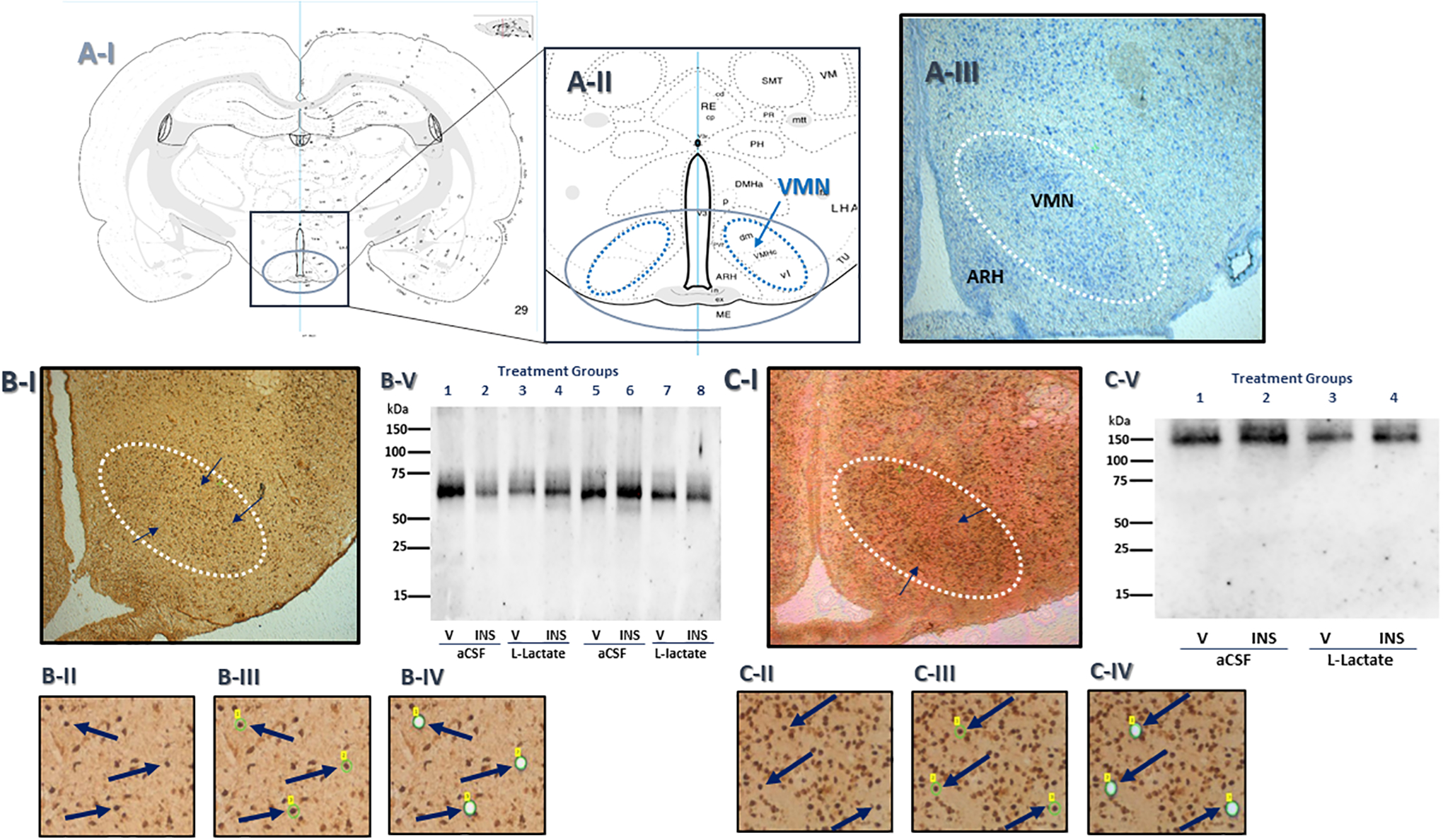
Background: The oxidizable glycolytic end-product L-lactate is a gauge of nerve cell metabolic fuel stability that metabolic-sensory hindbrain A2 noradrenergic neurons impart to the brain glucose-regulatory network. Current research investigated the premise that hindbrain lactate deficiency exerts sex-specific control of energy sensor and transmitter marker protein responses to hypoglycemia in ventromedial hypothalamic nucleus (VMN) glucose-regulatory nitrergic and γ-aminobutyric acid (GABA) neurons.

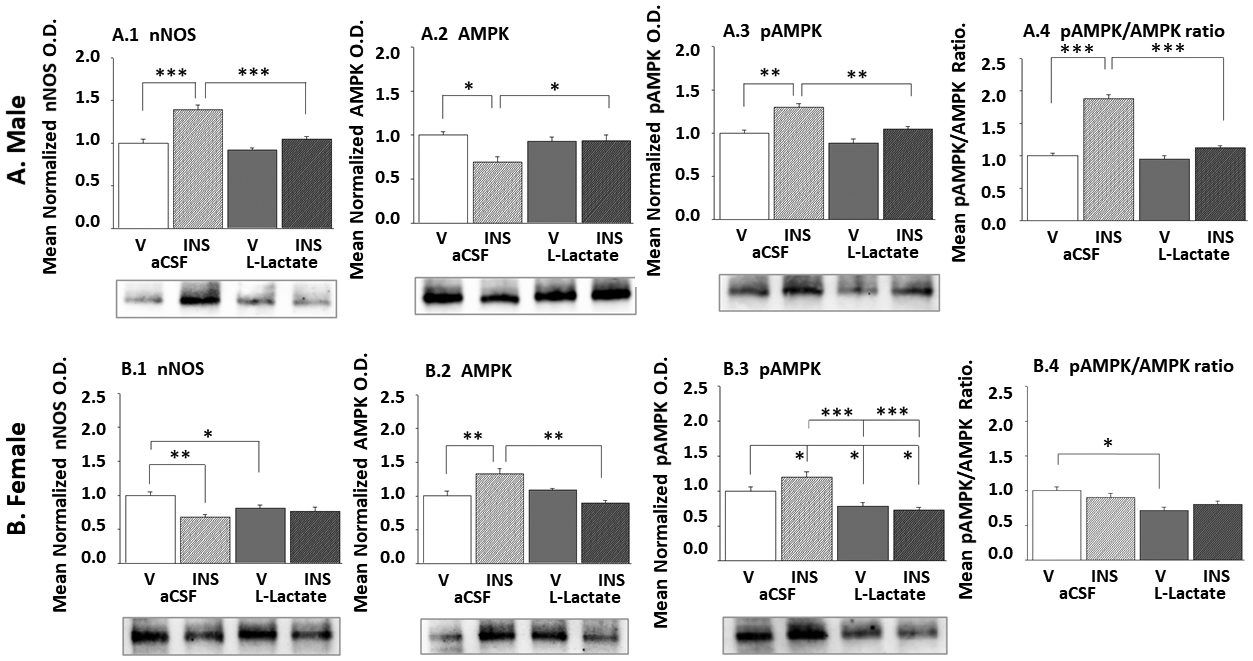
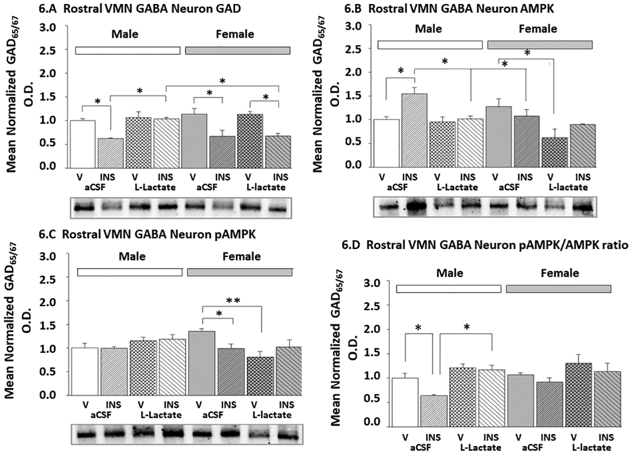
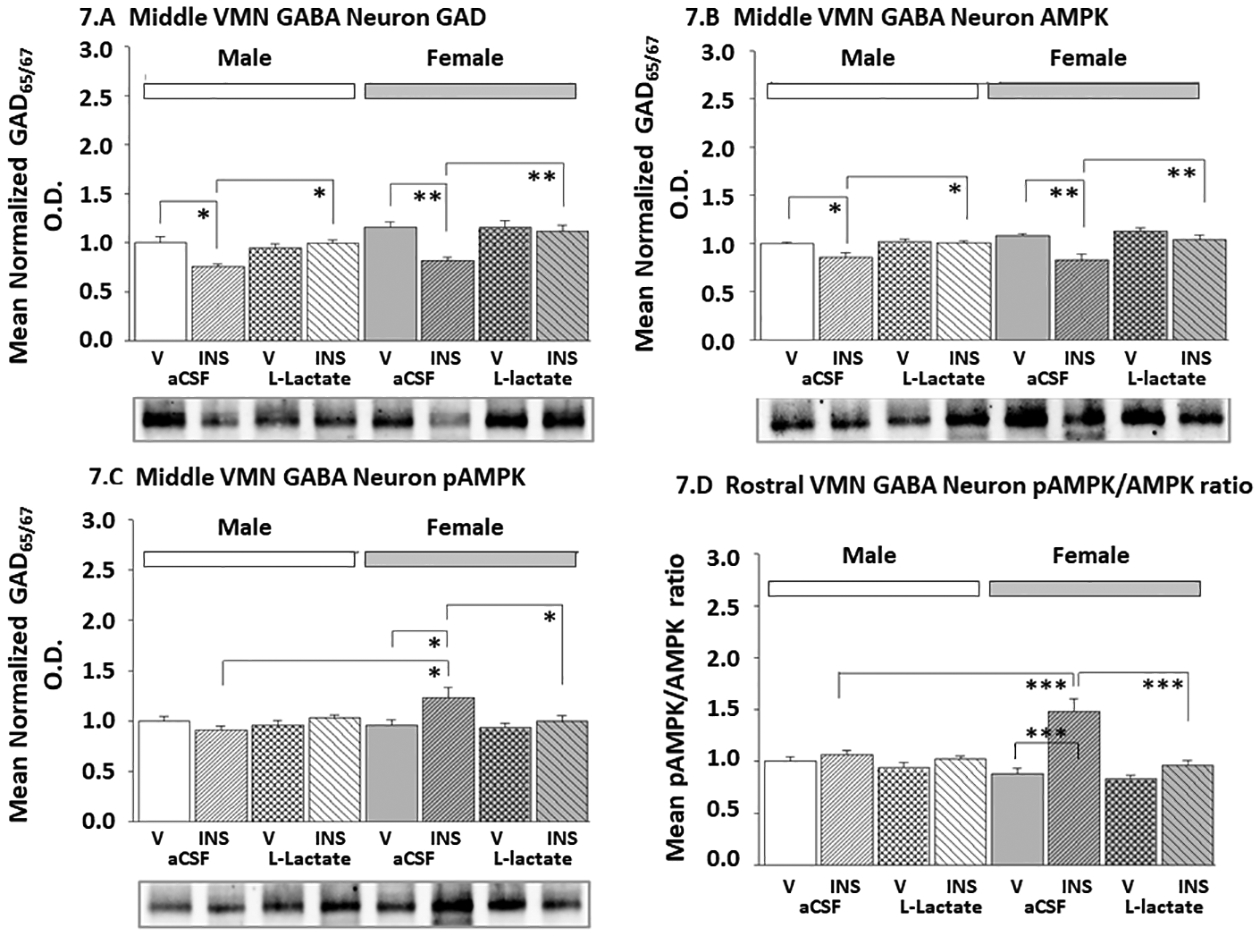
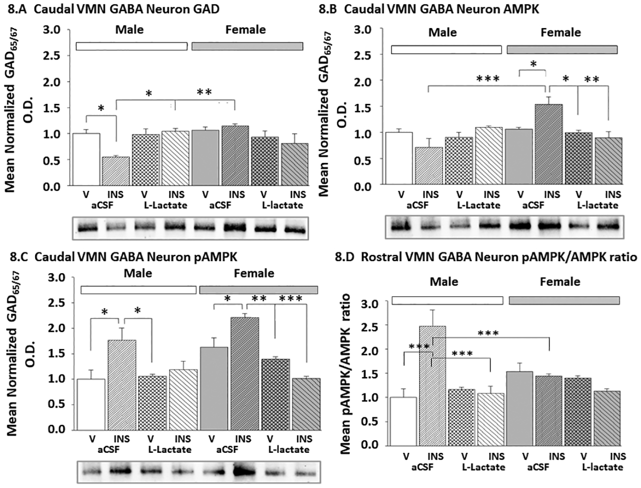
Methods: Nitric oxide synthase (nNOS)- or glutamate decarboxylase65/67 (GAD)-immunoreactive neurons were laser-catapult-microdissected from male and female rat VMN after subcutaneous insulin injection and caudal fourth ventricular L-lactate or vehicle infusion for Western blot protein analysis.

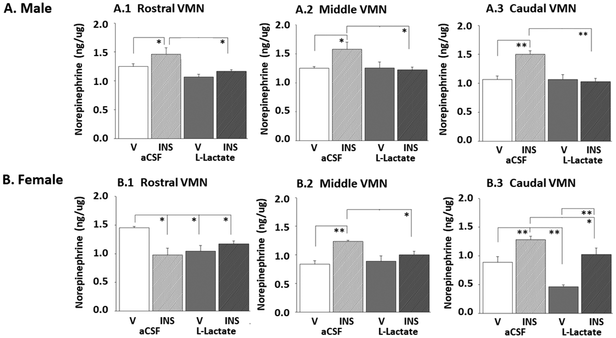
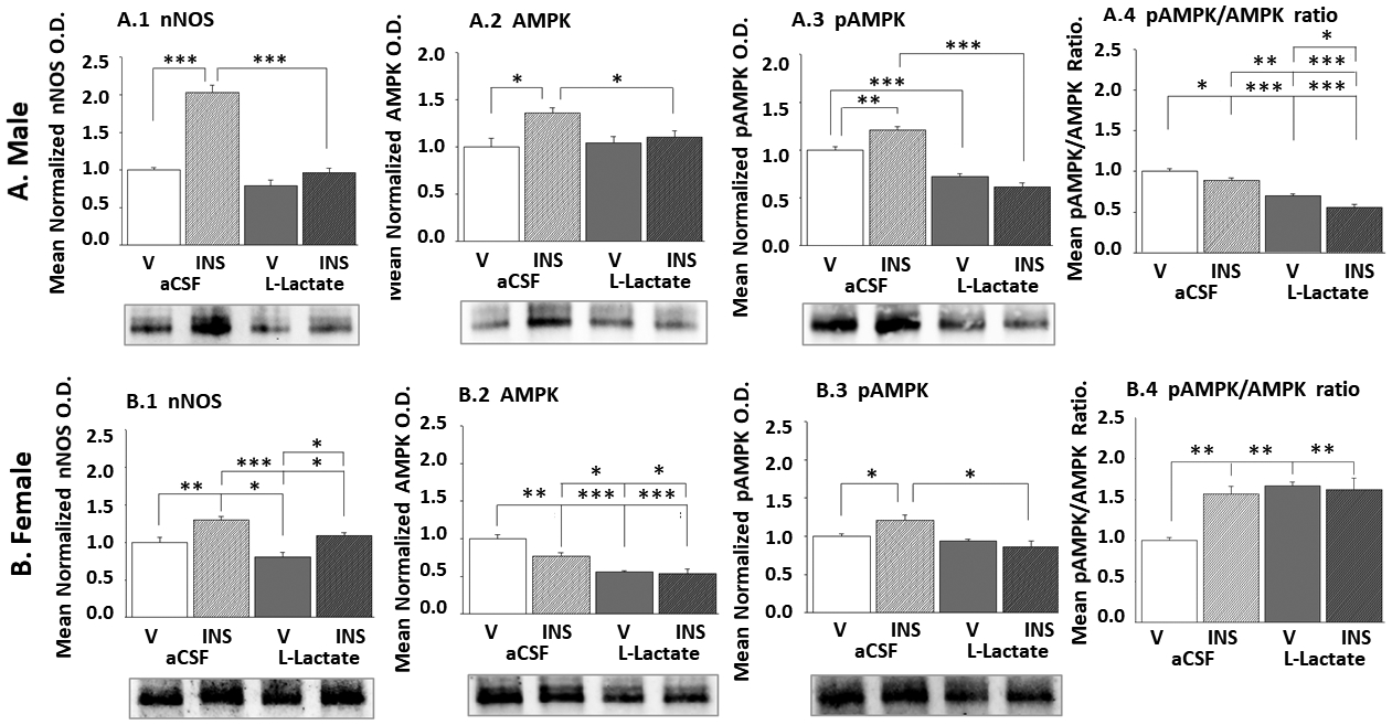
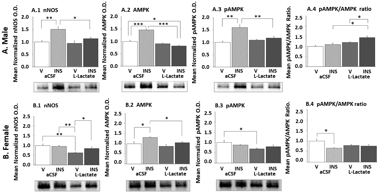
Results: Hindbrain lactate repletion reversed hypoglycemia-associated augmentation (males) or inhibition (females) of nitrergic neuron nNOS expression, and prevented up-regulation of phosphorylated AMPK 5'-AMP-activated protein kinase (pAMPK) expression in those neurons. Hypoglycemic suppression of GABAergic neuron GAD protein was averted by exogenous lactate over the rostro-caudal length of the male VMN and in the middle region of the female VMN. Lactate normalized GABA neuron pAMPK profiles in hypoglycemic male (caudal VMN) and female (all VMN segments) rats. Hypoglycemic patterns of norepinephrine (NE) signaling were lactate-dependent throughout the male VMN, but confined to the rostral and middle female VMN.

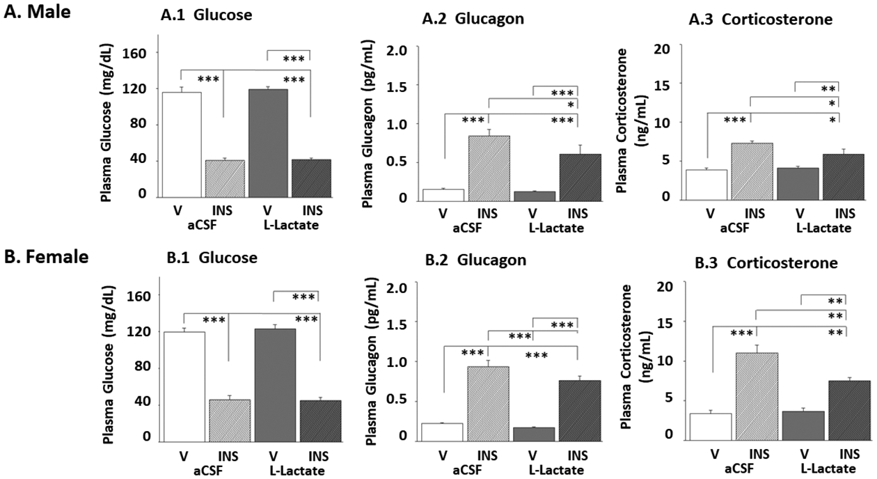
Conclusions: Results document, in each sex, regional VMN glucose-regulatory transmitter responses to hypoglycemia that are controlled by hindbrain lactate status. Hindbrain metabolic-sensory regulation of hypoglycemia-correlated nitric oxide or GABA release may entail AMPK-dependent mechanisms in specific VMN rostro-caudal segments in each sex. Additional effort is required to examine the role of hindbrain lactoprivic-sensitive VMN neurotransmitters in lactate-mediated attenuation of hypoglycemic hyperglucagonemia and hypercorticosteronemia in male and female rats.

Keywords: AMPK; Corticosterone; L-lactate; Nitric oxide; Norepinephrine; Sex differences.


Conflict of interest statement

Declaration of Competing Interest The authors have no conflicts of interests to declare.

Figures

Figure 1.
Figure 1.. Laser-Catapult Microdissection of Immunolabeled Ventromedial Hypothalamic Nucleus (VMN) γ-Aminobutyric Acid (GABA) or Nitric Oxide (NO) Neurons.
Rat brain coronal brain sections illustrated in Panels A-I and A-II depict the orientation of the VMN (bilateral oblong ovals; dotted blue lines) within the mediobasal hypothalamus (midline oval; solid blue line). The methylene blue-stained tissue section in Panel A-III shows the demarcation of the VMN (white dotted-line oval) from surrounding structures, including the arcuate hypothalamic nucleus (ARH). Characteristic distribution pattern of glutamate decarboxylase65/67 (GAD)-ir-positive neuron perikarya (black arrows) within the VMN is shown in Panel B-I. The area depicted in Panel B-II featuring three representative GAD-ir neuron (each indicated by a blue arrow) was re-photographed after positioning of a continuous laser track (depicted in green) [Panel B-III] around each of those nerve cells, followed by subsequent ejection of those individual GAD-ir neurons by laser pulse [Panel B-IV]. Note that this microdissection technique causes negligible destruction of surrounding tissue and minimal inclusion of adjacent tissue. The whole immunoblot in Panel B-V shows that the GAD primary antiserum used for immunostaining detects a single band of antigen of predicted molecule weight in laser-microdissected VMN GAD-ir nerve cell lysates. Panel CI illustrates immunostaining for neuronal nitric oxide synthase (nNOS) in the VMN. Panels CII-CIV depict laser-assisted collection of individual VMN nNOS-ir neurons (indicated by blue arrows). The whole immunoblot in Panel C-V denotes specific detection of nNOS protein of expected molecular weight in VMN nNOS-ir cell lysates by the primary antiserum used for immunocytochemical labeling.
Figure 2.
Figure 2.. Effects of Caudal Fourth Ventricular (CV4) L-Lactate Infusion on Ventromedial Hypothalamic Nucleus (VMN) Norepinephrine (NE) Tissue Content in Eu- and Hypoglycemic Male and Female Rats.
Bilateral micropunch-dissected VMN tissue was taken from 100 μm-thick sections cut through the rostral (Figures A.1, B.1), middle (Figures A.2, B.2), and caudal (Figures A.3, B.3) VMN of vehicle (V)- or insulin (INS)-injected male (top row) or female (bottom row) rats infused into the CV4 with L-lactate or vehicle (artificial cerebrospinal fluid, aCSF), and pooled with treatment groups for ELISA analysis of NE content. Data were analyzed by three-way ANOVA and Student-Newman-Kuels post-hoc test. Within each VMN segment, data show for each sex mean NE values ± SE for the following treatment groups: aCSF/V (white bars; n=6 males, n=6 females), aCSF/INS (diagonal-striped white bars; n=6 males, n=6 females), L-lactate/V (solid gray bars; n=6 males, n=6 females), L-lactate/INS (diagonal-striped gray bars; n=6 males, n=6 females). Post-hoc statistical comparison of treatment group means: *p<0.05; **p<0.01; ***p<0.001. Data show that lactate infusion suppressed baseline NE activity in a segment-specific manner in the euglycemic female, but not male rat VMN. IIH significantly increased NE levels in each segment of the male VMN, yet lactate prevented this stimulatory response in all regions. However, hypoglycemic females exhibited down- or up-regulation of NE content, according to VMN region; in this sex, rostral and middle, but not caudal VMN patterns of hypoglycemia-associated NE activity were abolished by hindbrain lactate infusion.
Figure 3.
Figure 3.. Impact of Hindbrain L-Lactate on Rostral VMN NO Neuron Transmitter Biomarker and 5’-AMP-Activated Protein Kinase (AMPK) Protein Expression in Male and Female Rats.
Pooled lysates of laser-catapult-microdissected rostral VMN neuronal nitric oxide synthase (nNOS)-immunopositive neurons were analyzed by Western blot for nNOS (Figures A.1, B.2), AMPK (Figures A.2, B.2), or phosphoAMPK (pAMPK; Figures A.3, B.3) protein expression. Data were analyzed by three-way ANOVA and Student-Newman-Kuels post-hoc test. Results show mean normalized protein O.D. measures ± SE for aCSF/V (white bars; n=6 males, n=6 females), aCSF/INS (diagonal-striped white bars; n=6 males, n=6 females), L-lactate/V (solid gray bars; n=6 males, n=6 females), L-lactate/INS (diagonal-striped gray bars; n=6 males, n=6 females) groups of male (top row) or female (bottom row) rats. Figures A.4 and B.4 show mean values ± SE for the nitrergic nerve cell pAMPK/AMPK ratio for male and female rat treatment groups, respectively (n=6 males, n=6 females). *p<0.05; **p<0.01; ***p<0.001. Data show that each sex, insulin increased nNOS protein levels in rostral VMN NO neurons; this stimulatory response was abolished by hindbrain lactate infusion in male, but not female rats. Rostral VMN nitrergic neurons from hypoglycemic males or females exhibited lactate-reversible up- or down-regulation of AMPK protein content. Hypoglycemia also produced hindbrain lactoprivic-dependent augmentation of pAMPK expression in these cells
Figure 4.
Figure 4.. Transmitter Biomarker and AMPK Protein Expression in Nitrergic Neurons Acquired from the Middle Region of Male or Female Rat VMN.
Data were analyzed by three-way ANOVA and Student-Newman-Kuels post-hoc test. Outcomes are presented as mean normalized nNOS (Figures A.1, B.1), AMPK (Figures A.2, B.2) or pAMPK (Figures A.3, B.3) protein O.D. measures ± SE for male (top row) or female (bottom row) rat aCSF/V (n=6 males, n=6 females); aCSF/INS (n=6 males, n=6 females); L-lactate/V (n=6 males, n=6 females);, and L-lactate/INS treatment groups (n=6 males, n=6 females). Figures A.4 and B.4 show mean nitrergic nerve cell pAMPK/AMPK ratio values ± SE for male and female rat treatment groups, respectively. *p<0.05; **p<0.01; ***p<0.001. Data show that lactate infusion to euglycemic female, but not male rats suppressed nNOS and pAMPK proteins in middle VMN NO neurons. Effects of hypoglycemia on nNOS profiles in this VMN segment varied between sexes, as levels were increased in the male (an effect reversed by lactate), yet decreased in the female. Middle VMN nitrergic neurons exhibited lactate-reversible up-regulation of AMPK by hypoglycemia in each sex, but lactate-dependent enhancement of pAMPK expression in males only.
Figure 5.
Figure 5.. Hindbrain L-Lactate-Sensitive Patterns of nNOS and AMPK Protein Expression in Caudal VMN NO Neurons of Each Sex.
Lysate pools of caudal VMN nNOS-immunopositive neurons were analyzed by Western blot for nNOS (Figures A.1, B.2), AMPK (Figures A.2, B.2), or phosphoAMPK (pAMPK; Figures A.3, B.3) protein expression. Data were evaluated by three-way ANOVA and Student-Newman-Kuels post-hoc test. Figures depict mean normalized protein O.D. measures ± SE for groups of male (top row) or female (bottom row) rats treated by aCSF/V (n=6 males, n=6 females); aCSF/INS (n=6 males, n=6 females); L-lactate/V (n=6 males, n=6 females); or L-lactate/INS (n=6 males, n=6 females). Figures A.4 and B.4 show mean values for the nitrergic nerve cell pAMPK/AMPK ratio ± SE for male and female rat treatment groups, respectively. *p<0.05; **p<0.01; ***p<0.001. Data show that during hypoglycemia, caudal VMN nitrergic nerve cell nNOS content was increased in males, yet decreased in females due to hindbrain lactate shortage. Lactate deprivation down-(male) or up-(female) regulated AMPK profiles during hypoglycemia, yet amplified pAMPK expression in NO neurons in the caudal VMN segment in each sex.
Figure 6.
Figure 6.. Hindbrain L-Lactate Infusion Effects on Rostral VMN GABAergic Neuron Transmitter Biomarker and AMPK Protein Expression in Rats of Each Sex.
Rostral VMN glutamate decarboxylase 5/67 (GAD)-immunolabeled nerve cell GAD (Figure A.1), AMPK (Figure A.2), or pAMPK (Figure A.3) protein content was measured after V or INS injection of male and female rats. Data were analyzed by three-way ANOVA and Student-Newman-Kuels post-hoc test. Results are depicted as mean normalized protein O.D. measures ± SE for male (white bars) or female (gray bars) rats treated by aCSF/V (n=6 males, n=6 females); aCSF/INS (n=6 males, n=6 females); L-lactate/V (n=6 males, n=6 females); or L-lactate/INS (n=6 males, n=6 females). Figure A.4 depicts mean values ± SE for the nitrergic nerve cell pAMPK/AMPK ratio for the above treatment groups. *p<0.05; **p<0.01; ***p<0.001. Data show that baseline GAD protein levels did not differ between V-injected euglycemic male versus female rats in the rostral VMN. In each sex, hypoglycemia suppressed GAD protein levels in GABA neuron in this VMN segment; this inhibitory response was averted by lactate in males only. Hypoglycemia-associated lactate scarcity increased AMPK content in these cells in male, yet inhibited pAMPK content in the female.
Figure 7.
Figure 7.. Patterns of Middle VMN GABAergic Neuron Transmitter Biomarker and AMPK Protein Expression in Hindbrain L-Lactate-Infused Rats.
Middle VMN GABAergic nerve cell GAD (Figure A.1), AMPK (Figure A.2), or pAMPK (Figure A.3) protein content was measured after V or INS injection of male and female rats. Data were analyzed by three-way ANOVA and Student-Newman-Kuels post-hoc test, and are presented as mean normalized protein O.D. measures ± SE for male (white bars) or female (gray bars) rats treated by aCSF/V (n=6 males, n=6 females); aCSF/INS (n=6 males, n=6 females); L-lactate/V (n=6 males, n=6 females); or L-lactate/INS (n=6 males, n=6 females). Figure A.4 depicts mean values ± SE for the nitrergic nerve cell pAMPK/AMPK ratio for the above treatment groups. *p<0.05; **p<0.01; ***p<0.001. Results show that hypoglycemia caused hindbrain lactate-reversible suppression of GAD protein expression in GABA neuron in this segment of the VMN. Lactoprivic signaling decreased AMPK content in these cells in each sex, yet up-regulated pAMPK profiles in the female alone.
Figure 8.
Figure 8.. Caudal VMN GABAergic Neuron Transmitter Biomarker and AMPK Protein Expression in Hindbrain L-Lactate-Infused Rats.
Outcomes, analyzed by three-way ANOVA and Student-Newman-Kuels post-hoc test, are presented as mean normalized GAD (Figure A.1), AMPK (Figure A.2), or pAMPK (Figure A.3) protein content protein O.D. measures ± SE for caudal VMN neurons collected from male (white bars) or female (gray bars) rats treated by aCSF/V (n=6 males, n=6 females); aCSF/INS (n=6 males, n=6 females); L-lactate/V (n=6 males, n=6 females); or L-lactate/INS (n=6 males, n=6 females). Figure A.4 depicts mean values ± SE for the nitrergic nerve cell pAMPK/AMPK ratio for the above treatment groups. *p<0.05; **p<0.01; ***p<0.001. Data show that hypoglycemia decreased caudal VMN GABAergic nerve cell GAD content in male, but not female, and that associated lactate deficiency mediates this sex-specific suppressive effect. During hypoglycemia, these neurons exhibit lactate-reversible up-regulation of AMPK levels in females and pAMPK expression in each sex.
Figure 9.
Figure 9.. Plasma Glucose and Counterregulatory Hormone Responses to INS Injection in L-Lactate-Infused Male and Female Rats.
Results, analyzed by three-way ANOVA and Student-Newman-Kuels post-hoc test, are depicted as mean glucose (Figures A.1, B.1), glucagon (Figures A.2, B.2), and corticosterone (Figures A.3, B.3) concentrations ± SE for male (top row) and female (bottom row) rats treated by aCSF/V (white bars; n=6 males, n=6 females); aCSF/INS (diagonal-striped white bars; n=6 males, n=6 females), L-lactate/V (solid gray bars; n=6 males, n=6 females), or L-lactate/INS (diagonal-striped gray bars; n=6 males, n=6 females). *p<0.05; **p<0.01; ***p<0.001. Outcomes reveal that in each sex, INS injection suppressed circulating glucose levels to an equivalent extent in lactate- versus V-infused animals. Hindbrain lactate infusion did not alter glucagon or corticosterone release in euglycemic male or female rats, but attenuated stimulatory effects of hypoglycemia on hormone output in each sex.

Similar articles

Cited by

References

    1. Laming PR, Kimelberg H, Robinson S, Salm A, Hawrylak N, Muller C, et al. Neuronal-glial interactions and behavior. Neurosci. Biobehav. Rev 2000; 24: 295–340. doi: 10.1016/s0149-7634(99)00080-9. - DOI - PubMed
    1. Bröer S, Rahman B, Pellegri G, Pellerin L, Martin JL, Verleysdonk S, et al. Comparison of lactate transport in astroglial cells and monocarboxylate transporter 1 (MCT 1) expressing Xenopus laevis oocytes. Expression of two different monocarboxylate transporters in astroglial cells and neurons. J. Biol. Chem 1997; 272: 30096–30102. doi: 10.1074/jbc.272.48.30096. - DOI - PubMed
    1. Patil GD, Briski KP. Lactate is a critical ‘sensed’ variable in caudal hindbrain monitoring of CNS metabolic stasis. Amer. J. Physiol. Regul. Integr. Comp. Physiol 2005; 289: R1777–86. - PubMed
    1. Vavaiya KV, Briski KP. Effects of caudal hindbrain lactate infusion on insulin-induced hypoglycemia and neuronal substrate transporter glucokinase and sulfonylurea receptor-1 gene expression in the ovariectomized female rat dorsal vagal complex: Impact of estradiol. J. Neurosci Res 2008; 86(3): 694–701. doi: 10.1002/jnr.21530. - DOI - PubMed
    1. Vavaiya KV, Briski KP. Caudal hindbrain lactate infusion alters glucokinase, SUR1, and neuronal substrate fuel transporter gene expression in the dorsal vagal complex, lateral hypothalamic area, and ventromedial nucleus hypothalamus of hypoglycemic male rats. Brain Res 2007; 1176: 62–70. doi: 10.1016/j.brainres.2007.08.010. - DOI - PubMed