. 2023 Mar 15;435(6):168012.
doi: 10.1016/j.jmb.2023.168012. Epub 2023 Feb 13.

Self-inhibited State of Venezuelan Equine Encephalitis Virus (VEEV) nsP2 Cysteine Protease: A Crystallographic and Molecular Dynamics Analysis

Affiliations

Self-inhibited State of Venezuelan Equine Encephalitis Virus (VEEV) nsP2 Cysteine Protease: A Crystallographic and Molecular Dynamics Analysis

Gyula Hoffka et al. J Mol Biol. .

Abstract

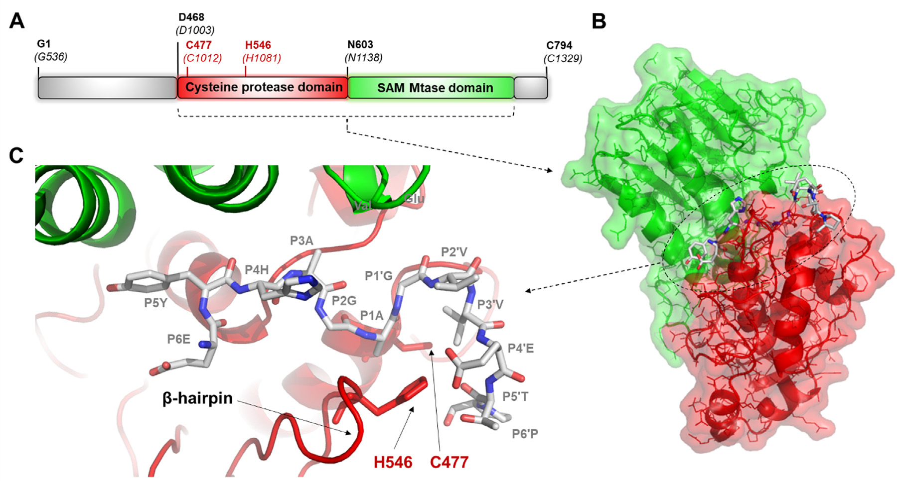
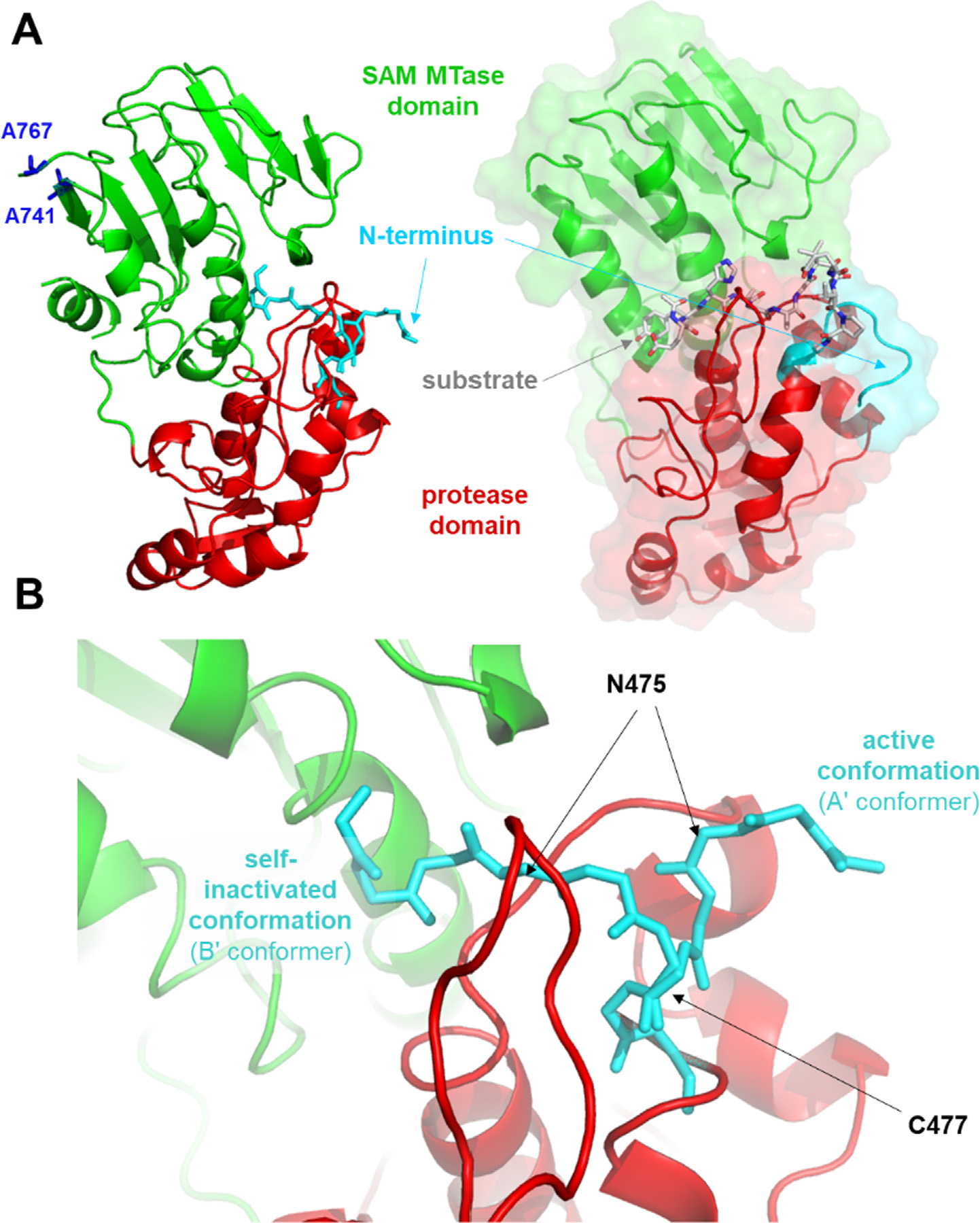
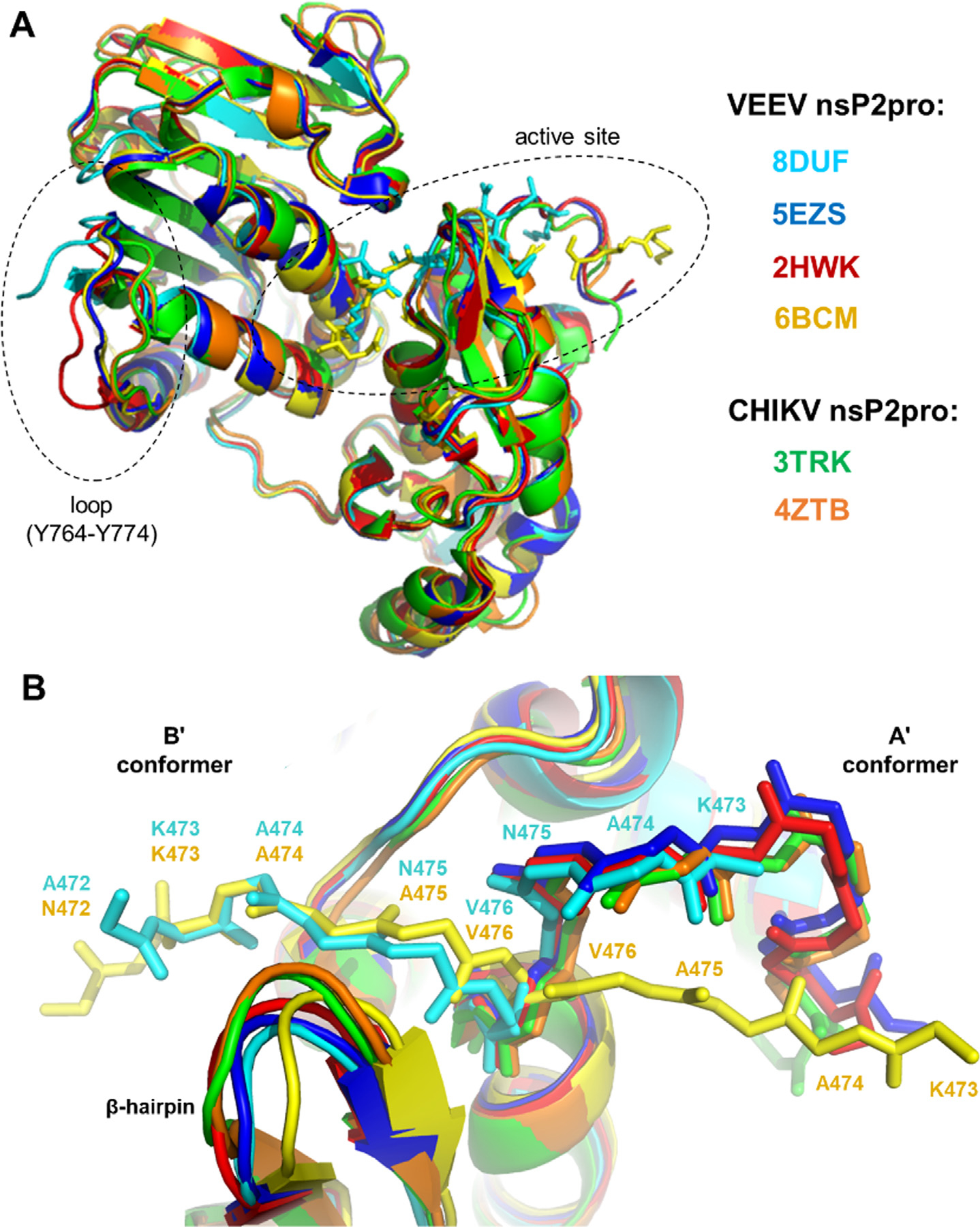
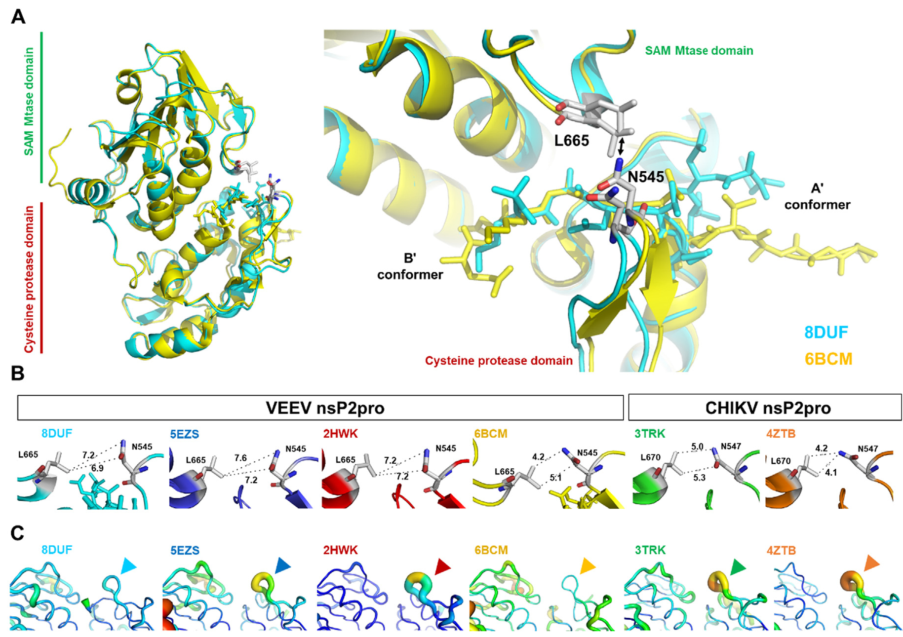
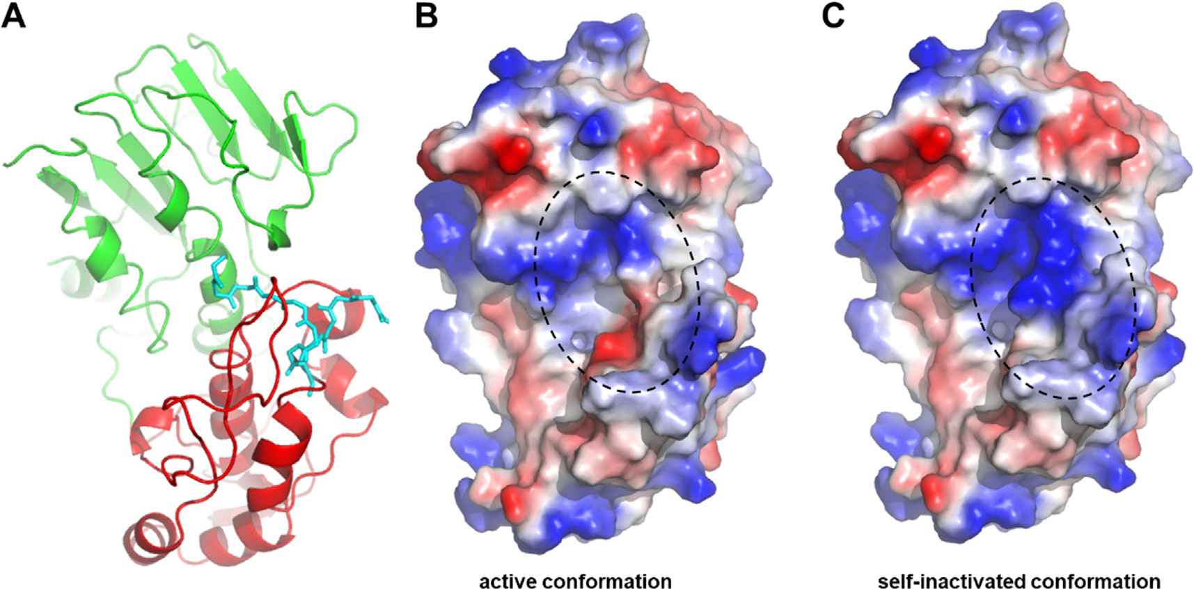
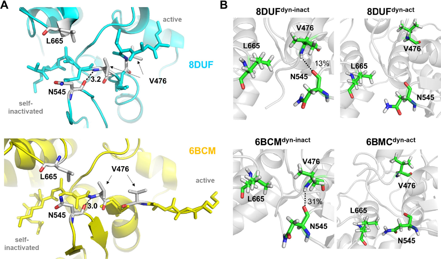
The Venezuelan equine encephalitis virus (VEEV) belongs to the Togaviridae family and is pathogenic to both humans and equines. The VEEV non-structural protein 2 (nsP2) is a cysteine protease (nsP2pro) that processes the polyprotein and thus it is a drug target for inhibitor discovery. The atomic structure of the VEEV nsP2 catalytic domain was previously characterized by both X-ray crystallography and computational studies. A modified nsP2pro harboring a N475A mutation in the N terminus was observed to exhibit an unexpected conformation: the N-terminal residues bind to the active site, mimicking binding of a substrate. The large conformational change of the N terminus was assumed to be induced by the N475A mutation, as N475 has an important role in stabilization of the N terminus and the active site. This conformation was first observed in the N475A mutant, but we also found it while determining a crystal structure of the catalytically active nsP2pro containing the wild-type N475 active site residue and K741A/K767A surface entropy reduction mutations. This suggests that the N475A mutation is not a prerequisite for self-inhibition. Here, we describe a high resolution (1.46 Å) crystal structure of a truncated nsP2pro (residues 463-785, K741A/K767A) and analyze the structure further by molecular dynamics to study the active and self-inhibited conformations of nsP2pro and its N475A mutant. A comparison of the different conformations of the N-terminal residues sheds a light on the interactions that play an important role in the stabilization of the enzyme.

Keywords: Venezuelan equine encephalitis virus; alphavirus; crystallography; molecular dynamics; protease.


Conflict of interest statement

Declaration of Competing Interest The authors declare that they have no known competing financial interests or personal relationships that could have appeared to influence the work reported in this paper.

Figures

Figure 1.
Figure 1.. Complex of VEEV nsP2pro and EYHAGAGVVETP oligopeptide substrate.
(a) The domain organization of the VEEV nsP2pro. Residues are labeled according to the numbering of nsP2 and that of the nsP1234 polyprotein (in parentheses) (UniProt ID: P27282). Aligned sequences of VEEV, SINV and CHIKV polyproteins are shown in Figure S1. (b) The overall structure of the protease bound to a peptide substrate is shown based on the enzyme-substrate complex that was modeled previously. (c) Enlarged view of the active site with the modeled position of the EYHAGAGVVETP oligopeptide substrate (P6-P6′ residues).
Figure 2.
Figure 2.. Overall structure and active site of the K741A/K767A mutant VEEV nsP2pro.
(a) The active and self-inhibited conformations of the K741A/K767A mutant VEEV nsP2pro (PDB code: 8DUF) are shown. For comparison, the modeled complex of the protease and the EYHAGAGVVETP oligopeptide substrate (P6-P6′ residues) is also shown. (b) Enlarged view of the active site showing the different conformational states of the N terminus. The main chain atoms of the 472AKANVCW478 residues are shown.
Figure 3.
Figure 3.. Comparison of nsP2pro structures.
(a) Alignment of overall nsP2pro structures. The different structures are shown by different colors, the color code is indicated in the figure. The active site and the loop for which the electron density was visible only in part are circled by dashed lines. (b) Comparison of N termini in the A’ (active) and B’ (self-inactivated) conformers. The backbone of 472–477 sequence motif is shown by sticks, the residues are labeled.
Figure 4.
Figure 4.. Comparison of subdomain interfaces in nsP2pro crystal structures.
(a) Overall structure of VEEV nsP2pro representing the A’ and B’ conformers, the L665 and N545 residues belonging to the respective SAM MTase and protease domains are labeled (according to VEEV nsP2pro numbering). The arrow indicates the distances between the residues shown in figure part B. (b) The interatomic distances (Å) between Asn (in β-hairpin) and Leu (in loop between β7-strand and α9-helix) residues are shown. (c) Comparison of B-factors of subdomain interfaces in nsP2pro structures. Arrowheads show the b-hairpins (encompassing N545 and H546 residues, according to VEEV nsP2pro numbering) in VEEV and CHIKV nsP2pro crystal structures.
Figure 5.
Figure 5.. Surface of the K741A/K767A mutant VEEV nsP2pro in the active and self-inactivated state, colored by surface electrostatic potential.
The structure of VEEV nsP2pro is shown based on 8DUF.pdb, the structure is shown by ribbon (a) and surface representations (b, c). The N-terminal residues are shown by cyan sticks, both in the open and closed conformations (a). The charge-smoothed surface is shown based on automated qualitative electrostatic representation of PyMOL. Color code of surface electrostatics: blue, basic; red, acidic; and white, neutral. The active site is circled by dashed line (b, c).
Figure 6.
Figure 6.. R.m.s.d. values during trajectories.
R.m.s.d. values (Å) are shown for the 8DUF, 2HWK and 6BCM. Both main and side chain atoms were included while calculating r.m.s.d. values. Values are colored differentially for the two different enzyme forms (blue and red for dyn-act and dyn-inact, respectively), the different shades represent the results obtained from three individual simulations.
Figure 7.
Figure 7.. Intramolecular interactions of N-terminal residues.
(a) Only those H-bond interactions are shown which are present in ≥10% of trajectories. N-terminal residues are shown by grey background, the 475th residue is highlighted by orange. The arrows show interactions between the residues of the active site and the N terminus, in the case of the A’ and B’ conformers. Red and blue arrows indicate backbone- and side chain-mediated H-bonds, respectively to the N-terminal residue. The numbers above the arrows indicate the number of same type of interaction between the residues. The active site residues that form interactions with the same N-terminal residue in A’ (8DUFdyn-act and 6BCMdyn-act) and B’ (8DUFdyn-act and 6BCMdyn-act) conformers are shown by green background, or are marked with a red circle in the case of residues of the N terminus. (b) Representative interactions involving the residue at the 475th position are also shown for each structure. The dashed lines indicate H-bonds, the bonds are labeled by the percentage of the bond being present during the trajectory (if ≥10%).
Figure 8.
Figure 8.. Changes of interdomain distances during trajectories.
(a) The changes of the distances (Å) between the center of mass of N545 and L665 residues during the trajectories are shown for the 8DUF, 2HWK and 6BCM structures. (b) The changes of the distance (A) measured between the center of mass of V476 and N545 residues during the trajectories. Values are colored differentially for the two different enzyme forms (with blue and red for dyn-act dyn-inact, respectively), the different shades represent the results obtained from three individual simulations.
Figure 9.
Figure 9.. Interaction of N545 residue with the N terminus.
(a) Distances (Å) between the main chain atoms of N545 and V476 residues are shown for wild-type (N475) and N475A mutant (A475) residue-containing enzymes based on crystal structures (PDB IDs: 8DUF and 6BCM, respectively). (b) The presence of H-bonds between the main chain atoms of N545 and V476 residues, the percentage values were determined based on the trajectories.
Figure 10.
Figure 10.. Distances between the N475-R662 and catalytic C477-H546 residues.
(a) The distances (Å) between the center of mass of N475 and R662 residues during the trajectories. (b) The changes of the distance (Å) measured between the center of mass of the catalytic residues (C477 and H546) during the trajectories. Values are colored differentially for the A’ and B’ conformers (with blue and red for dyn-act dyn-inact, respectively), the different shades represent the results obtained from three individual simulations. The spatial positions of these residues at the active site are represented in Figure S4.

Similar articles

Cited by

References

    1. Powers AM, Brault AC, Shirako Y, Strauss EG, Kang W, Strauss JH, Weaver SC, (2001). Evolutionary relationships and systematics of the alphaviruses. J. Virol. 75 (21), 10118–10131. - PMC - PubMed
    1. Zacks MA, Paessler S, (2010). Encephalitic alphaviruses. Vet. Microbiol. 140 (3–4), 281–286. - PMC - PubMed
    1. Garmashova N, Gorchakov R, Volkova E, Paessler S, Frolova E, Frolov I, (2007). The Old World and New World alphaviruses use different virus-specific proteins for induction of transcriptional shutoff. J. Virol. 81 (5), 2472–2484. - PMC - PubMed
    1. Bronze MS, Huycke MM, Machado LJ, Voskuhl GW, Greenfield RA, (2002). Viral Agents as Biological Weapons and Agents of Bioterrorism. Am. J. Med. Sci. 323, 316–325. - PubMed
    1. Carey BD, Bakovic A, Callahan V, Narayanan A, Kehn-Hall K, (2019). New World alphavirus protein interactomes from a therapeutic perspective. Antiviral Res. 163, 125–139. - PubMed

Publication types

LinkOut - more resources