

Cognitive and neuroimaging outcomes in individuals with benign and low-grade brain tumours receiving radiotherapy: a protocol for a prospective cohort study
- PMID: 36792323
- PMCID: PMC9933762
- DOI: 10.1136/bmjopen-2022-066458
Cognitive and neuroimaging outcomes in individuals with benign and low-grade brain tumours receiving radiotherapy: a protocol for a prospective cohort study
Abstract
Introduction: Radiation-induced cognitive decline (RICD) occurs in 50%-90% of adult patients 6 months post-treatment. In patients with low-grade and benign tumours with long expected survival, this is of paramount importance. Despite advances in radiation therapy (RT) treatment delivery, better understanding of structures important for RICD is necessary to improve cognitive outcomes. We hypothesise that RT may affect network topology and microstructural integrity on MRI prior to any gross anatomical or apparent cognitive changes. In this longitudinal cohort study, we aim to determine the effects of RT on brain structural and functional integrity and cognition.
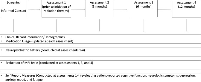
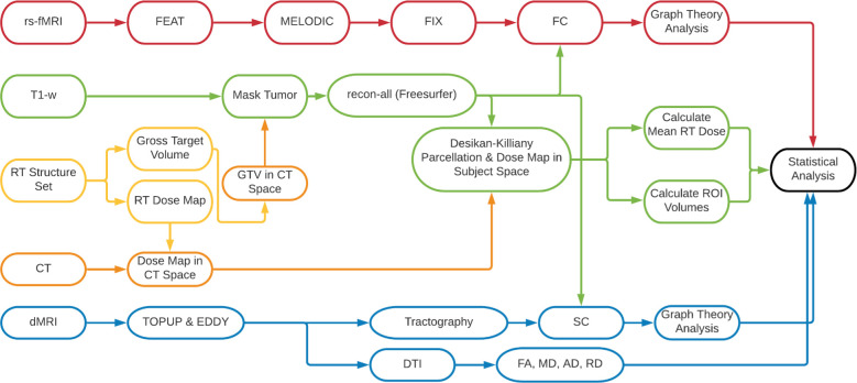
Methods and analysis: This study will enroll patients with benign and low-grade brain tumours receiving partial brain radiotherapy. Patients will receive either hypofractionated (>2 Gy/fraction) or conventionally fractionated (1.8-2 Gy/fraction) RT. All participants will be followed for 12 months, with MRIs conducted pre-RT and 6-month and 12 month post-RT, along with a battery of neurocognitive tests and questionnaires. The study was initiated in late 2018 and will continue enrolling through 2024 with final follow-ups completing in 2025. The neurocognitive battery assesses visual and verbal memory, attention, executive function, processing speed and emotional cognition. MRI protocols incorporate diffusion tensor imaging and resting state fMRI to assess structural connectivity and functional connectivity, respectively. We will estimate the association between radiation dose, imaging metrics and cognitive outcomes.
Ethics and dissemination: This study has been approved by the Research Subjects Review Board at the University of Rochester (STUDY00001512: Cognitive changes in patients receiving partial brain radiation). All results will be published in peer-reviewed journals and at scientific conferences.
Trial registration number: ClinicalTrials.gov NCT04390906.
Keywords: magnetic resonance imaging; neurological oncology; radiation oncology; radiotherapy.
© Author(s) (or their employer(s)) 2023. Re-use permitted under CC BY-NC. No commercial re-use. See rights and permissions. Published by BMJ.
Conflict of interest statement
Competing interests: None declared.
Figures
Similar articles
-
Diffusion tensor imaging predicts cognitive function change following partial brain radiotherapy for low-grade and benign tumors.Radiother Oncol. 2016 Aug;120(2):234-40. doi: 10.1016/j.radonc.2016.06.021. Epub 2016 Jul 11. Radiother Oncol. 2016. PMID: 27418525 Free PMC article.
-
Identifying early diffusion imaging biomarkers of regional white matter injury as indicators of executive function decline following brain radiotherapy: A prospective clinical trial in primary brain tumor patients.Radiother Oncol. 2019 Mar;132:27-33. doi: 10.1016/j.radonc.2018.11.018. Epub 2018 Dec 20. Radiother Oncol. 2019. PMID: 30825966 Free PMC article.
-
Microstructural Cerebellar Injury Independently Associated With Processing Speed in Adult Patients With Primary Brain Tumors: Implications for Cognitive Preservation.Int J Radiat Oncol Biol Phys. 2023 Dec 1;117(5):1107-1117. doi: 10.1016/j.ijrobp.2023.06.013. Epub 2023 Jul 4. Int J Radiat Oncol Biol Phys. 2023. PMID: 37414262 Clinical Trial.
-
Radiotherapy of high-grade gliomas: current standards and new concepts, innovations in imaging and radiotherapy, and new therapeutic approaches.Chin J Cancer. 2014 Jan;33(1):16-24. doi: 10.5732/cjc.013.10217. Chin J Cancer. 2014. PMID: 24384237 Free PMC article. Review.
-
Longitudinal assessment of chemotherapy-induced changes in brain and cognitive functioning: A systematic review.Neurosci Biobehav Rev. 2018 Sep;92:304-317. doi: 10.1016/j.neubiorev.2018.05.019. Epub 2018 May 20. Neurosci Biobehav Rev. 2018. PMID: 29791867
Cited by
-
Association of Radiation Dose to the Amygdala-Orbitofrontal Network with Emotion Recognition Task Performance in Patients with Low-Grade and Benign Brain Tumors.Cancers (Basel). 2023 Nov 23;15(23):5544. doi: 10.3390/cancers15235544. Cancers (Basel). 2023. PMID: 38067248 Free PMC article.
References
Publication types
MeSH terms
Associated data
Grants and funding
LinkOut - more resources
Full Text Sources
Medical