

Review
doi: 10.4070/kcj.2022.0247.
Q Fever Endocarditis Combined With Thrombus and Antiphospholipid Syndrome
Affiliations
- PMID: 36792561
- PMCID: PMC9932221
- DOI: 10.4070/kcj.2022.0247
Item in Clipboard
Review
Q Fever Endocarditis Combined With Thrombus and Antiphospholipid Syndrome
Korean Circ J.
2023 Feb.
No abstract available
Conflict of interest statement
The authors have no financial conflicts of interest.
Figures
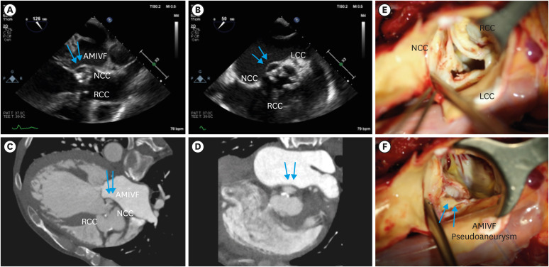
Pre-operative TEE, CT images and Intra-op findings. (A-D) Severe degenerative aortic stenosis with regurgitation and left ventricular dilatation. Outpouching structure at AMIVF with systolic expansion (blue arrows) suggesting pseudoaneurysm. (E) Thickened aortic valve leaflets were noted, especially on NCC and LCC. (F) When extracting the degenerative aortic valve leaflets, subannular pouching was seen between NCC and LCC sides without evidence of active inflammation or infection (blue arrows). AMIVF = aortomitral intervalvular fibrosa; CT = computed tomography; LCC = left coronary cusp; NCC = noncoronary cusp; TEE = transesophageal echocardiography.
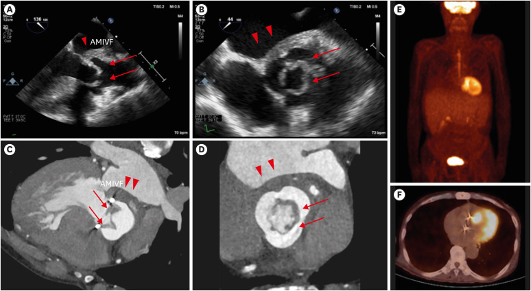
One-year follow-up TEE, CT and 18F-FDG PET-CT images. (A-D) Symmetric nodular thickening of bioprosthetic aortic valve leaflets with opening limitation (red arrows), suggestive of valve thrombus, combined with thickened left atrial wall on AMIVF side (red arrows heads); (E, F) There was no definite abnormal hypermetabolic lesion at the periprosthetic valve in 18F-FDG PET-CT, and there was only mild uptake on hepatosplenomegaly. 18F-FDG = 18F-fluorodeoxyglucose; AMIVF = aortomitral intervalvular fibrosa; CT = computed tomography; LCC = left coronary cusp; NCC = noncoronary cusp; PET = positron emission tomography.
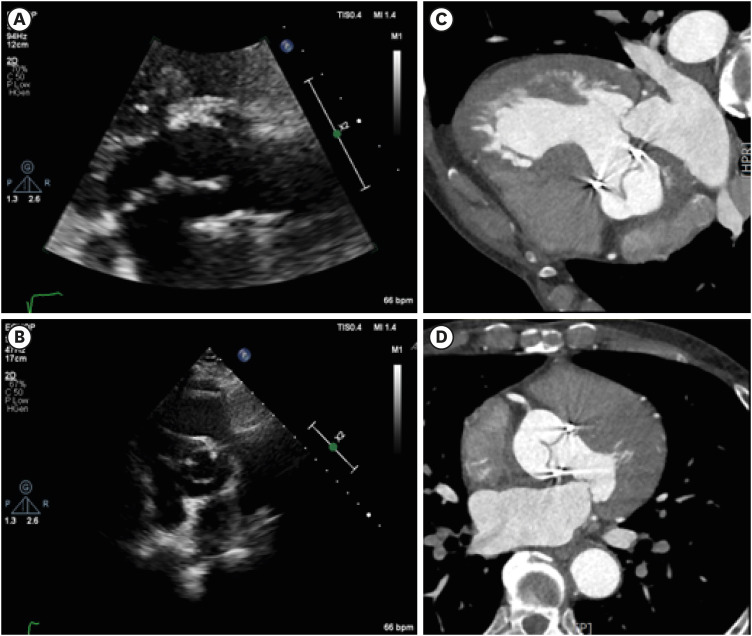
Follow-up TTE and CT images after anticoagulation and antibiotics. (A-D) Symmetric nodular thickening of bioprosthetic AV leaflets were disappeared, suggesting complete resolution of prosthetic AV thrombus. AV = aortic valve; CT = computed tomography; TTE = transthoracic echocardiography.
Publication types
LinkOut - more resources
Full Text Sources
