. 2023 Feb 15;13(1):2703.
doi: 10.1038/s41598-023-28149-3.

Anti-DEspR antibody treatment improves survival and reduces neurologic deficits in a hypertensive, spontaneous intracerebral hemorrhage (hsICH) rat model

Affiliations

Anti-DEspR antibody treatment improves survival and reduces neurologic deficits in a hypertensive, spontaneous intracerebral hemorrhage (hsICH) rat model

Victoria L M Herrera et al. Sci Rep. .

Abstract

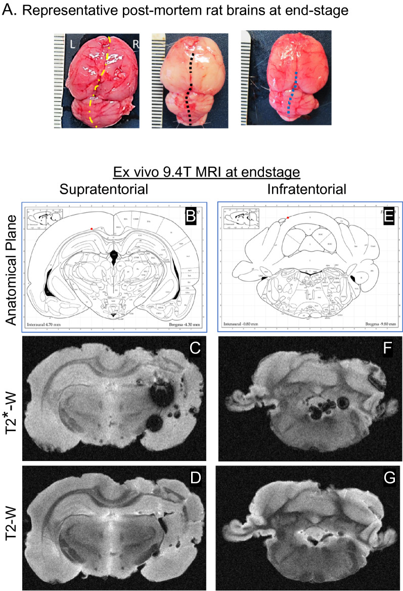
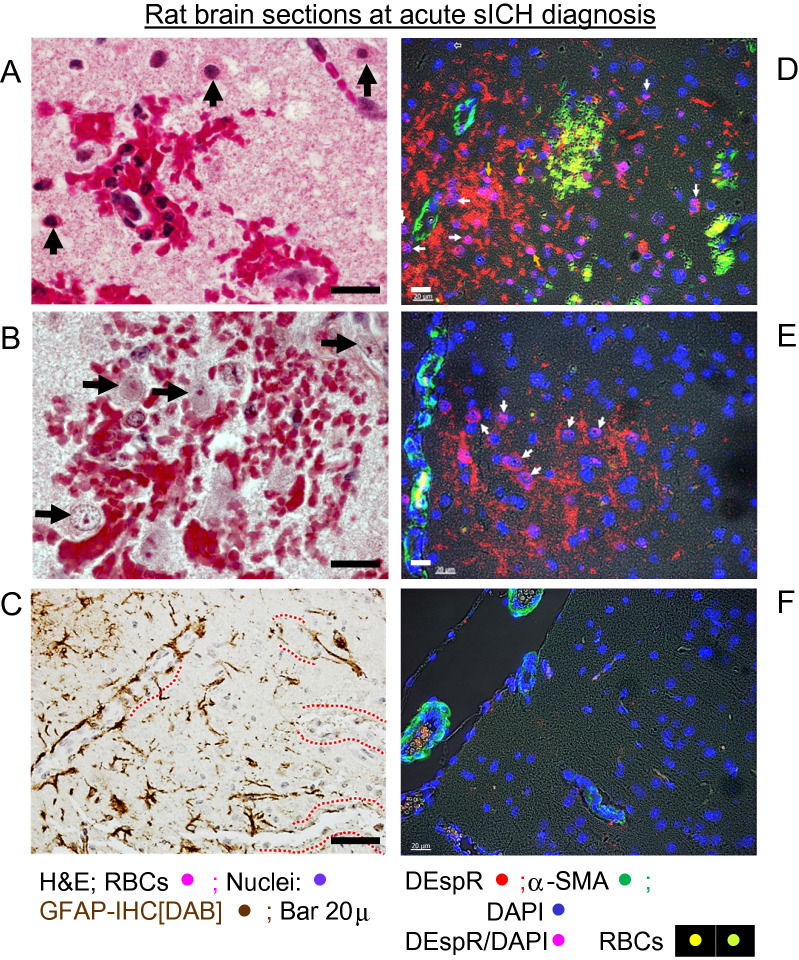
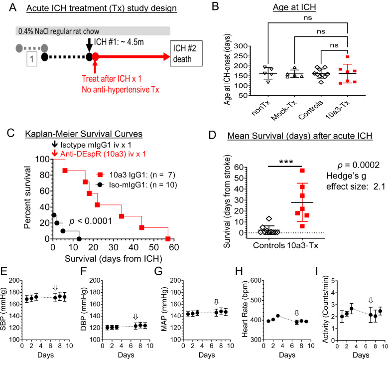
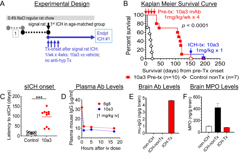
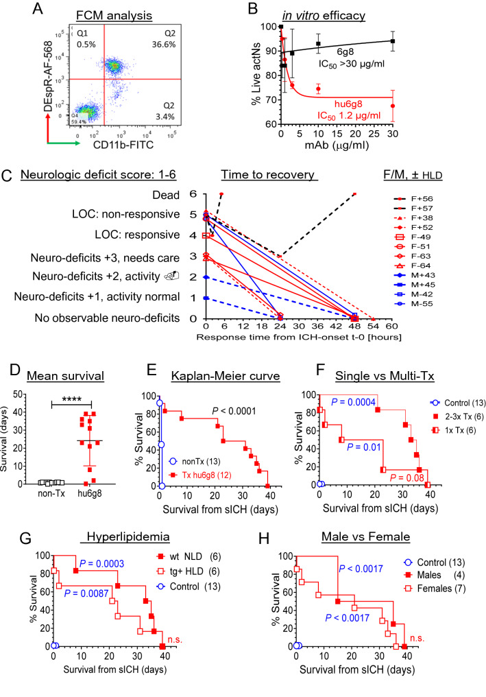
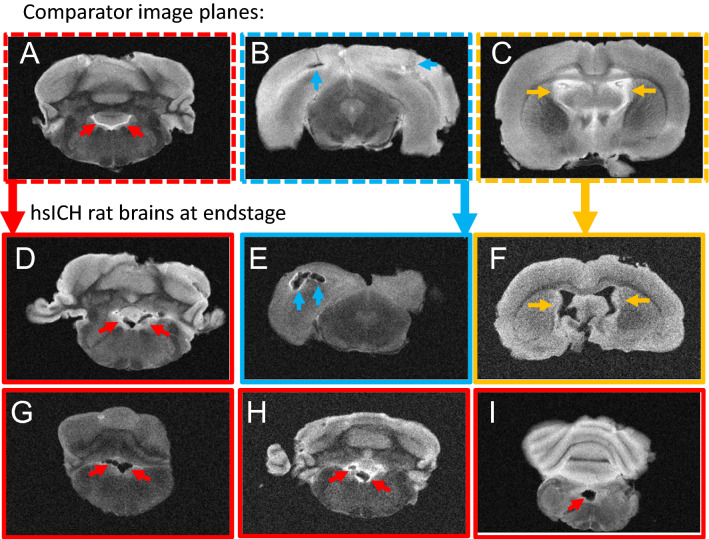
Progressive secondary brain injury-induced by dysregulated neuroinflammation in spontaneous intracerebral hemorrhage (sICH)-underlies high sICH-mortality and remains without FDA-approved pharmacotherapy. Clinical insight that hematoma-directed interventions do not improve mortality prioritizes resolving acute secondary brain injury in sICH. As neutrophils are implicated in sICH secondary brain injury, we tested whether inhibition of a rogue neutrophil-subset expressing the dual endothelin-1/signal peptide receptor (DEspR) and associated with secondary tissue injury, DEspR+ CD11b+ immunotype, will attenuate mortality in a hypertensive-sICH (hsICH) rat model. We confirmed sICH-related deaths in hsICH-rats by T2*-weighted 9.4 T MRI and DEspR+ neutrophils in hsICH-rat brain perihematomal areas by immunostaining. At acute sICH, anti-DEspR muIgG1-antibody, mu10a3, treatment increased median survival in hsICH rats vs controls (p < 0.0001). In pre-stroke sICH, weekly 10a3-treatment did not predispose to infection and delayed sICH-onset vs controls (p < 0.0001). As potential sICH-therapeutic, we tested humanized anti-DEspR IgG4S228P-mAb, hu6g8. In vitro, hu6g8 reversed delayed-apoptosis in DEspR+ CD11b+ neutrophils. In vivo, hu6g8 increased median survival and reduced neurologic symptoms in male/female hsICH-rats vs controls (p < 0.0001). Altogether, preclinical efficacy of inhibition of DEspR+ CD11b+ neutrophils in acute sICH-without infection complications, supports the potential of anti-DEspR therapy in sICH. Data provide basis for clinical study of DEspR+ CD11b+ neutrophil-subset in sICH patients.


Conflict of interest statement

VLMH and NRO: co-inventors on BU patents on DEspR and DEspR-inhibition for stroke therapy. All other authors have no competing interests.

Figures

Figure 1
Figure 1
(A) Representative post-mortem images of hsICH rat brains isolated immediately after euthanasia depicts hemorrhages. (BG) Representative ex vivo 9.4 T MRI of hsICH rat brain after ICH-related death showing sICH phenotype in supratentorial site (BD) and infratentorial site (EG) in a control rat. (B) Diagram of anatomical plane through thalamus, dorsal 3rd ventricle and lateral ventricles. (C) T2* weighted (T2*-W) intraventricular hemorrhage (IVH) in the left lateral ventricle. (D) T2-weighted (T2-W) image showing areas of perihematomal edema (PHE). (E) Diagram of anatomical plane through medulla, cerebellum, 4th ventricle and recess of 4th ventricle. (F) T2*-W gradient echo MRI showing IPH in the plane of the cerebellum and medulla, IVH in the 4th ventricle. (G) T2-W image in the same plane as (F) showing edema and IVH in the 4th ventricle and recess. MRI right, rat left.
Figure 2
Figure 2
Representative histopathological analysis of rat brain sections at sICH detection. (A) H&E staining of microbleed area with neutrophil infiltrates; ↑ pyknotic, eosinophilic neurons. (B) H&E staining of microbleed area with no neutrophils: neurons not pyknotic nor eosinophilic. (C) Immunohistochemistry-DAB staining of GFAP; dashed red line (----) mark microvessels with non-contiguous GFAP immunostaining suggesting BBB disruption. (D,E) Representative immunofluorescence staining of sICH rat brain sections at acute ICH detection; white arrows → DEspR+ neutrophils examples. Red DEspR+ anuclear fragments noted. (F) IF-staining of age-matched non-ICH Dahl S rat brain section. DEspR+ (red); RBCs, red blood cells autofluorescence (yellow-green); αSMA, alpha smooth muscle alpha actin (green) serves as positive-IF control for microvessels; DAPI, nuclear stain; merged DEspR+ DAPI+, magenta. Bar 20 microns.
Figure 3
Figure 3
Preclinical efficacy testing of single-dose anti-DEspR murine mAb prototype, 10a3, at acute sICH. (A) Diagram of experimental design of onset of treatment (anti-DEspR mAb 10a3, 50 μg/kg/dose × 1) or mock treatment (isotype murine IgG1, 50 μg/kg/dose × 1) given after first ICH (#1) documented. Monitoring until the 2nd stroke with SBP remaining at ≥ 200 mmHg; no anti-hypertensive therapy given. (B) Graph of age at onset of sICH among study groups: treated rats (n = 7), total control group (n = 10), made up of non-treated (nonTx, n = 5) and muIgG1-isotype (mock-Tx, n = 5). Kruskall Wallis ANOVA on ranks and Dunn’s multiple pairwise comparison were not-significant. (C) Kaplan–Meier Survival curves comparing treated (n = 7) and all control (n = 10) hsICH rats: median overall survival (Control vs Treated: 0.5 vs 22 days); Mantel Cox log-rank test p < 0.0001. Log rank hazard ratio 3.5, (95% CI 1.23–10.05). (D) Two-tailed unpaired Mann Whitney rank test of survival to 2nd stroke comparing controls (n = 10), mean ± sd: 2.4 ± 3.96) vs treated (n = 7: 28.9 ± 17.5), ***p = 0.0002 with Hedge’s g effect size [− 4%] = 2.1. (EI) Average 24-h blood pressure and activity measurements obtained via radio-telemetry implant system before and after single-dose anti-DEspR 10a3 treatment (↓) in naïve 4 m-old Dahl S female rats (n = 6): (E) systolic blood pressure, SBP, (F) diastolic blood pressure, DPB, (G) mean arterial pressure (MAP), (H) heart rate, and (I) activity (number of times the single-housed rat crosses the midline section of the cage).
Figure 4
Figure 4
Preclinical testing for potential infection-risk or adverse events using multi-dose anti-DEspR mAb prototype, 10a3, in imminent pre-ICH stage. (A) Diagram of experimental design: monitor for 1st sICH-event identifying “signal” rat and onset of imminent pre-sICH stage in age-matched cohort, randomize to treatment or vehicle-control group; start 10a3-treatment: 1 mg/kg/dose iv/week × 4 weeks; daily monitoring; no anti-hypertensive meds. (B) Kaplan–Meier curve analysis to onset of 1st stroke comparing controls (n = 7) and age-matched 10a3-treated (n = 10) hsICH female rats: significant differences in time to 1st sICH, Mantel-Cox log rank test p < 0.0001 with log rank hazard ratio (95% CI) 5.46 (1.2–24.4). (C) Comparison of sICH-onset from day of signal rat stroke between treated and controls: two tailed, unpaired Mann Whitney rank test: ***p = 0.0001. (D) Analysis of plasma levels of single-dose anti-DEspR murine prototype 10a3 mAb and 6g8 mAb (murine precursor for humanized anti-DEspR) in imminent pre-hsICH rats (n = 2 rats/group), each at 1 mg/kg/dose iv. (E) Brain target engagement: mouse-specific IgG levels in brain membrane bound proteins (mu-IgG ng/ gram brain) of 6g8 in pre-hsICH rat brain (ICH-Tx) compared with control non-sICH (non-ICH) and control nontreated pre-hsICH (ICH-nonTx) rat brains. (F) Brain target bioeffect: reduction of brain levels of released myeloperoxidase (MPO) and MPO + neutrophils in ICH-Tx rat brain, compared with control non-ICH and ICH-nonTx.
Figure 5
Figure 5
Characterization and preclinical efficacy study of humanized anti-DEspR antibody, hu6g8, in acute sICH. (A) Flow cytometry analysis of DEspR+ CD11b+ neutrophils in Dahl S rats induced in vivo upon TLR-4 activation by endotoxin (LPS). Gated DEspR+ /CD11b+ neutrophils depicted in quadrant 2 (Q2). (B) Concentration-dependent neutrophil survival assay comparing murine precursor 6g8 and humanized anti-DEspR hu6g8 antibodies. Efficacy defined by decrease in % live neutrophils from untreated controls. Hu6g8 IC50 = 1.2 ± 0.3 μg/ml; 6g8 IC50 > 30 μg/ml. (C) Graph of treated rat profiles: neurologic deficit score before treatment (score 1–6), time to recovery (hours) after single hu6g8 treatment (3 mg/kg iv), sex and whether normal [–] or hyper [+] lipidemic (± HLD). (D) Two-tailed Mann Whitney rank test comparing survival (days) of controls non-treated (nonTx, n = 13) and hu6g8-treated (n = 12) p < 0.0001. Hedge’s g effect size [− 4% correction] for difference in means: 2.3. (E) Kaplan–Meier survival curve of hu6g8-treated (median overall survival, mOS, 27-days [d]) vs non-treated controls (mOS 0.75d), Mantel Cox Log Rank Sum test P < 0.0001, log rank hazard ratio (95% CI) 3.2 (1.3–8.0). (F) Kaplan–Meier survival curve Log rank test followed by Holm Sidak multiple comparison testing of hu6g8-treated rats stratified for number of treatments detected P = 0.01 for single treatment (mOS 15.5d), P = 0.0004 for repeat treatment upon sICH recurrence (mOS 34d); comparison of single vs repeat treatment groups detected P = 0.08. (G) Kaplan–Meier survival curve Log rank test followed by Holm Sidak multiple comparison testing of hu6g8-treated rats stratified for hyperlipidemia (HLD) : hu6g8-treated HLD + mOS 22d vs non-treated controls 0.75d (n = 13), P = 0.0087; hu6g8-treated normolipidemic (NLD) median survival 34d (n = 6) vs non-treated controls, P = 0.0003; comparison of HDL vs NLD groups was not significant. (H) Kaplan–Meier survival curve Log rank test followed by Holm Sidak multiple comparison testing of hu6g8-treated rats stratified for sex detected P = 0.0017 for both females (mOS 21d) and males (mOS 25d) respectively compared to control non-treated hsICH rats (mOS 0.75d); comparison of males vs females was not significant.
Figure 6
Figure 6
Ex vivo T2-weighted 9.4 T MR-images of hsICH rat brains at end-stage. (AC) Representative comparator 9.4 T MR-images at the (A) plane of medulla with no blood in the 4th ventricle ( →), CSF shows white on T2-W MRI; (B) at the plane of the midbrain showing smaller IPH and PHE; and (C) at the plane of CSF-filled (white) lateral ventricles ( →) and 3rd ventricle; no IVH. (DI) Representative hsICH rat brains (n = 6) obtained after sICH-death confirming sICH as cause of death showing (D) IVH in the 4th ventricle (red →), (E) severe IPH with PHE (blue →), (F) IVH in lateral and 3rd ventricles (yellow →), (GI) IVH in the 4th ventricles (red →). Panels (AC) show comparator MR-images for Panels (DI) as marked and outlined per color.

Similar articles

References

    1. An SJ, Kim TJ, Yoon BW. Epidemiology, risk factors, and clinical features of intracerebral hemorrhage: An update. J. Stroke. 2017;19:3–10. doi: 10.5853/jos.2016.00864. - DOI - PMC - PubMed
    1. Kearns KN, et al. Neuroprotective therapies for spontaneous intracerebral hemorrhage. Neurocrit. Care. 2021;35:862–886. doi: 10.1007/s12028-021-01311-3. - DOI - PubMed
    1. Lapchak PA, Araujo DM. Advances in hemorrhagic stroke therapy: Conventional and novel approaches. Expert. Opin. Emerg. Drugs. 2007;12:389–406. doi: 10.1517/14728214.12.3.389. - DOI - PubMed
    1. Hanley DF, et al. Efficacy and safety of minimally invasive surgery with thrombolysis in intracerebral haemorrhage evacuation (MISTIE III): A randomised, controlled, open-label, blinded endpoint phase 3 trial. Lancet. 2019;393:1021–1032. doi: 10.1016/S0140-6736(19)30195-3. - DOI - PMC - PubMed
    1. The Hemorrhagic Stroke Academia Industry (HEADS) Roundtable Participants. Basic and Translational Research in Intracerebral Hemorrhage: Limitations, Priorities, and Recommendations. Stroke49, 1308–1314. 10.1161/STROKEAHA.117.019539 (2018). - PMC - PubMed

Publication types