

Evaluation of polyanionic cyclodextrins as high affinity binding scaffolds for fentanyl
- PMID: 36792632
- PMCID: PMC9932099
- DOI: 10.1038/s41598-023-29662-1
Evaluation of polyanionic cyclodextrins as high affinity binding scaffolds for fentanyl
Abstract
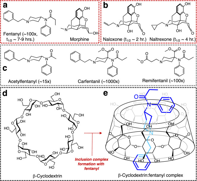
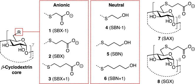
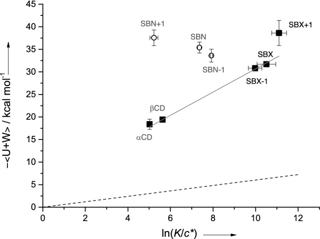
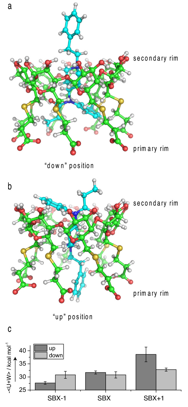
Cyclodextrins (CDs) have been previously shown to display modest equilibrium binding affinities (Ka ~ 100-200 M-1) for the synthetic opioid analgesic fentanyl. In this work, we describe the synthesis of new CDs possessing extended thioalkylcarboxyl or thioalkylhydroxyl moieties and assess their binding affinity towards fentanyl hydrochloride. The optimal CD studied displays a remarkable affinity for the opioid of Ka = 66,500 M-1, the largest value reported for such an inclusion complex to date. One dimensional 1H Nuclear Magnetic Resonance (NMR) as well as Rotational Frame Overhauser Spectroscopy (2D-ROESY) experiments supported by molecular dynamics (MD) simulations suggest an unexpected binding behavior, with fentanyl able to bind the CD interior in one of two distinct orientations. Binding energies derived from the MD simulations work correlate strongly with NMR-derived affinities highlighting its utility as a predictive tool for CD candidate optimization. The performance of these host molecules portends their utility as platforms for medical countermeasures for opioid exposure, as biosensors, and in other forensic science applications.
© 2023. This is a U.S. Government work and not under copyright protection in the US; foreign copyright protection may apply.
Conflict of interest statement
The authors declare no competing interests.
Figures

Similar articles
-
Solution-State Structure and Affinities of Cyclodextrin:Fentanyl Complexes by Nuclear Magnetic Resonance Spectroscopy and Molecular Dynamics Simulation.J Phys Chem B. 2016 Mar 10;120(9):2423-33. doi: 10.1021/acs.jpcb.5b12333. Epub 2016 Feb 18. J Phys Chem B. 2016. PMID: 26842886
-
Interaction of fentanyl with various cyclodextrins in aqueous solutions.J Pharm Pharmacol. 2016 May;68(5):588-97. doi: 10.1111/jphp.12437. Epub 2015 Jun 16. J Pharm Pharmacol. 2016. PMID: 26077960
-
Investigation of the complexation between cyclodextrins and medetomidine enantiomers by capillary electrophoresis, NMR spectroscopy and molecular modeling.J Chromatogr A. 2018 Sep 14;1567:198-210. doi: 10.1016/j.chroma.2018.06.010. Epub 2018 Jun 6. J Chromatogr A. 2018. PMID: 30055912
-
Conformational landscape of selective mu-opioid agonists in gas phase and in aqueous solution: the fentanyl series.Drug Des Discov. 2000;17(1):55-67. Drug Des Discov. 2000. PMID: 10928449 Review.
-
Combined approach using capillary electrophoresis and NMR spectroscopy for an understanding of enantioselective recognition mechanisms by cyclodextrins.Chem Soc Rev. 2004 Jul 30;33(6):337-47. doi: 10.1039/b111412n. Epub 2004 Jul 5. Chem Soc Rev. 2004. PMID: 15280966 Review.
Cited by
-
The Utility of Cyclodextrin for Countering μ-Opioid Receptor Drug Overdoses.ACS Cent Sci. 2024 Dec 10;10(12):2182-2184. doi: 10.1021/acscentsci.4c01990. eCollection 2024 Dec 25. ACS Cent Sci. 2024. PMID: 39735313 Free PMC article. No abstract available.
-
A systematic review of sensors to combat crime and routes to further sensor development.Front Chem. 2025 Jun 12;13:1568867. doi: 10.3389/fchem.2025.1568867. eCollection 2025. Front Chem. 2025. PMID: 40575464 Free PMC article. Review.
-
Cyclodextrin-Enabled Enantioselective Complexation Study of Cathinone Analogs.Molecules. 2024 Feb 16;29(4):876. doi: 10.3390/molecules29040876. Molecules. 2024. PMID: 38398627 Free PMC article.
-
Evaluation of Subetadex-α-methyl, a Polyanionic Cyclodextrin Scaffold, as a Medical Countermeasure against Fentanyl and Related Opioids.ACS Cent Sci. 2024 Oct 23;10(12):2200-2212. doi: 10.1021/acscentsci.4c00682. eCollection 2024 Dec 25. ACS Cent Sci. 2024. PMID: 39735318 Free PMC article.
References
-
- Clotz MA, Nahata MC. Clinical uses of fentanyl, sufentanil, and alfentanil. Clin. Pharm. 1991;10:581–593. - PubMed
Publication types
MeSH terms
Substances
Grants and funding
LinkOut - more resources
Full Text Sources
