. 2023 Feb 15;13(1):2685.
doi: 10.1038/s41598-023-29439-6.

Classification of electrically-evoked potentials in the parkinsonian subthalamic nucleus region

Affiliations

Classification of electrically-evoked potentials in the parkinsonian subthalamic nucleus region

Joshua Rosing et al. Sci Rep. .

Abstract

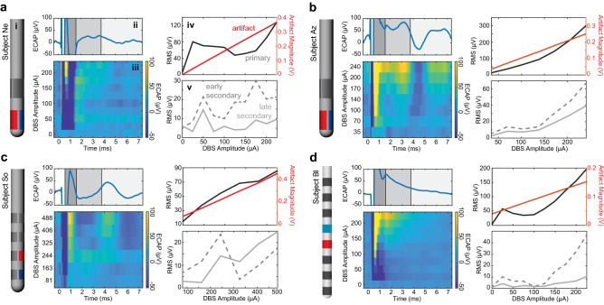
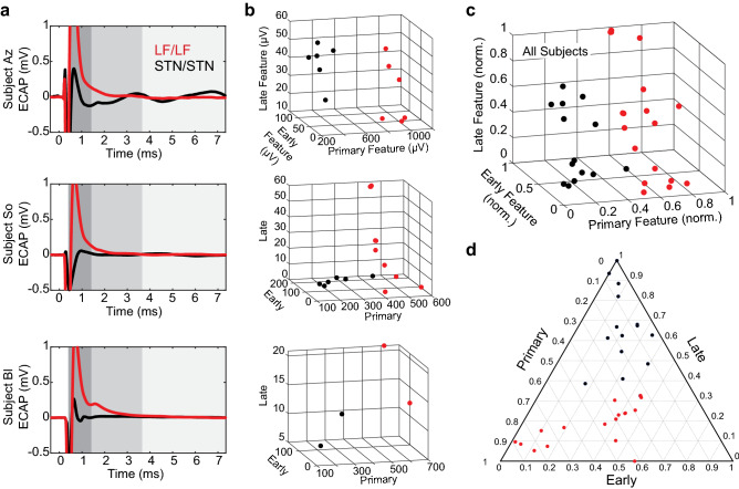
Electrically evoked compound action potentials (ECAPs) generated in the subthalamic nucleus (STN) contain features that may be useful for titrating deep brain stimulation (DBS) therapy for Parkinson's disease. Delivering a strong therapeutic effect with DBS therapies, however, relies on selectively targeting neural pathways to avoid inducing side effects. In this study, we investigated the spatiotemporal features of ECAPs in and around the STN across parameter sweeps of stimulation current amplitude, pulse width, and electrode configuration, and used a linear classifier of ECAP responses to predict electrode location. Four non-human primates were implanted unilaterally with either a directional (n = 3) or non-directional (n = 1) DBS lead targeting the sensorimotor STN. ECAP responses were characterized by primary features (within 1.6 ms after a stimulus pulse) and secondary features (between 1.6 and 7.4 ms after a stimulus pulse). Using these features, a linear classifier was able to accurately differentiate electrodes within the STN versus dorsal to the STN in all four subjects. ECAP responses varied systematically with recording and stimulating electrode locations, which provides a subject-specific neuroanatomical basis for selecting electrode configurations in the treatment of Parkinson's disease with DBS therapy.


Conflict of interest statement

The authors declare no competing interests.

Figures

Figure 1
Figure 1
DBS lead localization within the STN. (a) Directional and non-directional DBS leads used in this study. (b, c) High-field 7 T or 10.5 T MRI and CT co-registration were used to approximate the position of each lead within the subthalamic nucleus. The exact electrode position (all leads) and orientation (for the directional leads) was identified through post-mortem block-face histology and a post-mortem bubble test. These positions are shown in the figure.
Figure 2
Figure 2
Signal processing of ECAPs within and adjacent to the STN. (a) An alternating sequence of cathodic-leading and anodic-leading waveforms was applied through one electrode while ECAP recordings were collected through all adjacent electrodes. (b) Raw ECAP data were grouped, averaged, filtered, and separated into primary, early secondary, and late secondary features.
Figure 3
Figure 3
Distributions of all ECAP stimulation/recording experiments across subjects. (a) Pooled recordings across tested stimulation pulse widths (subjects Az and So only), amplitudes (all subjects), and electrode configurations (all subjects) for a single recording day. (b) Variance across pooled recordings across the ECAP window. Black lines indicate edges of selected feature windows (primary: 0.6–1.6 ms, early secondary 1.6–3.8 ms, late secondary 3.8–7.4 ms).
Figure 4
Figure 4
ECAP responses to increasing stimulation amplitudes across all subject (ad). (subpanel i) Lead diagrams for each subject with stimulation electrode shown in red and recording site shown in blue. Example ECAP response to (subpanel ii) the highest stimulation amplitude tested in the MPTP-treated state and (subpanel iii) varying stimulation current amplitudes. RMS values across stimulation amplitudes from epochs containing the (subpanel iv) stimulation artifact and primary features and (subpanel part v) secondary features.
Figure 5
Figure 5
Classification of ECAP responses within and dorsal to the STN. (a) Example ECAP responses for stimulation/recording configurations with STN/STN or LF/LF from subjects Az, So, and Bl. Subject Ne was not included in this figure as all directional DBS electrodes were within the STN. Stimulation amplitudes in each case are similar but not identical within each subject. ECAP responses are shown at a scale for visualizing primary features (dark grey). Early (middle gray) and late (light gray) secondary features were considerably smaller and played a smaller role in differentiating brain regions. (b) Plots of the three RMS features for each subject used by the classifier showing separability of spatial categories (STN/STN, LF/LF) in the feature space. (c, d) RMS values (normalized within subjects) that are plotted together to show separability of spatial categories in feature space.

References

    1. Kent AR, Grill WM. Neural origin of evoked potentials during thalamic deep brain stimulation. J. Neurophysiol. 2013;110:826–843. doi: 10.1152/jn.00074.2013. - DOI - PMC - PubMed
    1. Schmidt SL, Brocker DT, Swan BD, Turner DA, Grill WM. Evoked potentials reveal neural circuits engaged by human deep brain stimulation. Brain Stimul. 2020;13:1706–1718. doi: 10.1016/j.brs.2020.09.028. - DOI - PMC - PubMed
    1. Buzsáki G, Anastassiou CA, Koch C. The origin of extracellular fields and currents: EEG, ECoG, LFP and spikes. Nat. Rev. Neurosci. 2012;13:407–420. doi: 10.1038/nrn3241. - DOI - PMC - PubMed
    1. Kent AR, Grill WM. Recording evoked potentials during deep brain stimulation: Development and validation of instrumentation to suppress the stimulus artefact. J. Neural Eng. 2012;9:036004. doi: 10.1088/1741-2560/9/3/036004. - DOI - PMC - PubMed
    1. Cohen LT, Richardson LM, Saunders E, Cowan RSC. Spatial spread of neural excitation in cochlear implant recipients: Comparison of improved ECAP method and psychophysical forward masking. Hear. Res. 2003;179:72–87. doi: 10.1016/S0378-5955(03)00096-0. - DOI - PubMed

Publication types