. 2023 Feb 15;14(1):850.
doi: 10.1038/s41467-023-36486-0.

SVEP1 is an endogenous ligand for the orphan receptor PEAR1

Affiliations

SVEP1 is an endogenous ligand for the orphan receptor PEAR1

Jared S Elenbaas et al. Nat Commun. .

Erratum in

  • Author Correction: SVEP1 is an endogenous ligand for the orphan receptor PEAR1.
    Elenbaas JS, Pudupakkam U, Ashworth KJ, Kang CJ, Patel V, Santana K, Jung IH, Lee PC, Burks KH, Amrute JM, Mecham RP, Halabi CM, Alisio A, Di Paola J, Stitziel NO. Elenbaas JS, et al. Nat Commun. 2023 Mar 17;14(1):1511. doi: 10.1038/s41467-023-37005-x. Nat Commun. 2023. PMID: 36932061 Free PMC article. No abstract available.

Abstract

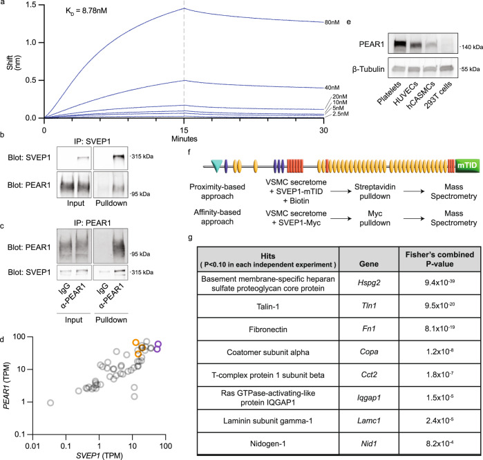
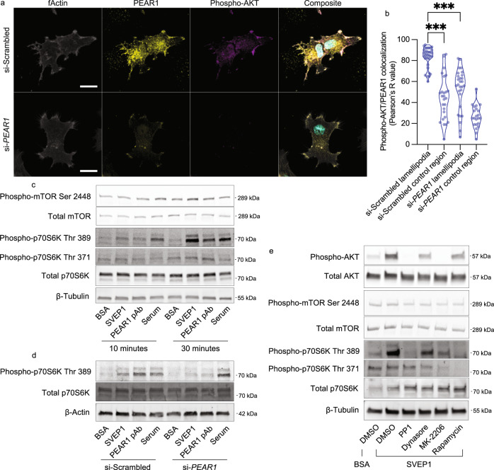
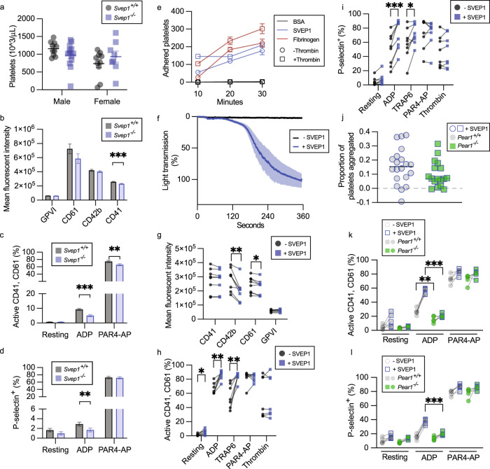
Sushi, von Willebrand factor type A, EGF and pentraxin domain containing 1 (SVEP1) is an extracellular matrix protein that causally promotes vascular disease and associates with platelet reactivity in humans. Here, using a human genomic and proteomic approach, we identify a high affinity, disease-relevant, and potentially targetable interaction between SVEP1 and the orphan receptor Platelet and Endothelial Aggregation Receptor 1 (PEAR1). This interaction promotes PEAR1 phosphorylation and disease associated AKT/mTOR signaling in vascular cells and platelets. Mice lacking SVEP1 have reduced platelet activation, and exogenous SVEP1 induces PEAR1-dependent activation of platelets. SVEP1 and PEAR1 causally and concordantly relate to platelet phenotypes and cardiovascular disease in humans, as determined by Mendelian Randomization. Targeting this receptor-ligand interaction may be a viable therapeutic strategy to treat or prevent cardiovascular and thrombotic disease.


Conflict of interest statement

N.O.S. has received investigator-initiated research funds from Regeneron Pharmaceuticals unrelated to the content of this study. J.S.E., I.H.J., A.A., and N.O.S. are co-inventors on a patent application filed by Washington University focused on SVEP1 and PEAR1. The other authors declare no competing interests.

Figures

Fig. 1
Fig. 1. PEAR1 alters plasma levels of SVEP1.
a, b Schematic of SVEP1 (a) and PEAR1 (b) proteins. Domains were identified using the Simple Modular Architecture Research Tool (SMART). Teal, von Willebrand factor type A domain; purple, putative ephrin-receptor like; yellow, complement control protein/SUSHI repeat; orange, epidermal growth factor (EGF)-like domain or calcium-binding EGF-like domain or laminin-type EGF-like domain; scissors, putative cleavage site; P, representative phosphorylation of the PEAR1 intracellular domain. Protein coding variants discussed in the main text are denoted at the corresponding peptide. c Plasma SVEP1 (aptamer 11109.56.3) as a function of allelic copies of rs147639000 (PEAR1 p.D343N) in the INTERVAL study (N = 3,301). Beta = 0.67, P = 6.5 × 10−16. Boxes (c, d) depict upper and lower quartiles with median (center line); whiskers represent maximum and minimum values. (d) Plasma PEAR1 (aptamer 8275.31.3) as a function of allelic copies of rs147639000 (PEAR1 p.D343N) in the INTERVAL study (N = 3,301). Beta = −0.18, P = 0.03. e Manhattan plot of associations between PEAR1 D343N and 2,994 plasma proteins measured in INTERVAL. Each point represents the genomic location of the gene coding for a measured protein. f Two-sample MR of estimated SNP effects (with 95% confidence intervals) on PEAR1 in deCODE (x-axis) and either PEAR1, green, or SVEP1, blue, in INTERVAL (y-axis). The causal estimate is designated by a line of the corresponding color. PEAR1 Beta = 0.86, P = 2.4 × 10−37; SVEP1 Beta = −0.66, P = 3.5 × 10−20. g Two-sample MR of estimated SNP effects (with 95% confidence intervals) on SVEP1 in deCODE (x-axis) and either PEAR1, green, or SVEP1, blue, in INTERVAL (y-axis). The causal estimate is designated by a line of the corresponding color. PEAR1 Beta = −0.07, P = 0.002; SVEP1 Beta = 0.84, P = 2.5 × 10−54.
Fig. 2
Fig. 2. SVEP1 and PEAR1 physically interact and colocalize in tissue.
a Biolayer interferometry sensorgrams. A dilution series of SVEP1 was analyzed by sensors loaded with PEAR1ECD. The dashed line represents the end of the association step and the beginning of the dissociation step. b, c Immunoblots of the indicated proteins after co-immunoprecipitation. Negative controls included no SVEP1 (b), or non-specific IgG (c). Additional details listed in “Methods” section. d Expression of PEAR1 and SVEP1 in transcripts per million (TPM) in tissues from the GTEx database. Purple circles designate adipose tissues. Orange circles designate arterial tissues. Pearson r correlation = 0.74, P = 3.5 × 10−10. e Immunoblot analysis of PEAR1 levels using cell lysates from the indicated cell-type. β-Tubulin served as a loading control. f Schematic of proximity or affinity-based proteomics experiments. g List of proteins enriched in experiments represented in f. Hits were identified as those proteins enriched at a confidence level of P < 0.10 in each experiment. Fisher’s combined p-value is a meta-analysis of the four experiments. Additional details listed in “Methods” section.
Fig. 3
Fig. 3. SVEP1 activates AKT signaling through PEAR1.
a Isolated human platelets were exposed to immobilized BSA, SVEP1, or soluble PEAR1 pAb for 15 min prior to lysis. Lysates were subjected to immunoprecipitation with an anti-PEAR1 antibody and analyzed by immunoblot assays for PEAR1 and pTyrosine. The pTyrosine signal directly overlapped with the PEAR1 signal at approximately 140 kDa. b Platelets were exposed to stimuli as described in (a). Lysates were analyzed by immunoblot assays for the indicated proteins. ce HUVECs (c), hCASMCs (d), and 293T cells (e) were exposed to stimuli before lysis and analysis by immunoblot assays for the indicated proteins. f hCASMCs were transfected with scrambled siRNA or anti-PEAR1 siRNA prior to exposure to the listed stimuli. Lysates were analyzed by immunoblot assays for the indicated proteins. g, h HUVECs were pretreated with DMSO (carrier), PP1 (SFK inhibitor, g), or Dynasore (dynamin inhibitor, h) prior to exposure to the listed stimuli. Lysates were analyzed by immunoblot assays for the indicated proteins.
Fig. 4
Fig. 4. mTOR signaling is activated by SVEP1-induced PEAR1 signaling.
a Images of hCASMCs pre-treated with scrambled siRNA or anti-PEAR1 siRNA seeded on immobilized SVEP1 for 60 min. Scale bar = 20 μm. Composite image includes DAPI (teal). b Quantification of PEAR1 and pAKT colocalization in a, as determined by the Pearson correlation coefficient. Lamellipodia were identified as bundles of fActin on the periphery of cells. Cellular regions not containing lamellipodia were used as control regions. N = 36, 26, 28, and 22 regions. ***P = 1.5 × 10−15 or P = 2.8 × 10−11 from ANOVA with post hoc unpaired, two-sided t-test. c HUVECs exposed to immobilized BSA or SVEP1, soluble PEAR1 pAb, or serum for 10 or 30 min. Lysates were analyzed by immunoblot assays for the indicated proteins. d hCASMCs were transfected with scrambled siRNA or anti-PEAR1 siRNA prior to exposure to the listed stimuli. Lysates were analyzed by immunoblot assays for the indicated proteins. e Platelets were pretreated with DMSO (carrier), PP1 (SFK inhibitor), Dynasore (dynamin inhibitor), MK-2206 (AKT inhibitor), or Rapamycin (mTOR inhibitor) prior to exposure to BSA or SVEP1. Lysates were analyzed by immunoblot assays for the indicated proteins.
Fig. 5
Fig. 5. SVEP1 activates platelets.
a Platelet counts in whole blood from Svep1+/+ and Svep1-/- mice. Data are presented as mean values ± SEM. b Platelet receptor density in whole blood from Svep1+/+ and Svep1-/- mice. Bars represent least square means and error bars represent the standard error of difference (bd). ***P = 0.0006. c Percentage of activated CD41/61+ platelets from Svep1+/+ and Svep1-/- mice at rest or upon exposure to the indicated stimulant. **P = 0.0032, ***P = 1.0 × 10−6. d Percentage of P-selectin+ platelets from Svep1+/+ and Svep1-/- mice at rest or upon exposure to the indicated stimulant. **P = 0.0038. e Adherence of platelets to BSA (negative control), SVEP1, or Fibrinogen (positive control) coated coverslips for 10–30 min with or without thrombin. Data are presented as mean values ± SEM (el). f Aggregometry of platelet rich plasma in response to SVEP1 or carrier buffer. The shaded region corresponds to ±SEM. g Human platelet receptor density in freshly isolated platelets before and after exposure to SVEP1. *P = 0.016, **P = 0.0020. h Percentage of activated human CD41/61+ platelets before and after exposure to SVEP1 at rest or upon exposure to the indicated stimulant. *P = 0.029, **P = 0.0020 or P = 0.0043. i Percentage of P-selectin+ human platelets before and after exposure to SVEP1 at rest or upon exposure to the indicated stimulant. *P = 0.027, ***P = 0.0005. j The proportion of aggregated platelets in whole blood collected from Pear1+/+ and Pear1-/- mice exposed to soluble SVEP1. k Percentage of activated CD41/61+ platelets from Pear1+/+ and Pear1-/- mice before and after exposure to SVEP1 at rest or upon exposure to the indicated stimulant. **P = 0.0051, ***P = 1.5 × 10−6. l Percentage of P-selectin+ platelets from Pear1+/+ and Pear1-/- mice before and after exposure to SVEP1 at rest or upon exposure to the indicated stimulant. ***P = 8.1 × 10−5. N for groups = 10, 10, 13, 8 mice in a; N = 21, 18, 21, 17, 21, 18, 21, 18 mice in b; N = 17, 19, 20, 19, 20, 19 mice in c; N = 20, 16, 20, 17, 20, 18 mice in d; N = 3, 5, 8, 8, 8 independent human samples in ei; N = 20 and 17 mice in j; N = 5 mice for all groups in k and l. Statistical significance calculated by two-way ANOVA (ad) or two-sided t-test (paired for gi, unpaired for k, l).
Fig. 6
Fig. 6. SVEP1 and PEAR1 causally and concordantly relate to human platelet traits and cardiovascular disease.
a Two-sample MR of estimated SNP effects (with 95% confidence intervals) on MPV (y-axis) and either PEAR1, green, or SVEP1, blue, in deCODE (x-axis). The causal estimate is designated by a line of the corresponding color. SVEP1 Beta = 0.018, P = 5.1 × 10−6; PEAR1 Beta = 0.11, P = 1.8 × 10−8. b Two-sample MR of estimated SNP effects (with 95% confidence intervals) on platelet count (y-axis) and either PEAR1, green, or SVEP1, blue, in deCODE (x-axis). The causal estimate is designated by a line of the corresponding color. SVEP1 Beta = −0.0075, P = 0.015; PEAR1 Beta = −0.048, P = 2.3 × 10−5. c Two-sample MR of estimated SNP effects (with 95% confidence intervals) on cardiovascular disease (y-axis) and either PEAR1, green, or SVEP1, blue, in deCODE (x-axis). The causal estimate is designated by a line of the corresponding color. SVEP1 Beta = 0.058, P = 4.5 × 10−12; PEAR1 Beta = 0.067, P = 0.0051. d Model of the vascular wall and lumen. Solid lines represent experimentally tested relationships. Dashed lines represent relationships supported by indirect evidence.

References

    1. Zhang Y, et al. Glyco-CPLL: an integrated method for in-depth and comprehensive N-glycoproteome profiling of human plasma. J. Proteome Res. 2020;19:655–666. doi: 10.1021/acs.jproteome.9b00557. - DOI - PubMed
    1. Gilges D, et al. Polydom: a secreted protein with pentraxin, complement control protein, epidermal growth factor and von Willebrand factor A domains. Biochem. J. 2000;352:49–59. doi: 10.1042/bj3520049. - DOI - PMC - PubMed
    1. Stitziel NO, et al. Coding variation in ANGPTL4, LPL, and SVEP1 and the risk of coronary disease. N. Engl. J. Med. 2016;374:1134–1144. doi: 10.1056/NEJMoa1507652. - DOI - PMC - PubMed
    1. Jung I-H, et al. SVEP1 is a human coronary artery disease locus that promotes atherosclerosis. Sci. Transl. Med. 2021;13:eabe0357. doi: 10.1126/scitranslmed.abe0357. - DOI - PMC - PubMed
    1. Nakada T-a, Russell JA, Boyd JH, Thair SA, Walley KR. Identification of a nonsynonymous polymorphism in the SVEP1 gene associated with altered clinical outcomes in septic shock*. Crit. Care Med. 2015;43:101–108. doi: 10.1097/CCM.0000000000000604. - DOI - PubMed

Publication types